. 2006 Apr 28;98(8):1098-105.
doi: 10.1161/01.RES.0000218454.76784.66. Epub 2006 Mar 23.

Postinfarct cytokine therapy regenerates cardiac tissue and improves left ventricular function

Affiliations

Postinfarct cytokine therapy regenerates cardiac tissue and improves left ventricular function

Buddhadeb Dawn et al. Circ Res. .

Abstract

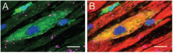
We systematically investigated the comparative efficacy of three different cytokine regimens, administered after a reperfused myocardial infarction, in regenerating cardiac tissue and improving left ventricular (LV) function. Wild-type (WT) mice underwent a 30-minute coronary occlusion followed by reperfusion and received vehicle, granulocyte colony-stimulating factor (G-CSF)+Flt-3 ligand (FL), G-CSF+stem cell factor (SCF), or G-CSF alone starting 4 hours after reperfusion. In separate experiments, chimeric mice generated by reconstitution of radioablated WT mice with bone marrow from enhanced green fluorescent protein (EGFP) transgenic mice underwent identical protocols. Mice were euthanized 5 weeks later. Echocardiographically, LV function was improved in G-CSF+FL- and G-CSF+SCF-treated but not in G-CSF-treated mice, whereas LV end-diastolic dimensions were smaller in all three groups. Morphometrically, cytokine-treated hearts had smaller LV diameter and volume. Numerous EGFP-positive cardiomyocytes, capillaries, and arterioles were noted in the infarcted region in cytokine-treated chimeric mice treated with G-CSF+FL or G-CSF+SCF, but the numbers were much smaller in G-CSF-treated mice. G-CSF+FL therapy mobilized bone marrow-derived cells exhibiting increased expression of surface antigens (CD62L and CD11a) that facilitate homing. We conclude that postinfarct cytokine therapy with G-CSF+FL or G-CSF+SCF limits adverse LV remodeling and improves LV performance by promoting cardiac regeneration and probably also by exerting other beneficial actions unrelated to regeneration, and that G-CSF alone is less effective.


Figures

Figure 1
Figure 1
A through D, Representative 2D (A and C) and M-mode (B and D) images from a vehicle-treated (A and B) and a G-CSF+FL–treated (C and D) mouse 35 days after coronary occlusion/reperfusion. Compared with the vehicle-treated heart, the G-CSF+FL–treated heart exhibits a smaller LV cavity and thicker infarct wall. Contractile activity in the infarct area is present in the G-CSF+FL–treated mouse (arrowheads) and absent in the vehicle-treated mouse. E through G, Echocardiographic assessment of LV systolic function. Echocardiographic measurements in control and cytokine-treated mice (groups I through IV) are illustrated. The administration of G-CSF+FL and G-CSF+SCF improved LV systolic function 35 days after MI. Data are mean ± SEM. n = 14 to 19 mice per group. *P < 0.05 vs group I.
Figure 2
Figure 2
Cytokine therapy and postinfarct LV remodeling. Representative Masson’s trichrome-stained myocardial sections from a control (A) and a G-CSF+FL–treated (B) WT mouse. Scar tissue and viable myocardium are identified in blue and purple, respectively. The LV cavity is smaller, and the infarcted as well as the noninfarcted walls are thicker in the G-CSF+FL–treated mouse compared with the control mouse. Panels C through E illustrate measurements of LV structural parameters in diastole by echocardiography (C and D) and morphometry (E) in groups I through IV. Administration of G-CSF+FL and G-CSF+SCF attenuated adverse LV remodeling and improved cardiac anatomy, whereas G-CSF alone had marginal effects. Data are mean ± SEM. n = 14 to 19 mice per group. *P < 0.05 vs group I.
Figure 3
Figure 3
Postinfarct cytokine therapy induces cardiomyocyte regeneration in EGFP chimeric mice. BMCs and myocytes are identified by EGFP (green) and α-sarcomeric actin (red), respectively. A through C, Low-magnification images of the infarct area in a vehicle-treated mouse show a small number of EGFP+ BMCs and rare transdifferentiation into myocytes (C, yellow, arrowhead). D through F, In contrast, images of the infarct area in a G-CSF+FL–treated mouse show numerous small myocytes (F, yellow) that are positive for both EGFP and α-sarcomeric actin. Arrowheads indicate small myocytes that are EGFP+, possibly derived from resident stem cells. G through I, Higher magnification images of the infarct area in a G-CSF+FL–treated mouse show several newly formed myocytes (I, yellow, arrowheads) positive for EGFP and α-sarcomeric actin. Nuclei are stained with 4′,6-diamidino-2-phenylindole (blue). Bar = 20 μm.
Figure 4
Figure 4
Quantitative measurement of myocyte regeneration in the infarct area and in the nonischemic area in EGFP chimeric mice (groups V through VIII). Solid bars show EGFP+ myocytes (identified by the colocalization of EGFP and α-sarcomeric actin) and hatched bars EGFP+ nonmyocytes (cells positive for EGFP but not for α-sarcomeric actin). In the ischemic zone, the majority of EGFP+ cells were myocytes; in the nonischemic zone, the reverse was observed. Data are mean ± SEM. n = 3 to 7 mice per group. *P < 0.05 vs group V; #P < 0.05 vs group VIII.
Figure 5
Figure 5
EGFP+ (A and B, green) and α-sarcomeric actin+ (B, red) small newly formed myocyte in the peri-infarct region expresses connexin 43 (A and B, magenta). Bar = 10 μm.
Figure 6
Figure 6
Assessment of cardiomyocyte proliferation after MI. Representative images of BrdU+ (A; bright blue) and Ki67+ (B; yellow) cardiomyocyte nuclei. Positivity for α-sarcomeric actin (red) identifies cardiomyocytes. C and D, Quantitative measurement of proliferating myocytes positive for BrdU (C) and Ki67 (F) at 14 days and 35 days after MI. Nuclei are stained with propidium iodide (dark blue). A and B, bar = 10 μm. C and D, data are mean ± SEM. n = 3 to 6 mice per group. *P < 0.05 vs group I; #P < 0.05 vs group IV.
Figure 7
Figure 7
Postinfarct cytokine therapy induces regeneration of vascular structures. A, A newly formed coronary arteriole within the infarcted myocardium is identified by the expression of EGFP (green) and α-smooth muscle actin (red) in smooth muscle cells (yellow-green). B, A newly formed capillary profile (arrowhead) within infarcted myocardium is identified by the expression of EGFP (green) and PECAM-1 (red) in capillary endothelial cells (yellow-green). A pre-existing capillary is identified by the arrow. Myocytes are stained in purple (α-sarcomeric actin). Nuclei are stained with 4′,6-diamidino-2-phenylindole (blue). Bar = 10 μm.
Figure 8
Figure 8
Flow cytometric assessment of adhesion molecule expression in lin/Sca-1+/c-kit+ cells from untreated bone marrow and peripheral blood of mice treated with G-CSF+FL, G-CSF+SCF, or G-CSF alone. Shown is the overlay of the adhesion molecules (red) and the isotype control (green). PB indicates peripheral blood.

Comment in

References

    1. Orlic D, Kajstura J, Chimenti S, Limana F, Jakoniuk I, Quaini F, Nadal-Ginard B, Bodine DM, Leri A, Anversa P. Mobilized bone marrow cells repair the infarcted heart, improving function and survival. Proc Natl Acad Sci U S A. 2001;98:10344–10349. - PMC - PubMed
    1. Fukuhara S, Tomita S, Nakatani T, Ohtsu Y, Ishida M, Yutani C, Kitamura S. G-CSF promotes bone marrow cells to migrate into infarcted mice heart, and differentiate into cardiomyocytes. Cell Transplant. 2004;13:741–748. - PubMed
    1. Kawada H, Fujita J, Kinjo K, Matsuzaki Y, Tsuma M, Miyatake H, Muguruma Y, Tsuboi K, Itabashi Y, Ikeda Y, Ogawa S, Okano H, Hotta T, Ando K, Fukuda K. Nonhematopoietic mesenchymal stem cells can be mobilized and differentiate into cardiomyocytes after myocardial infarction. Blood. 2004;104:3581–3587. - PubMed
    1. Wollert KC, Drexler H. Clinical applications of stem cells for the heart. Circ Res. 2005;96:151–163. - PubMed
    1. Deten A, Volz HC, Clamors S, Leiblein S, Briest W, Marx G, Zimmer HG. Hematopoietic stem cells do not repair the infarcted mouse heart. Cardiovasc Res. 2005;65:52–63. - PubMed

Publication types

MeSH terms