. 2006 Apr;133(8):1575-85.
doi: 10.1242/dev.02322.

Isl1Cre reveals a common Bmp pathway in heart and limb development

Affiliations

Isl1Cre reveals a common Bmp pathway in heart and limb development

Lei Yang et al. Development. 2006 Apr.

Abstract

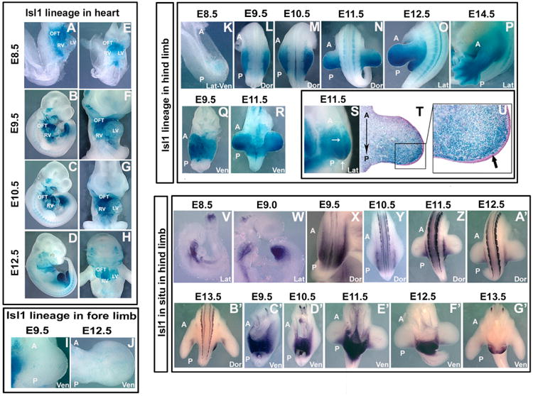
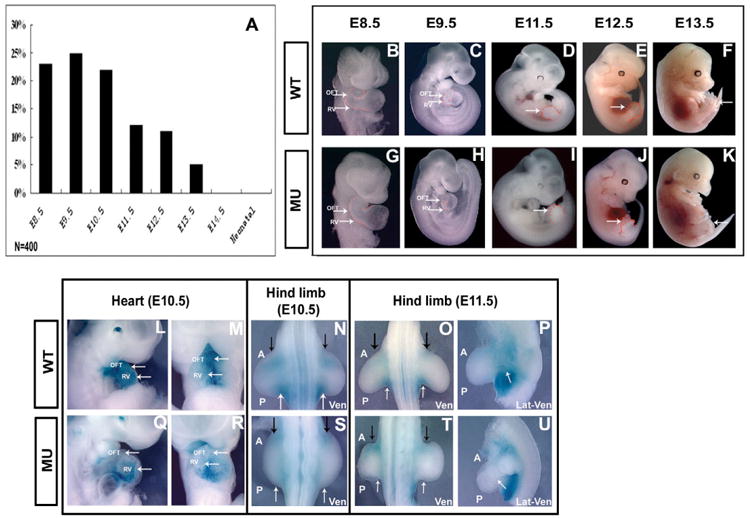
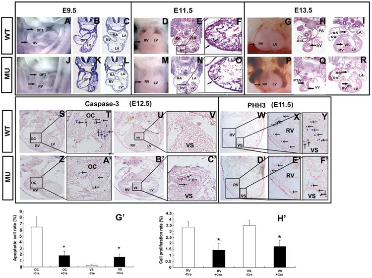
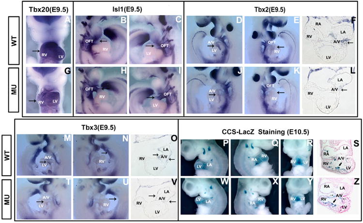
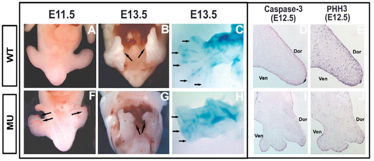
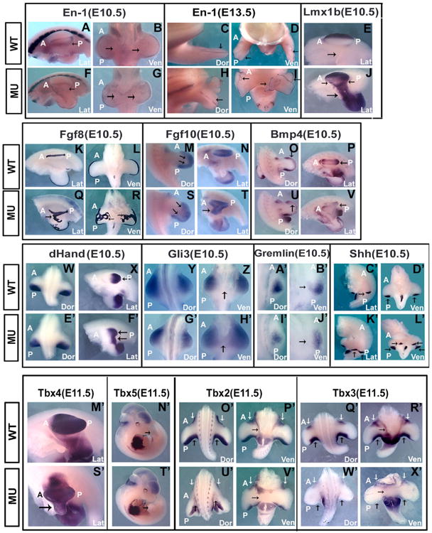
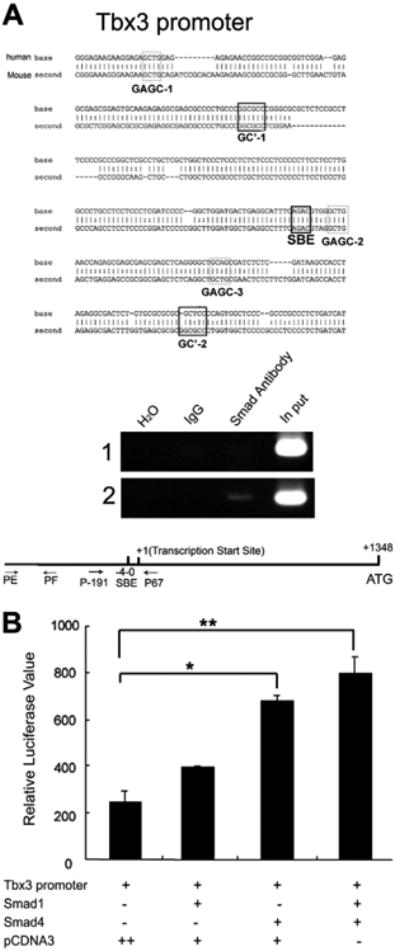
A number of human congenital disorders present with both heart and limb defects, consistent with common genetic pathways. We have recently shown that the LIM homeodomain transcription factor islet 1 (Isl1) marks a subset of cardiac progenitors. Here, we perform lineage studies with an Isl1Cre mouse line to demonstrate that Isl1 also marks a subset of limb progenitors. In both cardiac and limb progenitors, Isl1 expression is downregulated as progenitors migrate in to form either heart or limb. To investigate common heart-limb pathways in Isl1-expressing progenitors, we ablated the Type I Bmp receptor, Bmpr1a utilizing Isl1Cre/+. Analysis of consequent heart and limb phenotypes has revealed novel requirements for Bmp signaling. Additionally, we find that Bmp signaling in Isl1-expressing progenitors is required for expression of T-box transcription factors Tbx2 and Tbx3 in heart and limb. Tbx3 is required for heart and limb formation, and is mutated in ulnar-mammary syndrome. We provide evidence that the Tbx3 promoter is directly regulated by Bmp Smads in vivo.


Figures

Fig. 1
Fig. 1. Isl1 marks a subset of progenitors in heart and limb
(A-H) Isl1 lineages in heart. Lineage studies with a new Isl1Cre/+ mouse line give results consistent with those observed utilizing an Isl1-IRES-Cre mouse line (Srinivas et al., 1999; Cai et al., 2003). Excision with the new Isl1Cre/+ occurred more efficiently. Isl1-expressing progenitors contribute to most cells of the outflow tract, right ventricle and atria, and also to some cells in the left ventricle (Cai et al., 203). (I,J) Isl1 lineages in forelimb. Isl1 lineages do not contribute in any significant number to the forelimbs. (K-U) Isl1 lineages in hindlimb. A majority of cells in the hindlimb derive from Isl1-expressing cells. Isl1 cells contribute in an anterior-posterior gradient, with posterior domains deriving almost entirely from Isl1-expressing lineages. Isl1 lineages contribute to hindlimb mesoderm, not ectoderm (S-U). (V-G′) Isl1 mRNA expression in lateral plate mesoderm adjacent to and within the limb bud. A comparison of Isl1 mRNA expression to Isl1 lineage results demonstrated that Isl1 mRNA expression is downregulated as hindlimb progenitors migrate into the limb. OFT, outflow tract; RV, right ventricle; LV, left ventricle; A, anterior; P, posterior; Lat, lateral view; Dor, dorsal view; Ven, ventral view.
Fig. 2
Fig. 2. Ablation of Bmpr1a with Isl1Cre/+ results in embryonic lethality, malformations of heart and limb, and specific reduction of Bmp signaling
(A) Recovery of embryos with the Isl1Cre/+;Bmpr1a f/f genotype. Mendelian frequencies of Isl1Cre/+;Bmpr1a mutants were recovered at E10.5, but began to be lost by E11.5. No mutants were recovered at E14.5. In total 400 embryos were collected and ∼50 embryos at each stage. (B-K) Whole-mount morphological analysis of wild-type and mutant littermates. Abnormalities of outflow tract and right ventricle were evident by E8.5 (arrows, B,C,G,H); Hindlimb abnormalities were evident by E10 (N,S; arrows D-F,I-K). (L-U) Bmp signaling as monitored by Bmp-lacZ indicator genetic background. In Isl1Cre/+;Bmpr1a mutants, Bmp signaling was selectively reduced in Isl1-expressing lineages. Bmp signaling was reduced in the outflow tract and right ventricle of Isl1Cre/+;Bmpr1a mutants relative to control littermates (L,M,Q,R). In the hindlimb, Bmp signaling was strongly downregulated in the posterior limb margins (N,O,S,T) and inter hindlimb region (P,U) in Isl1Cre/+;Bmpr1a mutants relative to control littermates. OFT, outflow tract; RV, right ventricle; LV, left ventricle; A, anterior; P, posterior; Lat, lateral view; Dor, dorsal view; Ven, ventral view.
Fig. 3
Fig. 3. Aberrant cardiac morphology, apoptosis, and proliferation in Isl1Cre/+;Bmpr1a mutants
(A-R) Whole mount and section analysis of cardiac morphology. Isl1Cre/+ mutants exhibited abnormal looping of the outflow tract at E9.5 (A-CJ-L), thinning of the ventricular wall by E11.5 (D-F,M-O), and outflow tract, ventricular, and atrial septal defects by E13.5 (G-I,P-R). (S-V,Z-C′,G′) Analysis of apoptosis. Staining with antibody to activated caspase-3 revealed less cell death in the outflow tract cushions and increased cell death in the ventricular septum in Isl1Cre/+:Bmpr1a mutants relative to wild-type littermates. Arrows in T,V,A′ and C′ indicate apoptotic cells. *P<0.05. (W-Y,D′-F′,H′) Analysis of cell proliferation. Staining with antibody to phosphorylated histone H3 (PHH3) demonstrated decreased proliferation in ventricular myocardium, including the septum in Isl1Cre/+;Bmpr1a mutants relative to wild-type control littermates. Arrows in X,Y,E′ and F′ indicate proliferating cells. *P<0.05. OFT, outflow tract; RV, right ventricle; LA, left atrium; RA, right atrium; AO, aorta; VS, ventricular septum; AS, atrial septum; PTA, persistent truncus arteriosus; OC, outflow tract cushions; VV, valve; −Cre, control embryos; +Cre, mutant embryos. Arrows in F and O, indicate the ventricular wall.
Fig. 4
Fig. 4. Analysis of potential downstream effector targets of Bmp signaling in Isl1Cre/+;Bmpr1a mutants and wild-type littermate controls
(A-O,T,U,V) Whole-mount and section RNA in situ analysis of transcription factors required for cardiac development. In Isl1Cre/+;Bmpr1a mutants, expression of Tbx20 is down (A,G), Isl1 is upregulated in outflow tract (B,C,H,I); Tbx2 is downregulated in the heart and Tbx3 is downregulated both in heart and hindlimb bud (D-F,J-L,M-O,T-V), relative to somite-matched littermate controls. (P-S,W-Z) X-gal staining of a conduction system marker, CCS-lacZ in wild-type and Isl1Cre/+;Bmpr1a mutant backgrounds. Conduction system cells are formed and present in Isl1Cre/+;Bmpr1a mutants, suggesting that severe reduction of Tbx3 staining does not reflect absence of Tbx3-expressing cells. OFT, outflow tract; RV, right ventricle; LV, left ventricle; A/V, atrioventricular canal; LA, left atrium; RA, right atrium. Arrows in A,G indicate the heart; in B,C,E,H,I,K indicate the OFT; in D,F,J,L,M,O,T,V indicate A/V; and in N,U indicate the hind limb bud. In S and Z, thin arrows indicate A/V, thick arrows indicate ventricular septum.
Fig. 5
Fig. 5. Aberrant hindlimb morphology and proliferation in Isl1Cre/+;Bmpr1a mutants
(A,B,F,G) Whole-mount views of hindlimbs of wild-type and mutant embryos. Mutant limbs had ectopic outgrowths on ventral surface at right angles to endogenous limb (arrows, F,G). (C,H) Alcian Blue staining for cartilage. Cartilage structures were severely abnormal in mutant hindlimbs, with a reduced number of digit elements compared with wild-type (arrows C,H). (D,I) Apoptosis analysis. No increases in apoptosis were evident in mutant relative to wild-type hindlimb. (E,J) Cell proliferation analysis. Staining for phosphorylated histone H3 (PHH3) revealed reduced rates of proliferation in mutant hindlimbs relative to controls. Arrows in B indicate the hind limbs.
Fig. 6
Fig. 6. Gene expression analysis in hindlimbs of Isl1Cre/+;Bmpr1a mutants and wild-type littermate controls
Aberrant dorsal ventral patterning in mutants is evidenced by decreased expression of En1 in ventral ectoderm (A-D,F-I), and ventral expression of Lmx1b (E,J). En1 continues to be expressed in AER of mutants, but AER expression is sometimes disrupted or split (arrows H,I). Fgf8 expression in AER is broadened and ectopically expressed in mutant hindlimbs (K,L,Q,R). Fgf10 expression appears normal (M,N,S,T), whereas Bmp4 expression is decreased in mutants (O,P,U,V). Anterior-posterior patterning and the zone of polarizing activity appear normal in both endogenous and ectopic hindlimb structures, as evidenced by expression of Hand2 (dHand), Gli3, gremlin and Shh (W-L′). In Isl1Cre/+;Bmpr1a mutants, expression of Tbx4 and Tbx5 is unaffected (M′,N′,S′,T′); Tbx2 expression is somewhat reduced in limb and interlimb regions (arrows O′,P′,U′,V′); and Tbx3 expression is severely reduced throughout the limb (arrows Q′,R′,W′,X′). A, anterior; P, posterior; Lat, lateral view; Ven, ventral view; Dor, dorsal view. Arrows in E, Z and H′ indicate tissue between the two hind limbs.
Fig. 7
Fig. 7. The Tbx3 promoter is directly activated by Bmp Smads
(A) Bmp Smads bind to the Tbx3 promoter in vivo. (Top) A comparison of the human and mouse sequences showing a conserved consensus Smad binding site approximately 1.3 kb upstream of the ATG site within the Tbx3 promoter. Two other conserved elements (GAGC sequences and GC' sequences), previously found to be required for Smad activation of the Id1 promoter (Korchynskyi et al., 2002), were identified within the Tbx3 promoter region. (Bottom) In vivo ChIP analysis utilizing extracts from embryonic hindlimb and an antibody specific for Bmp Smads demonstrated specific binding of Bmp Smads to a region containing the Smad binding element (SBE; 2). Control primers flanking an unrelated region demonstrated no specific binding with Bmp Smads (A; 1). No recruitment was found with IgG. (B) Transient transfection analysis in P19 cells. A 2 kb fragment of the Tbx3 promoter region driving luciferase expression was cotransfected into P19 cells with control vector alone (pCDNA3), or expression constructs for Smad1, Smad4, or both Smad1 and Smad4. The Tbx3 promoter exhibited a significant increase in relative luciferase activity in response to Smad1/Smad4. **P<0.01, *P<0.05, paired t-test.

References

    1. Ahn K, Mishina Y, Hanks MC, Behringer RR, Crenshaw EB., 3rd BMPR-IA signaling is required for the formation of the apical ectodermal ridge and dorsal-ventral patterning of the limb. Development. 2001;128:4449–4461. - PubMed
    1. Ahn S, Joyner AL. Dynamic changes in the response of cells to positive hedgehog signaling during mouse limb patterning. Cell. 2004;118:505–516. - PubMed
    1. Bardot B, Lecoin L, Fliniaux I, Huillard E, Marx M, Viallet JP. Drm/Gremlin, a BMP antagonist, defines the interbud region during feather development. Int J Dev Biol. 2004;48:149–156. - PubMed
    1. Barr M, Jr, Blackburn WR, Cooley NR., Jr Human fetal somatic and visceral morphometrics. Teratology. 1994;49:487–496. - PubMed
    1. Basson CT, Bachinsky DR, Lin RC, Levi T, Elkins JA, Soults J, Grayzel D, Kroumpouzou E, Traill TA, Leblanc-Straceski J, et al. Mutations in human TBX5 [corrected] cause limb and cardiac malformation in Holt-Oram syndrome. Nat Genet. 1997;15:30–35. - PubMed

Publication types

MeSH terms

Substances