. 2006 Apr;116(4):1016-24.
doi: 10.1172/JCI21545. Epub 2006 Mar 23.

Induction of myasthenia by immunization against muscle-specific kinase

Affiliations

Induction of myasthenia by immunization against muscle-specific kinase

Kazuhiro Shigemoto et al. J Clin Invest. 2006 Apr.

Abstract

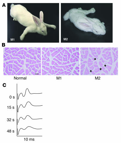
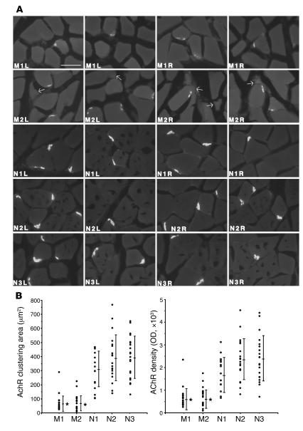
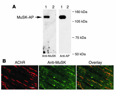
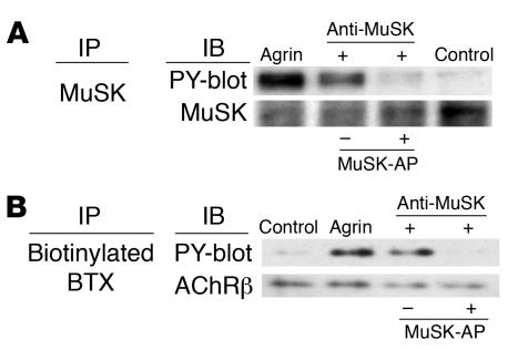
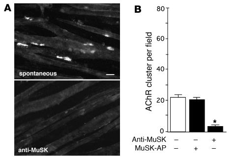
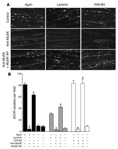
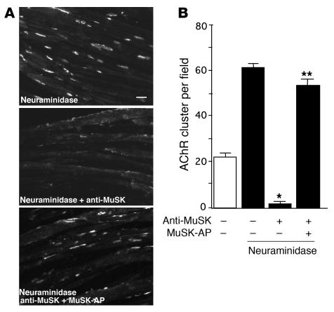
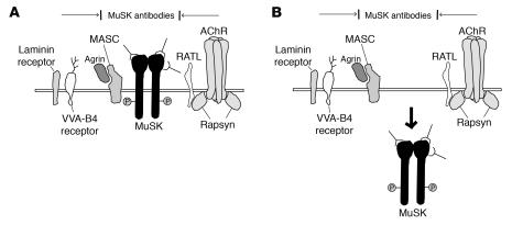
Muscle-specific kinase (MuSK) is critical for the synaptic clustering of nicotinic acetylcholine receptors (AChRs) and plays multiple roles in the organization and maintenance of neuromuscular junctions (NMJs). MuSK is activated by agrin, which is released from motoneurons, and induces AChR clustering at the postsynaptic membrane. Although autoantibodies against the ectodomain of MuSK have been found in a proportion of patients with generalized myasthenia gravis (MG), it is unclear whether MuSK autoantibodies are the causative agent of generalized MG. In the present study, rabbits immunized with MuSK ectodomain protein manifested MG-like muscle weakness with a reduction of AChR clustering at the NMJs. The autoantibodies activated MuSK and blocked AChR clustering induced by agrin or by mediators that do not activate MuSK. Thus MuSK autoantibodies rigorously inhibit AChR clustering mediated by multiple pathways, an outcome that broadens our general comprehension of the pathogenesis of MG.


Figures

Figure 1
Figure 1. Rabbits manifest MG-like paresis after immunization with MuSK protein.
(A) Two rabbits, representative of the 4 animals that showed myasthenia, manifested myasthenic weakness after immunization with the recombinant MuSK protein. After 3 injections of MuSK protein, M1 and M2 rabbits manifested flaccid weakness within 3 and 9 weeks, respectively. The M2 rabbit developed severe exhaustion with muscle weakness. (B) Cross sections from the soleus muscles of 2 paretic (M1 and M2) and 1 normal rabbit were stained with H&E. Muscle fibers in the M1 paretic rabbit showed only subtle changes in shape and smallness, whereas an atrophy of muscle fibers in the M2 paretic rabbit was observed as small angular fibers (arrows). Scale bar: 50 μm. (C) Electromyograms recorded from the M1 paretic rabbit. The retroauricular branch of the facial nerve was continuously stimulated by a constant-current stimulator, which delivered square-wave pulses of 0.1 ms at 20 Hz, and the compound muscle action potential (the second peak observed on the oscilloscope screen was recorded at the indicated time points during stimulation) showed a decremental pattern, consistent with MG.
Figure 2
Figure 2. Reduction of the size and density of AChR clusters at the NMJs in paretic rabbits.
(A) Cross sections from the soleus muscles of 2 paretic (M1 and M2) and 3 normal rabbits (N1, N2, and N3) were stained with 10 nM rhodamine-conjugated BTX. Bright crescents of bound BTX, indicative of endplate AChR, were smaller and less intense in the paretic rabbits’ muscle fibers than in those of the normal rabbits. Arrows indicate the small angular fibers in M2 soleus muscles. L, left; R, right. Scale bar: 50 μm. (B) Images of 10 AChR clusters at NMJs in the right and 10 in the left soleus muscles of the paretic and normal rabbits were randomly recorded by a digital imaging camera. Quantification of the area and intensity of AChR clustering in the unprocessed images were measured using NIH Image software. Bars indicate mean ± SD. *P < 0.01 versus normal rabbits.
Figure 3
Figure 3. MuSK autoantibodies specifically bind MuSK.
(A) Detection of secreted MuSK-AP from COS-7 cells by Western blot analysis with MuSK autoantibodies. The culture supernatants from COS-7 cells transfected with MuSK-AP (lane 1) or vector alone (lane 2) were immunoblotted with the MuSK antibodies, and the same immmunoblots were reprobed with an anti–alkaline phosphatase (anti-AP) antibody. (B) Specific binding of MuSK autoantibodies to MuSK in C2C12 myotubes. Myotubes were treated with agrin (1 nM for 18 h) and double labeled, first with rhodamine-conjugated BTX (left), and then with MuSK antibodies followed by fluorescein-conjugated secondary antibodies (middle). AChRs and MuSK colocalized, as shown by the overlay of red and green fluorescence (right). Green blotches are artifact. Scale bar: 20 μm.
Figure 4
Figure 4. MuSK antibodies specifically activate MuSK and the downstream cascade.
Specific phosphorylation of MuSK and AChR β subunit (AChRβ) by MuSK antibodies. C2C12 myotubes were treated with agrin or MuSK antibodies for 30 minutes and then immunoprecipitated with (A) MuSK antibodies or (B) biotinylated BTX using streptavidin-Sepharose. Immunoblots of immunoprecipitates were probed with antibodies to MuSK (A), AChRβ (B), or phosphotyrosine (PY, A and B). Both MuSK and AChRβ were phosphorylated after the treatment of agrin or MuSK antibodies. Phosphorylation of MuSK and AChRβ was inhibited by the absorption of MuSK antibodies with MuSK-AP protein.
Figure 5
Figure 5. Inhibition of spontaneous AChR clustering by MuSK antibodies.
(A) AChR clusters on C2C12 myotubes were stained with rhodamine-conjugated BTX with or without MuSK antibodies. Scale bar: 20 μm. (B) Quantification of the inhibitory activity of the MuSK antibodies showed that these antibodies provided significant inhibition of spontaneous AChR clustering (*P < 0.01, ANOVA). Values represent mean ± SEM of 10–15 fields for each of the 2 experiments per treatment.
Figure 6
Figure 6. Inhibition of agrin-induced and agrin-independent AChR clustering by MuSK antibodies.
(A) C2C12 cells were treated with agrin, laminin-1, or VVA-B4. AChR clusters were stained with rhodamine-conjugated BTX. AChR clustering induced by agrin, laminin-1, and VVA-B4 was inhibited in the presence of MuSK antibodies. This inhibition was blocked by absorption of the MuSK antibodies with MuSK-AP before treatment of the cells. Scale bar: 20 μm. (B) Quantification of the inhibitory activity of the MuSK antibodies confirmed that they significantly inhibited agrin-, laminin-1–, and VVA-B4–induced AChR clustering. Preabsorption of the MuSK antibodies with MuSK-AP significantly blocked inhibition. Values represent mean ± SEM of 10–15 fields for each of the 2 experiments per treatment. *P < 0.01 versus similar treatment without MuSK antibodies; #P < 0.01 versus similar treatment without preabsorption; ANOVA.
Figure 7
Figure 7. Inhibition of neuraminidase-induced AChR clustering by MuSK antibodies.
(A) C2C12 cells were treated with Clostridium perfringens neuraminidase. AChR clusters were stained with rhodamine-conjugated BTX. AChR clustering induced by neuraminidase was inhibited in the presence of the MuSK antibodies. This inhibition was blocked by absorption of the MuSK antibodies with MuSK-AP before treatment of the cells. Scale bar: 20 μm. (B) Quantification of the significant inhibition of neuraminidase-induced AChR clustering by MuSK antibodies. Inhibition was significantly blocked by preabsorption of the MuSK antibodies with 6.5 nM MuSK-AP. Values represent mean ± SEM of 10–15 fields for each of the 2 experiments per treatment. *P < 0.01 versus treatment without MuSK antibodies; **P < 0.01 versus treatment without preabsorption; ANOVA.
Figure 8
Figure 8. Models of MuSK antibody inhibition of AChR clustering on myotubes.
(A) MuSK antibodies cover the ectodomain of MuSK and inhibit interactions with other scaffolding molecules required for AChR clustering, including myotube-associated component (MASC), rapsyn-associated transmembrane linker (RATL), and receptors for VVA-B4 and laminin. MASC is a hypothetical molecule that is required for agrin binding and the activation of MuSK. RATL, also a hypothetical molecule, mediates the association of rapsyn with the extracellular domain of MuSK. (B) MuSK antibodies induce the internalization of MuSK in the presence or absence of associated molecules. The lack of MuSK on the surfaces of myotubes inhibits the effects of scaffolding and AChR clustering.

References

    1. Lindstrom J.M., Lennon V.A., Seybold M.E., Whittingham S. Experimental autoimmune myasthenia gravis and myasthenia gravis: biochemical and immunochemical aspects. Ann. N. Y. Acad. Sci. 1976;274:254–274. - PubMed
    1. Hoch W., et al. Auto-antibodies to the receptor tyrosine kinase MuSK in patients with myasthenia gravis without acetylcholine receptor antibodies. Nat. Med. 2001;7:365–368. - PubMed
    1. Evoli A., et al. Clinical correlates with anti-MuSK antibodies in generalized seronegative myasthenia gravis. Brain. 2003;126:2304–2311. - PubMed
    1. McConville J., et al. Detection and characterization of MuSK antibodies in seronegative myasthenia gravis. Ann. Neurol. 2004;55:580–584. - PubMed
    1. Ohta K., et al. MuSK antibodies in AChR Ab-seropositive MG vs AChR Ab-seronegative MG. Neurology. 2004;62:2132–2133. - PubMed

Publication types

MeSH terms