. 2006 Apr;116(4):905-15.
doi: 10.1172/JCI26836. Epub 2006 Mar 23.

Induction and blockage of oligodendrogenesis by differently activated microglia in an animal model of multiple sclerosis

Affiliations

Induction and blockage of oligodendrogenesis by differently activated microglia in an animal model of multiple sclerosis

Oleg Butovsky et al. J Clin Invest. 2006 Apr.

Abstract

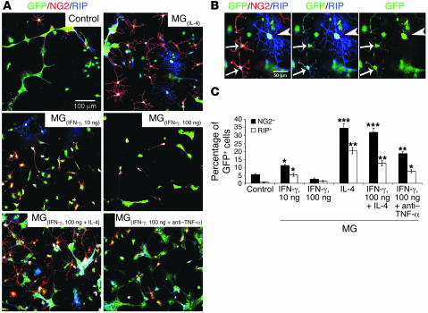
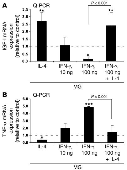
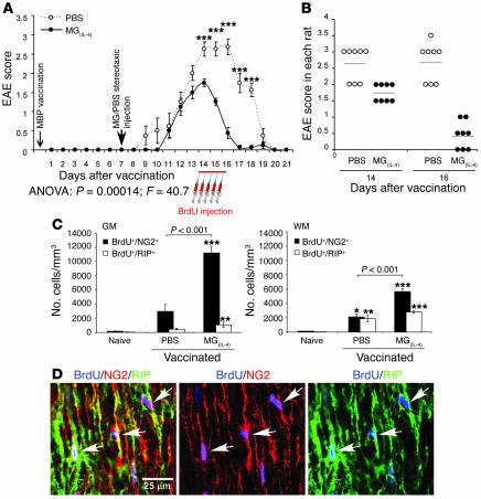
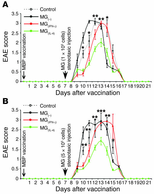
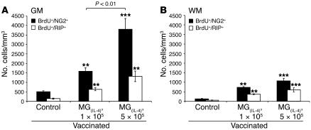
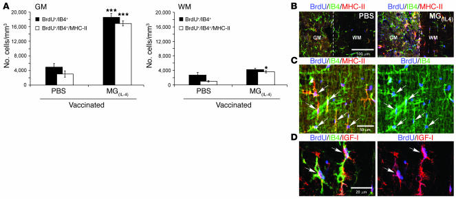
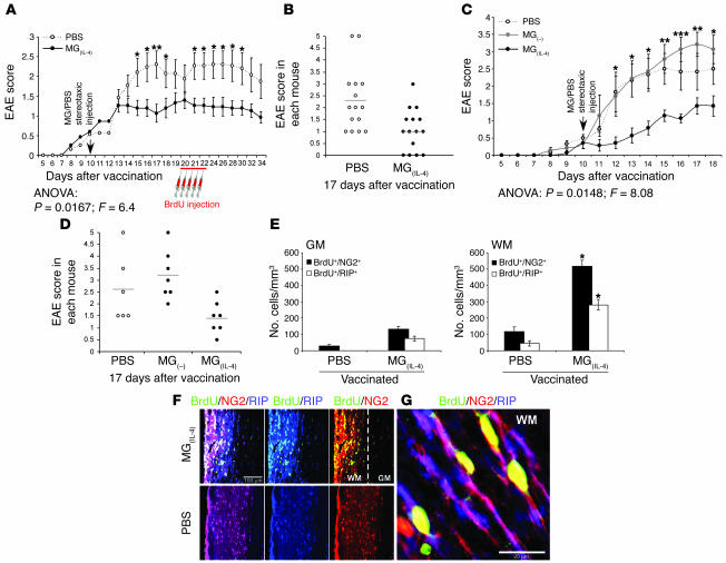
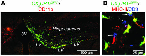
The role of activated microglia (MG) in demyelinating neurodegenerative diseases such as multiple sclerosis is controversial. Here we show that high, but not low, levels of IFN-gamma (a cytokine associated with inflammatory autoimmune diseases) conferred on rodent MG a phenotype that impeded oligodendrogenesis from adult neural stem/progenitor cells. IL-4 reversed the impediment, attenuated TNF-alpha production, and overcame blockage of IGF-I production caused by IFN-gamma. In rodents with acute or chronic EAE, injection of IL-4-activated MG into the cerebrospinal fluid resulted in increased oligodendrogenesis in the spinal cord and improved clinical symptoms. The newly formed oligodendrocytes were spatially associated with MG expressing MHC class II proteins and IGF-I. These results point to what we believe to be a novel role for MG in oligodendrogenesis from the endogenous stem cell pool.


Figures

Figure 1
Figure 1. High-dose IFN-γ, by inducing TNF-α, inhibits the ability of MG to support oligodendrogenesis, whereas MG activated by IL-4 can overcome the inhibition.
(A) GFP-expressing NPCs (green) were cultured for 10 days without MG (control) or cocultured for 10 days with MG(IL-4), MG(IFN-γ, 10 ng/ml), MG(IFN-γ, 100 ng/ml), MG activated by both IFN-γ (100 ng/ml) and IL-4 (10 ng/ml), or MG(IFN-γ, 100 ng/ml) in the presence of anti–TNF-α (1 ng/ml). (B) Colocalization of GFP, NG2, and pre-ensheathing marker of oligodendrocytes RIP. Arrows show the same cells expressing the indicated markers. Arrowhead shows a highly branched GFP+/RIP+ oligodendrocyte. Separate confocal channels are shown in 2 right panels. (C) Quantification of NG2+ or RIP+ cells (expressed as a percentage of GFP+ cells) obtained from confocal images. Data are from 2 independent experiments in replicate cultures; bars represent mean α SD. *P < 0.05, **P < 0.01, ***P < 0.001 versus control (2-tailed Student’s t test). The P values indicated in the figure represent a comparison of the groups as analyzed by ANOVA.
Figure 2
Figure 2. IL-4 partially reverses downregulation of IGF-I expression and upregulation of TNF-α expression in MG activated by 100 ng/ml IFN-γ.
(A) Q-PCR of MG identical to those described in Figure 1 24 hours after treatment. A significant increase in IGF-I was seen in MG activated by 10 ng/ml IL-4. (B) TNF-α transcripts in MG activated by 100 ng/ml IFN-γ concomitantly with 10 ng/ml IL-4 compared with IFN-γ–activated MG. Values represent the relative amounts of amplified mRNA normalized against β-actin and are expressed as fold of induction relative to untreated control MG (dashed line). Data are from 2 independent experiments in replicate cultures; bars represent mean α SD. *P < 0.05, **P < 0.01, ***P < 0.001 versus control; ANOVA.
Figure 3
Figure 3. Intraventricularly injected MG(IL-4) significantly improves the clinical symptoms of acute EAE and induces oligodendrogenesis in rats.
On day 7 after MBP vaccination, rat brain lateral ventricles were stereotaxically injected bilaterally with PBS or with syngeneic MG(IL-4) (n = 8 per group). From day 14, BrdU was injected for 2.5 days. Naive rats received the same course of BrdU injection (n = 4). Spinal cords were excised 7 days after the first BrdU injection (21 days after immunization). (A) EAE scores in rats treated either with MG(IL-4) or with PBS (n = 8 per group). Data are mean α SEM. ***P < 0.001; Student’s t test. (B) EAE scores of individual rats 14 and 16 days after vaccination. (C) NG2+ or RIP+ cells colabeled with BrdU+ cells were quantitatively analyzed both in gray matter (GM) and in white matter (WM) at 300-μm intervals along longitudinal 30-μm sagittal sections of spinal cord (T8–T9; n = 6–8 per group). Data are mean α SEM. *P < 0.05, **P < 0.01, ***P < 0.001 versus naive; ANOVA. (D) Representative confocal images of an area taken from the white matter of an MG(IL-4)-treated rat. Newly formed oligodendrocytes were identifiable by colocalization of BrdU and NG2 or RIP (arrows). Separate confocal channels are shown in 2 right panels.
Figure 4
Figure 4. Effect of MG on the clinical course of EAE depends on their number and activity.
On day 7 after induction of EAE in rats as described in Figure 3, their brain lateral ventricles were stereotaxically injected bilaterally with syngeneic MG(–), MG(IFN-γ), or MG(IL-4) (n = 6 per group). One group of rats with EAE remained untreated and served as a control (n = 6). From day 10, BrdU was injected for 2.5 days. Spinal cords were excised 11 days after the first BrdU injection, by which time disease in the rats of all groups was resolved. (A) EAE scores in rats injected stereotaxically with low-dose (1 × 10⁵ cells in 5 μl PBS for 5 minutes) of differently activated MG. (B) EAE scores in rats injected stereotaxically with high doses (5 × 10⁵ cells in 5 μl PBS for 5 minutes). Data are mean α SEM. *P < 0.05, **P < 0.01, ***P < 0.001, MG(IL-4) versus control; Student’s t test. Significant differences (2-factor repeated measures ANOVA) were found between the MG(–) and MG(IL-4) groups: (A) P = 0.0001, F = 61.1; (B) P = 0.0001, F = 79.4.
Figure 5
Figure 5. Dose dependency of oligodendrogenesis induced in MG(IL-4) -injected rats.
NG2+ or RIP+ cells colabeled with BrdU+ cells were quantitatively analyzed in (A) gray matter and (B) white matter at 300-μm intervals along longitudinal 30-μm sagittal sections of spinal cord (T8–T9) from MBP-vaccinated rats injected with low- or high-dose MG(IL-4) (n = 5–6 per group) as indicated. Data are expressed as mean α SEM. **P < 0.01, ***P < 0.001 versus control; ANOVA).
Figure 6
Figure 6. Rats injected intraventricularly with MG(IL-4) exhibit increased microglial proliferation and MHC class II (MHC-II) expression.
The spinal cords analyzed in Figure 3 were also examined for microgliogenesis. (A) Quantitative analysis of IB4+ and IB4+/MHC class II+ cells colabeled with BrdU+ (mean α SEM) from both gray matter and white matter (n = 8 per group). *P < 0.05, ***P < 0.001 versus PBS; 2-tailed Student’s t test. (B) Representative confocal microscopy of longitudinal sagittal sections of spinal cords (T8–T9) stained with BrdU and costained with IB4 for MG and MHC class II 21 days after injection with PBS or with MG(IL-4). The most abundant populations of BrdU+ cells were immunoreactive to IB4 in both control and MG(IL-4)-injected rats. Significantly more MHC class II+ cells were seen, especially in the gray matter, in slices from MG(IL-4)-injected rats than from those of PBS-injected control rats. Note that the images are from areas that include both gray matter and white matter; dashed line shows the border between the 2 areas. (C and D) Most of the newly formed MG (IB4+/BrdU+) in MG(IL-4)-treated rats coexpressed (C) MHC class II and (D) IGF-I (arrows).
Figure 7
Figure 7. In mice with chronic EAE, intraventricularly injected MG(IL-4) significantly improves clinical features and induces oligodendrogenesis.
Spinal cords were excised 12 days after the last BrdU injection. (A and B) EAE scores in mice injected with either MG(IL-4) or PBS (n = 15 per group). *P < 0.05, **P < 0.01; Student’s t test. (C and D) Lack of beneficial effect of MG(–). In an independent experiment, mice with EAE were injected with MG(IL-4) (n = 7), MG(–) (n = 7), or PBS (n = 6) on day 10 after MOG vaccination. *P < 0.05, **P < 0.01, ***P < 0.001, MG(–) versus MG(IL-4); Student’s t test. ANOVA revealed no significant effect of MG(–) injection relative to PBS injection, whereas MG(IL-4) had a significant effect. Shown are (A and C) mean α SEM and (B and D) individual EAE scores on day 17. (E) NG2+ and RIP+ cells colabeled with BrdU+ cells were quantitatively analyzed at 300-μm intervals in both gray matter and white matter of the spinal cord (n = 4 per group). Data are mean α SEM. *P < 0.05 versus PBS; 2-tailed Student’s t test. (F) Representative confocal images of longitudinal sections of spinal cords stained with BrdU and costained for NG2 and RIP 34 days after immunization in MG(IL-4)- and PBS-treated mice. Separate confocal channels are shown in 2 right panels. (G) Newly formed oligodendrocytes were identifiable by colocalization of BrdU, NG2, and RIP in the white matter of MG(IL-4)-treated mice.
Figure 8
Figure 8. Distribution of CX3 CR1/GFP/+ MG 9 days after stereotaxic injection.
(A) Localization of GFP+ cells colabeled with the microglial marker CD11b in the right lateral ventricle (LV) after injection of MG(IL-4) from CX3CR1/GFP/+ mice into mice with EAE. Note the hippocampal area in the MG(IL-4)-injected mice was heavily populated by MG(IL-4)-GFP+ cells. (B) MG(IL-4)-GFP+ cells coexpressing MHC class II populated the spinal cord at level T8–T9 adjacent to the ependyma of the central canal associated with CD3+ cells (arrows). 3V, third ventricle.

References

    1. Eriksson P.S., et al. Neurogenesis in the adult human hippocampus. Nat. Med. 1998;4:1313–1317. - PubMed
    1. Horner P.J., et al. Proliferation and differentiation of progenitor cells throughout the intact adult rat spinal cord. J. Neurosci. 2000;20:2218–2228. - PMC - PubMed
    1. Monje M.L., Toda H., Palmer T.D. Inflammatory blockade restores adult hippocampal neurogenesis. Science. 2003;302:1760–1765. - PubMed
    1. Ekdahl C.T., Claasen J.H., Bonde S., Kokaia Z., Lindvall O. Inflammation is detrimental for neurogenesis in adult brain. Proc. Natl. Acad. Sci. U. S. A. 2003;100:13632–13637. - PMC - PubMed
    1. Hartung H.P., et al. Inflammatory mediators in demyelinating disorders of the CNS and PNS. . J. Neuroimmunol. 1992;40:197–210. - PubMed

Publication types

MeSH terms