

Review
doi: 10.1002/jcp.20634.
Survivin study: an update of "what is the next wave"?
Affiliations
- PMID: 16557517
- PMCID: PMC2821201
- DOI: 10.1002/jcp.20634
Item in Clipboard
Review
Survivin study: an update of "what is the next wave"?
J Cell Physiol.
2006 Sep.
Abstract
Studies on survivin over the past 2-3 years have shown that survivin possesses multiple subcellular localizations and is a multifunctional molecule involved in many aspects of cellular processes and/or behaviors. The subcellular localization and function of the survivin splice variants, however, have not yet been well elucidated. We have, therefore, provided additional observations on several survivin splice variants for further exploration. This review article will update the role of survivin, and its splice variants in the mitosis/cell cycle, apoptosis, tumorigenesis, chemoprevention, drug/radiation resistance, and cancer therapeutics.
Figures

Structural diagram of the five known survivin splice variants. A: Diagram of the survivin gene/pre-mRNA and its mature mRNA structures. There are four dominant (E1, E2, E3, and E4) and three hidden (2a, 2b, and 3b) exons for the survivin gene. Differential splicing of survivin pre-mRNA produces five known survivin splice variant mRNA as shown. The orange “→” represents the translation start codon (ATG) and the red “▽” represents the translation stop codon for the corresponding protein. B: Diagram of the protein structures for the survivin splice variants. As shown, while survivin and survivin-3B possess the intact baculovirus IAP repeat (BIR) domain, the BIR domains are truncated in survivin-ΔEx3 and survivin-2α, and disrupted by the 2b exon (coding 23 aa) in the survivin-2B protein. aa, amino acid(s).

Structural diagram of the new/bizarre survivin-related splice variants. A: Comparison of the survivin mRNA structure with the mRNA structures of the new/bizarre survivin-related splice variants. A complete search of EST databases revealed several meaning new survivin-related splice variants. (1) SurvivinΔptEx2/3 (BP338529, BP338711): The last 14 bp from exon 2 and the first 107 bp from exon 3 were spliced into intron 2. (2) Survivin-ΔptEx1/2 (DA000819): The last 39 bp from exon 1 and the first 49 bp from exon 2 were spliced into intron 1. (3) Survivin-ΔptEx1/2G/T (CB149872): This variant is derived from survivin-ΔptEx1/2 with two G to T/U mutations at the position 120 and 135, and one A to T/U mutation at 209 as shown. (4) Survivin-ΔptEx2 (AU099904, BP273122): The RNA base sequence from the position 30 to 55 in exon 2 is spliced out as a small intron. (5) Survivin-image (AY830084): This is a hybrid splice variant of survivin with a part of the mRNA sequence from a clone IMAGE EST (349–503 bp, BC013858). The survivin-image contains multiple mutations in survivin exons 2 and 3 as well as a seven base sequence (GACGTTG, the “blue part”) from an unknown origin between the survivin exon 3 and the image exon as shown (Zheng et al., 2005). Translation start/stop sites, relative exon sizes/positions, and mutations in some of these survivin-related splice variants are indicated. B: Comparison of the survivin protein structure with the protein structures derived from the new/bizarre survivin-related splice variant mRNAs. Mutations and/or novel protein sequences/domains derived from the mRNAs of survivin-ΔptEx2/3, survivin-ΔptEx1/2, survivin-ΔptEx1/2G/T, survivin-ΔptEx2, and survivin-image are indicated, respectively.

Survivin-2B co-localizes to mitochondria and does not co-localize with the mitotic spindle, midbody, and chromosomal DNA. A: Survivin-2B localized to punctate structures in cytoplasm, which is in contrast to the evenly expressed EGFP proteins in the cells. HeLa cells were transfected with empty EGFP vectors (left part) or pEGFP-survivin-2B expression vectors (right part), and fixed with 4% paraformaldehyde in PBS 18 h after transfection. Images were captured under a Zeiss Axiovert 100M Fluorescence Microscopy System. B: Survivin-2B did not colocalize with the mitotic spindle, midbody, or chromosomal DNA. HeLa cells were transfected with pEGFP-survivin-2B (green), fixed as in A and stained with 4′, 6-diamidino-2-phenylindole (DAPI) for DNA (blue), or β-tubulin antibodies (red). Images were taken under a Leica confocal fluorescence microscopy. Arrows indicate the midbody, mitotic spindle, and chromosomal DNA, respectively. C: Survivin-2B co-localized with mitochondria in transfected cells. HeLa cells transfected with pEGFP-survivin-2B (green) were first incubated in 50 nM Mito Tracker Red CM-H2Xros for 1 h, then fixed as in A and stained with DAPI (DNA, blue) and Mito-Tracker (mitochondria, red). Images were taken as in B. D: Characterization of survivin-2B-specific antibodies. HeLa cells were transfected with or without various expression vectors as shown and analyzed by Western blotting using survivin-2B antibodies in the absence (left part) or presence (right part) of survivin-2B-specific peptides. The expression of various survivin splice variants was confirmed by Western blots shown at the bottom (left part). Actin is a loading control. E: Mitochondrial localization of endogenous survivin-2B. HeLa cells grown on circular coverslips were first incubated in 50 nM Mito Tracker Red CM-H2Xros for 1 h. Cells were then fixed with 4% paraformaldehyde in PBS, blocked/permeabilized with PBS containing 2% BSA and 0.2% Triton X-100, and then incubated in PBS containing 1% BSA, chicken anti-survivin-2B (1:500) antibodies for 60 min at 37°C. After washing with PBS, cells were incubated in PBS containing fluorescein-goat anti-chicken IgY (1:500) antibodies for 45 min at room temperature, followed by staining with DAPI at a final concentration of 0.5 μmg/ml in PBS for 10 min. Coverslips were then mounted on glass slides with Gel/Mount™ solution (Biomedia, Foster City, CA). Images were captured under a Zeiss Axiovert 100M Fluorescence Microscopy System. Note: DAPI (blue), Mito-Tracker (red), and survivin-2B (green). F. Survivin-2B existed in the mitochondrial fraction but not in the cytosol. HeLa cells were fractionated into cytosol and mitochondria 18 h after transfection. Survivin-2B and cytochrome c (internal control) were determined by immunoblotting. Note: Survivin-2B-specific IgY antibodies were purified by the corresponding peptide-specific affinity column from total IgY isolated from the KLH-conjugated peptides-immunized hen eggs (Aves Lab, Tigard, Oregon).

The subcellular localization of survivin-ΔEx3 in cancer cells. A: Characterization of the specificity of survivin-ΔEx3 IgY antibodies. HeLa cells were transfected with expression vectors for survivin-2B, survivin, survivin-ΔEx3, or empty vectors as shown, cells were lysed 36 h after transfection for Western blots using the survivin antibody (Santa Cruz, upper part, the weaker band at about 16 kD is the endogenous survivin), survivin-ΔEx3-specific antibody (middle part, the weak band with a similar size to the HA-survivin-ΔEx3 represents the endogenous survivin-ΔEx3), or actin antibody (lower part), respectively. Actin is the internal control. B: Subcellular localization of endogenous survivin-ΔEx3. HeLa cells were processed as described in Figure 3E with survivin-ΔEx3 antibodies. Note: Survivin-ΔEx3 (green), mitochondria (red), and DNA (blue). C: Subcellular localization of endogenous survivin-ΔEx3 in mitotic cells. Cells were processed as in (B). Note: Survivin-ΔEx3-specific IgY antibodies were purified through the peptide-specific affinity column from total IgY isolated from the KLH-conjugated peptides-immunized hen eggs (Aves Lab., Tigard, Oregon). D: Localization of exogenously expressed survivin-ΔEx3 in interphase cells. HeLa cells were transfected with pEGFP-survivin-ΔEx3. Cells were fixed with 4% paraformaldehyde in PBS and stained with DAPI 18 h after transfection. Images were taken under a Zeiss Axiovert 100M Fluorescence Microscopy System.
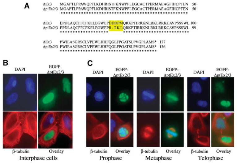
Subcellular localization of survivin-ΔptEx2/3. A: Protein sequence alignments of survivin-ΔptE2/3 with survivin-ΔEx3. B: Localization of survivin-ΔptEx2/3 in the interphase cells. HeLa cells were transfected with pEGFP-survivin-ΔptEx2/3. Cells were then fixed with 4% paraformaldehyde in PBS and stained with DAPI and β-tubulin antibodies 18 h after transfection. C: Localization of survivin-ΔptEx2/3 during mitosis (prophase, metaphase, and telophase). HeLa cells were transfected and stained as in (B). Images were taken under a Zeiss Axiovert 100M Fluorescence Microscopy System for both B and C. Note: Tubulin was stabilized by a brief taxol treatment in these experiments.
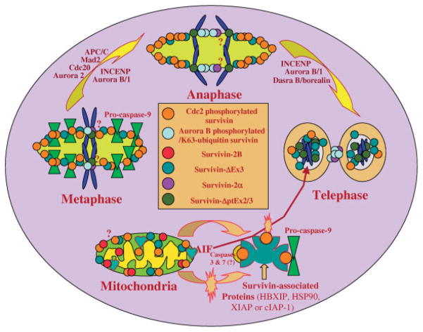
A diagrammatic model to summarize the role and subcellular localization of survivin and its splice variants in the regulation of apoptosis and control of mitosis in cancer cells. (1) The dynamic Cdc2-phosphorylated survivin localizes on the mitotic spindle and centrosome to inhibit a “default” apoptotic pathway during mitosis, and to facilitate microtubule polymerization and spindle formation. (2) The dynamic Aurora B-phosphorylated/K63-ubiquitin survivin complexes with Aurora B and INCENP, and plays a leading role in the binding of the chromosomal passenger protein complex to the centromere/kinetochore of chromosomes to control the precise completion of chromosome segregation and cytokinesis. (3) Survivin also localizes in mitochondria and is released into the cytoplasm upon apoptotic insult. The released survivin and/or other cytoplasmic survivin may interact with many other proteins to exert its role in apoptosis inhibition, G1/S transition/cell-cycle progression and possible other functions. Some of the survivin-associated proteins identified include HBXIP, HSP90, XIAP, and cIAP-1. Additionally, mitochondrial-associated survivin may block apoptosis-inducing factor (AIF) release and nuclear translocation as a mechanism for caspase-independent apoptosis control. (4) Survivin-2B localizes in mitochondria. One potential way for survivin-2B to induce cell death is that increased survivin-2B may be able to physically interact with and block the release of survivin from mitochondria for caspase inhibition. Alternatively, survivin-2B may also promote the AIF release and nuclear translocation. (5) It has been shown that survivin-ΔEx3 is localized in mitochondria and nuclei in interphase cells, and translocated to the mitotic spindle during mitosis. However, whether survivin-ΔEx3 plays a role in the spindle checkpoint and apoptosis control remains to be determined. It was shown that survivin-ΔEx3 is degraded in the nucleolus through the ubiquitination pathway although the significance of this phenomenon remains to be explored. (6) Survivin-ΔptEx2/3 and survivin-2 α were shown to be associated with the chromosomal DNA during mitosis in certain conditions. However, whether survivin-ΔptEx2/3 and survivin-2 α play a role during mitosis or whether the function of centromere-associated survivin can be modulated by survivin-ΔptEx2/3 and/or survivin-2 α remains to be determined in the future study. Note: The subcellular localization of survivin-3B is not clear currently and remains to be determined as well.
References
-
- Aziz M, Afaq F, Ahmad N. Prevention of ultraviolet B radiation damage by resveratrol in mouse skin is mediated via modulation in Survivin. Photochem Photobiol. 2005a;81(1):25–31. - PubMed
-
- Aziz MH, Reagan-Shaw S, Wu J, Longley BJ, Ahmad N. Chemoprevention of skin cancer by grape constituent resveratrol: Relevance to human disease? FASEB J. 2005b;19(9):1193–1195. - PubMed
-
- Badran A, Yoshida A, Ishikawa K, Goi T, Yamaguchi A, Ueda T, Inuzuka M. Identification of a novel splice variant of the human anti-apoptopsis gene survivin. Biochem Biophys Res Commun. 2004;314(3):902–907. - PubMed
-
- Beardmore VA, Ahonen LJ, Gorbsky GJ, Kallio MJ. Survivin dynamics increases at centromeres during G2/M phase transition and is regulated by microtubule-attachment and Aurora B kinase activity. J Cell Sci. 2004;117(Pt 18):4033–4042. - PubMed
-
- Belyanskaya LL, Hopkins-Donaldson S, Kurtz S, Simoes-Wust AP, Yousefi S, Simon HU, Stahel R, Zangemeister-Wittke U. Cisplatin activates Akt in small cell lung cancer cells and attenuates apoptosis by survivin upregulation. Int J Cancer. 2005;117(5):755–763. - PubMed
Publication types
MeSH terms
Substances
Grants and funding
LinkOut - more resources
Full Text Sources
Other Literature Sources
Miscellaneous
