Review
. 2006 Apr 3;45(15):2348-68.
doi: 10.1002/anie.200502794.

Synthetic multivalent ligands as probes of signal transduction

Affiliations
Review

Synthetic multivalent ligands as probes of signal transduction

Laura L Kiessling et al. Angew Chem Int Ed Engl. .

Abstract

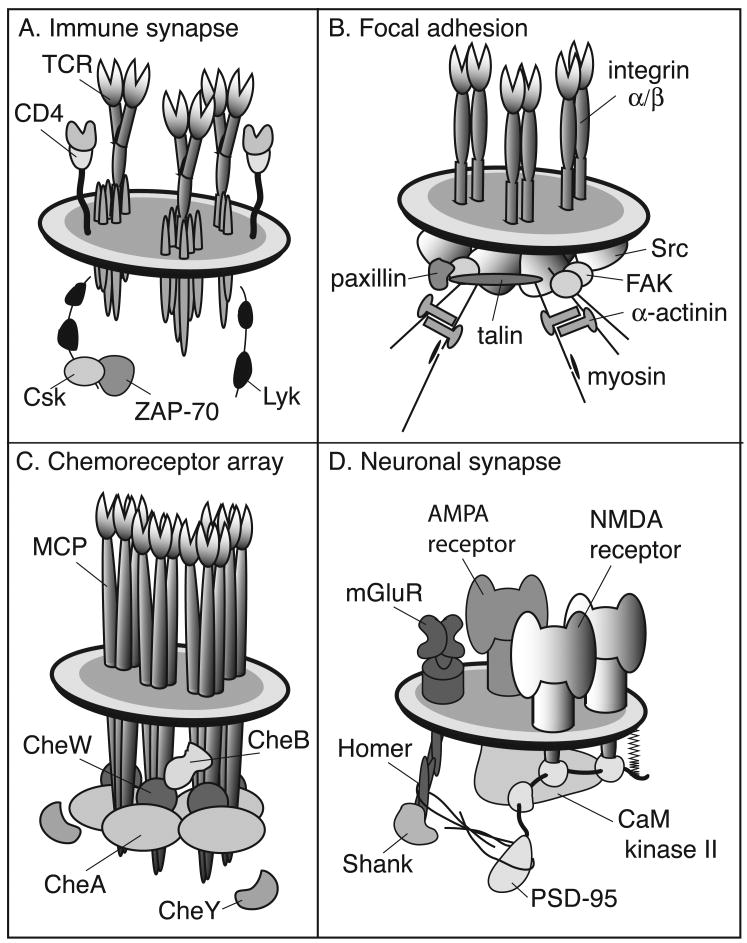
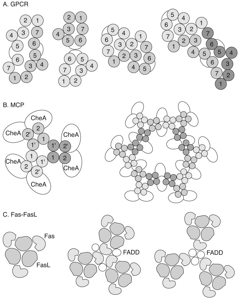
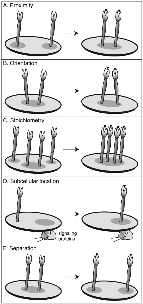
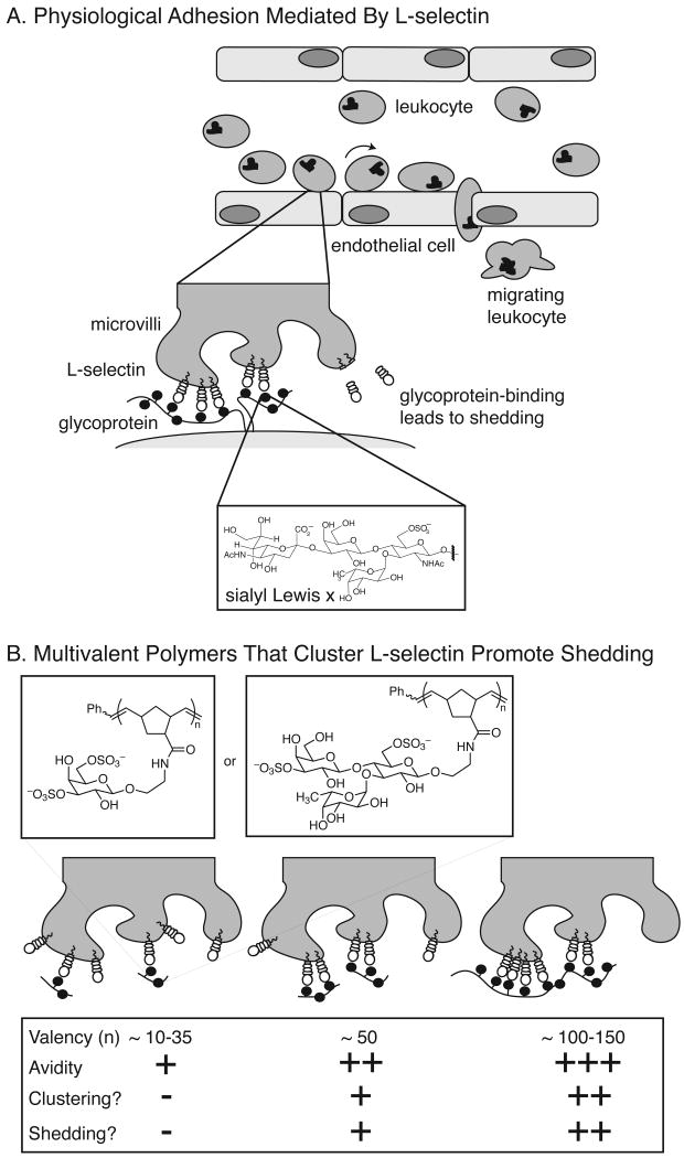
Cell-surface receptors acquire information from the extracellular environment and coordinate intracellular responses. Many receptors do not operate as individual entities, but rather as part of dimeric or oligomeric complexes. Coupling the functions of multiple receptors may endow signaling pathways with the sensitivity and malleability required to govern cellular responses. Moreover, multireceptor signaling complexes may provide a means of spatially segregating otherwise degenerate signaling cascades. Understanding the mechanisms, extent, and consequences of receptor co-localization and interreceptor communication is critical; chemical synthesis can provide compounds to address the role of receptor assembly in signal transduction. Multivalent ligands can be generated that possess a variety of sizes, shapes, valencies, orientations, and densities of binding elements. This Review focuses on the use of synthetic multivalent ligands to characterize receptor function.


Figures

Figure 1
Figure 1
Examples complexes of involved in signal transduction: (A) eukaryotic immune recognition; (B) eukaryotic cell adhesion; (C) prokaryotic chemotaxis; and (D) neuronal signaling. Although these schematic depictions are necessarily simplified, they have been designed to illustrate the complexity and elegance of biological multi-protein signaling complexes.
Figure 2
Figure 2
Proposed multi-receptor assemblies for some receptors. (A) Models of heptahelical GPCR dimers and a trimer based on mutagenesis results and the structure of bacteriorhodopsin. Each numbered circle represents a specific transmembrane helix. A variety of 1-7, 5-6, and 2-3 dimers and multimers have been proposed. (B) Bacterial chemoreceptors (methyl-accepting chemotaxis proteins or MCPs) are dimeric; and these can assemble into a trimer-of-dimers; this trimer of MCP dimers can further interact with signaling proteins, including the kinase CheA. Each MCP passes through the membrane twice (1 and 2) and coiled-coil interactions between these transmembrane domains (1 and 1′) mediate dimerization. A lattice model of MCP organization constructed of six of these trimers-of-dimers and 24 copies of CheA is shown. (C) The Fas-FasL interaction has been modeled using protein interfaces suggested by mutagenesis and crosslinking studies. Both the receptor Fas and its corresponding ligand, FasL, are trimers. The corresponding trimeric complex may be employed as a unit in the lattice stabilized by the adapter protein, FADD. Two models are shown with either Fas- or FADD-centered symmetries. See the text for the appropriate references.
Figure 3
Figure 3
Possible methods for regulating inter-receptor communication. (A) The distance between receptors can influence the transfer of information between receptors or other proteins. (B) The relative orientation of two receptors can influence the alignment of enzyme active sites and govern the rates of covalent modifications that result in signal generation. (C) The number of receptors in a complex can influence the intensity of a signal. Additionally, the likelihood that receptors will come into contact increases when the numbers of localized receptors is greater. (D) The subcellular location of a receptor controls the access of the receptor to some intracellular signaling proteins. Changes in position can govern the flow of information through a receptor or cluster of receptors. (E) When co-receptors act as negative regulators, ligand binding can lead to activation of receptors by separation. This receptor mechanism is conceptually related to proximity-induced activation, but the underlying molecular interactions are quite different.
Figure 4
Figure 4
Scaled diagram of the classes of multivalent ligand scaffolds. Although the actual size of ligands that are based on these scaffolds varies, representative examples of each class are pictured. To illustrate the importance of resolution, the sizes are shown relative to a mammalian lymphocyte. Size bars are as follows: cell and bead 1 nm; liposome and polymer 0.5 nm; antibody, dendrimer, and albumin 0.05 nm.
Figure 5
Figure 5
Receptor binding mechanisms that are unique to multivalent ligands.
Figure 6
Figure 6
Depiction of the intracellular-mediated alteration of receptor proximity by CID. A receptor fused to a binding protein is expressed in a target cell. Addition of a small molecule dimerizer (FK1012) induces the clustering of the receptor.
Figure 7
Figure 7
The density of REs presented influences the adhesion of cells to integrin-ligand-bearing surfaces. (A) Surfaces coated with multivalent ligands have a greater functional affinity for B cells. (B) Star polymers are depicted that display variable copies of the pentapeptide YGRGD, an integrin-binding RE. The relative potency of each surface is indicated by the number of cells bound.
Figure 8
Figure 8
Schematic of streptavidin-MHC-MCC complexes. Four complexes with one, two, three, or four biotinylated MHC-MCC moieties are shown on the left. A model for the activation of TCRs by multivalent engagement by the highest valency ligand is shown at the right.
Figure 9
Figure 9
Synthetic multivalent polymer-based investigation of receptor proximity effects in bacterial chemotaxis. Addition of a multivalent ligand with sufficient valency can induce the re-organization of MCPs. This potentiates signaling through these receptors and activates bacterial locomotion.
Figure 10
Figure 10
Orientation of ZAP-70 influences its kinase function. A CID strategy was used to explore the influence of ZAP-70 orientation on function of this kinase. The chemical structures of three dimerizer compounds are shown, FK1012, FK1012H2, and FK1012Z. These dimers present REs in three distinct relative orientations. The relative abilities of these compounds to induce ZAP-70 activity were similar.
Figure 11
Figure 11
Multivalent ligands for L-selectin mimic cell surface glycoproteins. (A) L-selectin expressed on lymphocytes binds to glycoproteins on the endothelium. This interaction slows lymphocyte progression through the vessel and triggers proteolytic release of L-selectin. (B) Multivalent polymers displaying sulfated carbohydrates also bind multiple copies of L-selectin, which leads to receptor clustering and proteolytic shedding. There is a direct relationship between the valency of the polymer, the number of L-selectin proteins bound, and the avidity of the interaction.

References

    1. Chaneux JP, Thiéry J, Tung Y, Kittel C. Proc Natl Acad Sci USA. 1966;57:335. - PMC - PubMed
    1. Heldin CH. Cell. 1995;80:213. - PubMed
    1. Schlessinger J. TIBS. 1988;13:443. - PubMed
    1. Klemm JD, Schreiber SL, Crabtree GR. Annu Rev Immunol. 1998;16:569. - PubMed
    1. Metzger H. J Immunol. 1992;149:1477. - PubMed