. 2006;8(3):R61.
doi: 10.1186/ar1929. Epub 2006 Mar 17.

The matrix-forming phenotype of cultured human meniscus cells is enhanced after culture with fibroblast growth factor 2 and is further stimulated by hypoxia

Affiliations

The matrix-forming phenotype of cultured human meniscus cells is enhanced after culture with fibroblast growth factor 2 and is further stimulated by hypoxia

Adetola B Adesida et al. Arthritis Res Ther. 2006.

Abstract

Human meniscus cells have a predominantly fibrogenic pattern of gene expression, but like chondrocytes they proliferate in monolayer culture and lose the expression of type II collagen. We have investigated the potential of human meniscus cells, which were expanded with or without fibroblast growth factor 2 (FGF2), to produce matrix in three-dimensional cell aggregate cultures with a chondrogenic medium at low (5%) and normal (20%) oxygen tension. The presence of FGF2 during the expansion of meniscus cells enhanced the re-expression of type II collagen 200-fold in subsequent three-dimensional cell aggregate cultures. This was increased further (400-fold) by culture in 5% oxygen. Cell aggregates of FGF2-expanded meniscus cells accumulated more proteoglycan (total glycosaminoglycan) over 14 days and deposited a collagen II-rich matrix. The gene expression of matrix-associated proteoglycans (biglycan and fibromodulin) was also increased by FGF2 and hypoxia. Meniscus cells after expansion in monolayer can therefore respond to chondrogenic signals, and this is enhanced by FGF2 during expansion and low oxygen tension during aggregate cultures.


Figures

Figure 1
Figure 1
Cell doubling for P1 and P2 meniscus cells in the presence and absence of FGF2. Results are expressed as means ± SD (n = 4). FGF2, fibroblast growth factor 2; P1, passage 1; P2, passage 2.
Figure 2
Figure 2
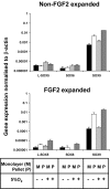
Sox gene expression in monolayer and cell aggregate cultures. Gene expression for monolayer cells (black bars) between P2 and P3 in standard medium (n = 3), for cell aggregate (white open bars) culture in chondrogenic differentiation medium after 14 days of culture under normoxia (n = 3). Gene expression for monolayer cells (light grey bars) between P2 and P3 in standard medium (n = 3) and for cell aggregate (dark grey bars) in chondrogenic differentiation medium after 14 days of culture under low oxygen tension (n = 3). *p < 0.05; **p < 0.001 (Student's unpaired t test) in cell aggregates from fibroblast growth factor 2 (FGF2)-expanded versus non-FGF2-expanded cells. P2, passage 2; P3, passage 3.
Figure 3
Figure 3
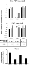
Collagen expression in monolayer and cell aggregate cultures, and in meniscus tissue with SOX9 expression (n = 3). Gene expression levels for monolayer cells (light grey bars) between P2 and P3 in standard medium (n = 3) and for cell aggregate (dark grey bars) cultures in chondrogenic differentiation medium (n = 3) under low oxygen tension after 14 days in culture. *p < 0.05; **p < 0.001 (Student's unpaired t test) in cell aggregates from fibroblast growth factor 2 (FGF2)-expanded versus non-FGF2-expanded cells.
Figure 4
Figure 4
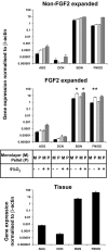
Proteoglycan gene expression in monolayer and aggregate cultures from non-FGF2 and FGF2 expanded meniscus cells. Gene expression levels for monolayer cells (black bars) between P2 and P3 in standard medium (n = 3) and for cell aggregate (white open bars) in chondrogenic differentiation medium (n = 3) under normal oxygen after 14 days in culture. Gene expression for monolayer cells (light grey bars) between P2 and P3 in standard medium (n = 3) and for cell aggregate (dark grey bars) in chondrogenic differentiation medium (n = 3) under hypoxia after 14 days in culture. AGG, aggrecan; BGN, biglycan; DCN, decorin; FGF2, fibroblast growth factor 2; FMOD, fibromodulin; P2, passage 2; P3, passage 3.
Figure 5
Figure 5
Weights and GAG per DNA content of cell aggregates. Wet weights of cell aggregate derived from non-FGF2-expanded (black bars) and FGF2-expanded (grey bars) meniscus cells under normoxia (-) or hypoxia (+), and GAG content of cell aggregate normalised to DNA content. FGF2, fibroblast growth factor 2; GAG, glycosaminoglycan.
Figure 6
Figure 6
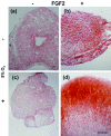
Safranin-O staining of cell aggregates. Histological analysis and matrix accumulation of cell aggregates derived from meniscus cells expanded in the absence ((a) normoxia; (c) hypoxia) and presence ((b) normoxia; (d) hypoxia) of fibroblast growth factor 2 (FGF2). Scale bar, 100 μm. FGF2, fibroblast growth factor 2.
Figure 7
Figure 7
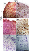
Immunolocalisation of type I collagen in cell aggregates after 14 days of culture. Cell aggregates derived from meniscus cells expanded in the absence ((a) normoxia; (c) hypoxia) and presence ((b) normoxia; (d) hypoxia) of fibroblast growth factor 2 (FGF2). No primary antibody control and non-specific IgG control. Scale bar, 100 μm.
Figure 8
Figure 8
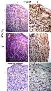
Immunolocalisation of type II collagen in cell aggregates after 14 days of culture. Cell aggregates derived from meniscus cells expanded in the absence ((a) normoxia; (c) hypoxia) and presence ((b) normoxia; (d) hypoxia) of fibroblast growth factor 2 (FGF2). No primary antibody control on cell aggregates derived from non-FGF2-expanded and FGF2-expanded cells after culture under hypoxia. Scale bar, 100 μm.

References

    1. Fithian DC, Kelly MA, Mow VC. Material properties and structure-function relationships in the menisci. Clin Orthop. 1990;252:19–31. - PubMed
    1. Ahmed AM. The load-bearing role of the knee meniscus. In: Mow VC, Jackson DW, editor. Knee Meniscus: Basic and Clinical Foundations. New York: Raven Press; 1992. pp. 59–73.
    1. Levy IM, Torzilli PA, Fisch ID. The contribution of the menisci to the stability of the knee. In: Mow VC, Jackson DW, editor. Knee Meniscus: Basic and Clinical Foundations. New York: Raven Press; 1992. pp. 107–115.
    1. Fairbank T. Knee joint changes after menisectomy. J Bone Joint Surg. 1948;30B:664–670. - PubMed
    1. Cox JS, Nye CE, Schaefer WW, Woodstein IJ. The degenerative effects of partial and total resection of the medial meniscus in dogs' knees. Clin Orthop Relat Res. 1975;109:178–183. - PubMed

Publication types

LinkOut - more resources