. 2006;7(3):R23.
doi: 10.1186/gb-2006-7-3-r23. Epub 2006 Mar 22.

Phylogenetic and structural analysis of centromeric DNA and kinetochore proteins

Affiliations

Phylogenetic and structural analysis of centromeric DNA and kinetochore proteins

Patrick Meraldi et al. Genome Biol. 2006.

Abstract

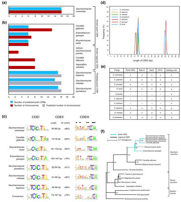
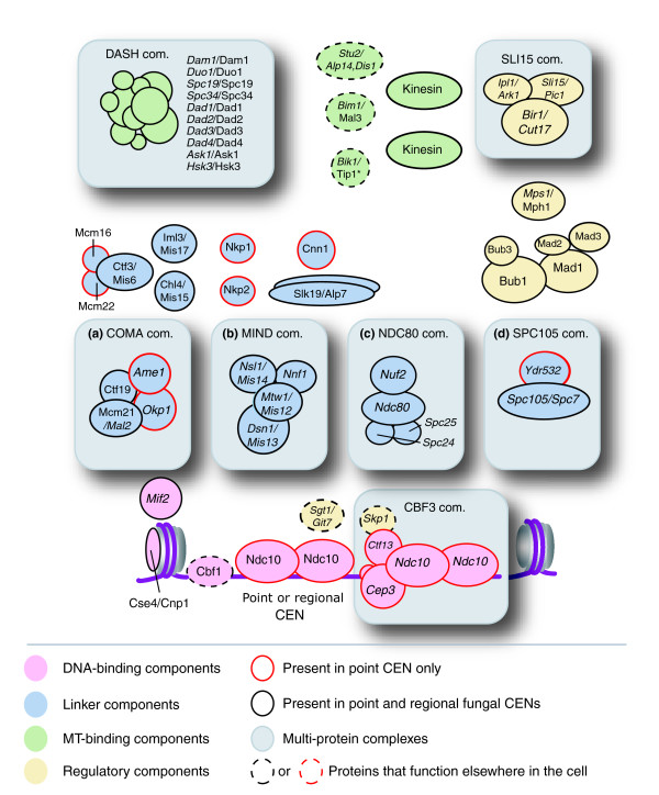
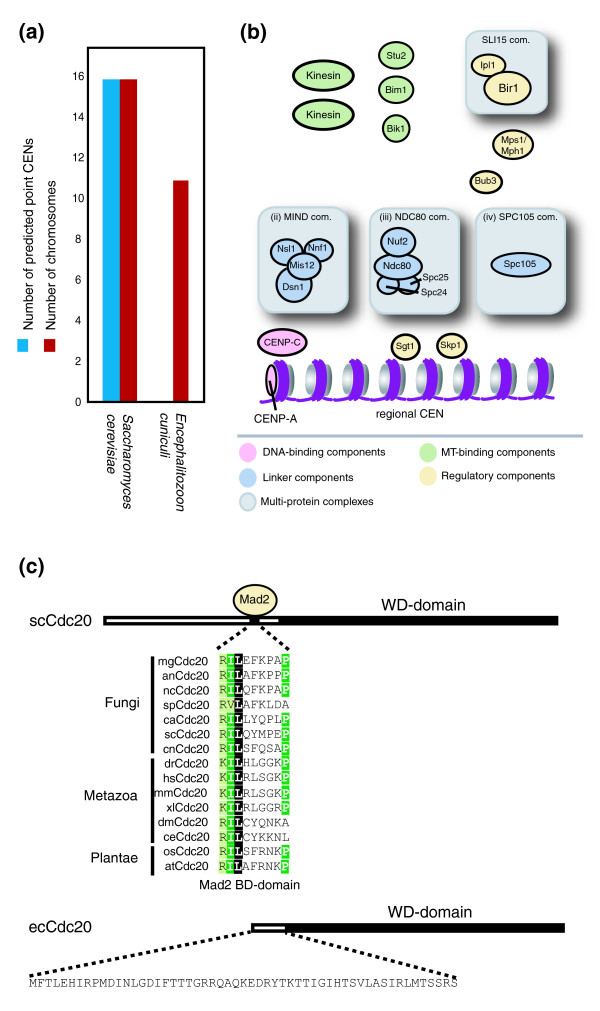
Background: Kinetochores are large multi-protein structures that assemble on centromeric DNA (CEN DNA) and mediate the binding of chromosomes to microtubules. Comprising 125 base-pairs of CEN DNA and 70 or more protein components, Saccharomyces cerevisiae kinetochores are among the best understood. In contrast, most fungal, plant and animal cells assemble kinetochores on CENs that are longer and more complex, raising the question of whether kinetochore architecture has been conserved through evolution, despite considerable divergence in CEN sequence.

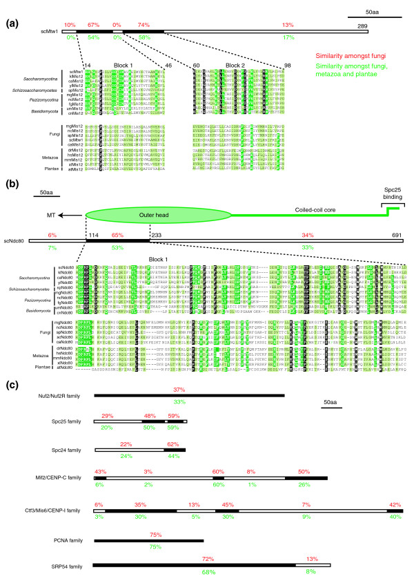
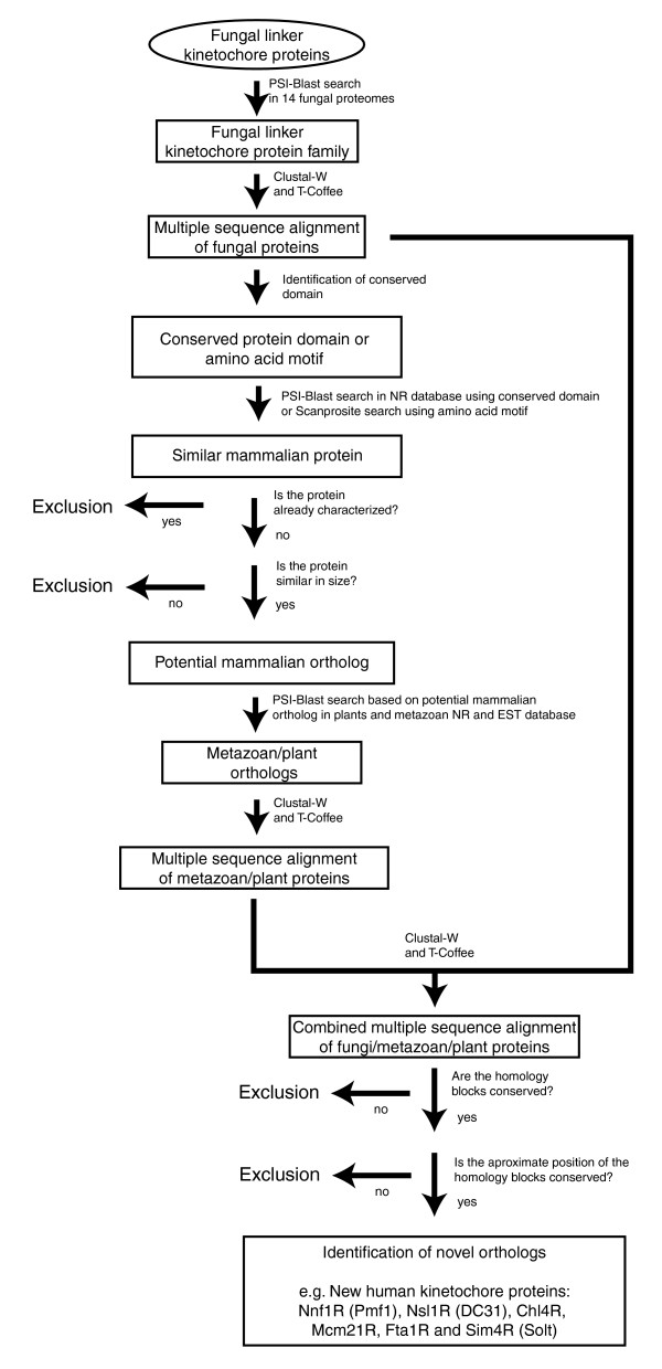
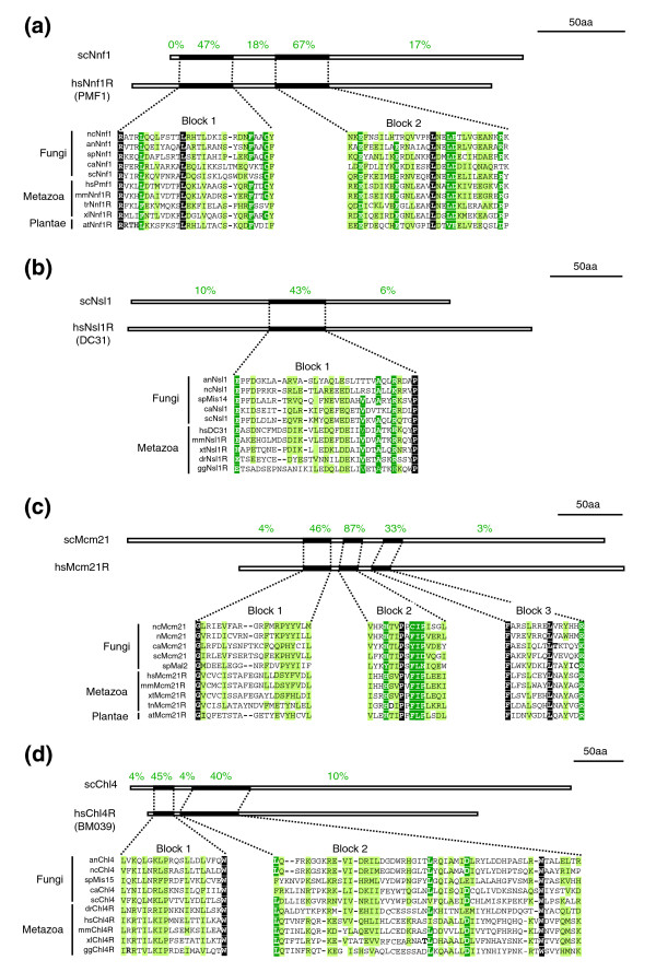
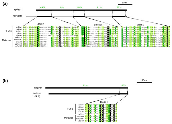
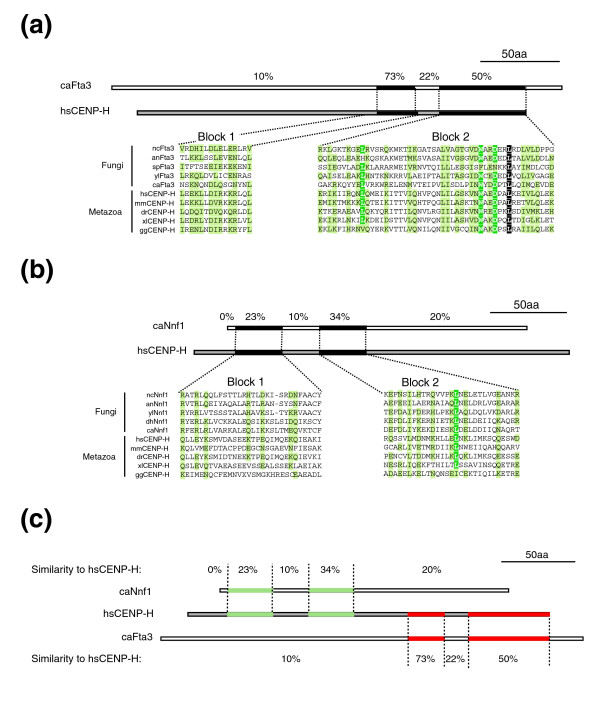
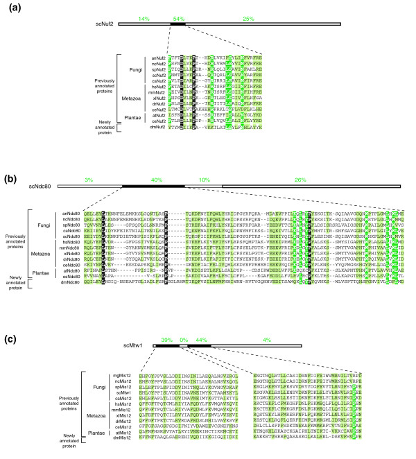
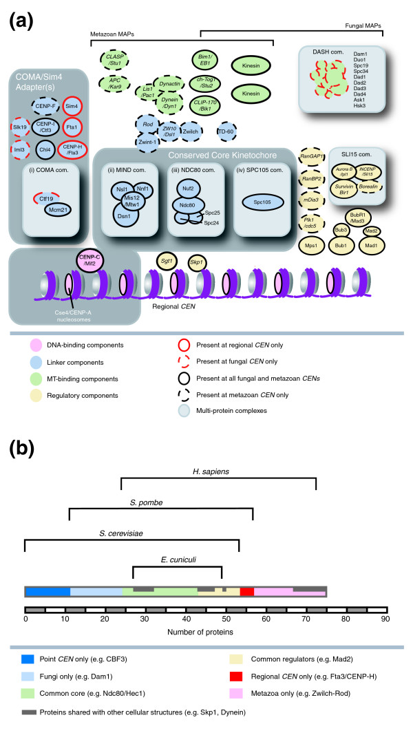
Results: Using computational approaches, ranging from sequence similarity searches to hidden Markov model-based modeling, we show that organisms with CENs resembling those in S. cerevisiae (point CENs) are very closely related and that all contain a set of 11 kinetochore proteins not found in organisms with complex CENs. Conversely, organisms with complex CENs (regional CENs) contain proteins seemingly absent from point-CEN organisms. However, at least three quarters of known kinetochore proteins are present in all fungi regardless of CEN organization. At least six of these proteins have previously unidentified human orthologs. When fungi and metazoa are compared, almost all have kinetochores constructed around Spc105 and three conserved multi-protein linker complexes (MIND, COMA, and the NDC80 complex).

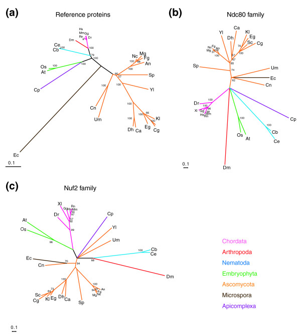
Conclusion: Our data suggest that critical structural features of kinetochores have been well conserved from yeast to man. Surprisingly, phylogenetic analysis reveals that human kinetochore proteins are as similar in sequence to their yeast counterparts as to presumptive Drosophila melanogaster or Caenorhabditis elegans orthologs. This finding is consistent with evidence that kinetochore proteins have evolved very rapidly relative to components of other complex cellular structures.


Figures

Figure 1
Figure 1
Point centromeres are derived from regional centromeres and appeared only once during evolution. (a) The 16 CENs from S. cerevisiae were used to train a HMM. The blue bar indicates the number of predicted point CENs in the genome and the red bar represents the number of known chromosomes. (b) HMM from (a) was used to search the genome of fungi with known point CENs, known regional CENs and predicted point CENs. Blue and red bars are as described in (a) except gray bars, which indicate the predicted number of chromosomes, based on synteny within other Saccharomyces species. (c) Sequence comparison of the CDEI, CDEII and CDEIII elements from budding yeast with point centromeres. (d) Frequency distribution of the CDEII length (measured in bp) in each budding yeast with point centromeres. (e) Evolutionary conservation of CBF3 subunits in fungi with point and regional CENs. (f) Phylogenetic analysis of 17 different fungi, including the 7 budding yeast with point centromeres and the 3 budding yeast with regional centromeres using 3 highly conserved reference proteins (α-tubulin, the signal recognition protein SRP54 and the DNA replication factor PCNA). Blue branches represent fungi with point centromeres and black branches those with regional centromeres.
Figure 2
Figure 2
Sequence similarity between kinetochore proteins is restricted to short stretches between orthologs. Multiple sequence alignments of the (a) Mis12Mtw1 and (b) Ndc80Hec1 families. Schematic drawing above the alignment indicate the length of the S. cerevisiae proteins and the percentages denote the degree of similarity of successive sequence blocks (black boxes) within fungi (red letters) or fungi, metazoa and plantae (green letters). The schematic drawing above the Ndc80 multiple sequence alignment also indicates the relative position of the globular and coiled-coil domain of Ndc80, as determined by electron-microscopy [32,33]. White letters on black denote identical residues, white letters on green, identical residues in ≥ 80% of the organisms and black letters on green, similar residues in ≥ 80% of the organisms. (c) Schematic drawings indicating the percentage similarity of successive sequence blocks (black boxes) within fungi (red letters) or fungi, metazoa and plantae (green letters) based on multiple sequence alignments of the Nuf2, Spc25, Spc24. CENP-CMif2 and Mis6Ctf3/CENP-I, PCNA and SRP54 protein families
Figure 3
Figure 3
Fungal kinetochores contain a set of point centromere specific components. Schematic model of kinetochore subunitorganization based on the architecture of the S. cerevisiae kinetochore. Kinetochore proteins can be roughly divided into DNA-binding (pink), linker (blue), MT-binding (green) and regulatory layers (yellow). Within each layer many proteins are organized into multi-protein complexes, for example, the linker layer is composed of at least four complexes (gray boxes (a) to (d)): COMA, NDC80, MIND and SPC105. Protein names are given for S. cervisiae first and S. pombe second, while essential genes (italic letters) and non-essential (normal letters) is indicated. Protein names followed by an asterisk indicate that this specific ortholog is known not to localize to kinetochores. The kinesins present at kinetochores in S. cerevisiae are Kip3 (Kinesin-8), Cin8 (Kinesin-5), Kip1 (Kinesin-5) and Kar3 (Kinesin-14), while in S. pombe they are Klp5 (Kinesin-8), Klp6 (Kinesin-8) and Klp2 (Kinesin-14) (for nomenclature see [38].
Figure 4
Figure 4
Schematic describing the sequence-search based approach used to identify fungal, metazoan, and plant orthologs of the kinetochore proteins scNnf1, scNsl1, scChl4, scMcm21, spSim4 and spFta1. Since such sequence-based searches can yield a significant number of false positives, strict exclusion criteria were applied to ensure the identification of orthologs.
Figure 5
Figure 5
Identification of potential orthologs of scNnf1, scNsl1, scMcm21 and scChl4 in humans. S. cerevisiae (a) Nnf1, (b) Nsl1, (c) Mcm21 and (d) Chl4 were aligned with five fungal, four metazoan and one plant sequence. White letters on black denote identical residues, white letters on green, identical residues in ≤ 80% of the organisms and black letters on green, similar residues in ≤ 80% of the organisms. Schematic drawings above the alignments indicate the length of the S. cerevisiae proteins and the percentages denote the degree of similarity of successive sequence blocks (black boxes).
Figure 6
Figure 6
Identification of potential orthologs of spFta1 and spSim4 in humans. S. pombe (a) Fta1 and (b) Sim4 were aligned with five fungal, and three to five metazoan sequences. White letters on black denote identical residues, white letters on green, identical residues in ≥ 80% of the organisms and black letters on green, similar residues in ≥ 80% of the organisms. Schematic drawings above the alignments indicate the length of the S. cerevisiae proteins and the percentages denote the degree of similarity of successive sequence blocks (black boxes).
Figure 7
Figure 7
The human kinetochore protein CENP-H is more closely related to a novel family of fungal proteins than the Nnf1 family. Multiple sequence alignments of metazoan CENP-H proteins and either (a) fungal Fta3 family proteins or (b) fungal Nnf1 family of proteins. Sequences were annotated as in Figure 5. (c) Comparison of sequence similarity between human conserved domains of CENP-H and C. albicans Nnf1 and C. albicans Fta3.
Figure 8
Figure 8
Phylogenetic analysis of kinetochore protein conserved domains. Radial phylogenetic trees were assembled for (a) reference proteins (α-tubulin, the signal recognition protein SRP54 and the DNA replication factor PCNA), (b) the Ndc80 family and (c) the Nuf2 family. For bootstrap analysis, sample size equals 100. Nodes with support less than 50% were collapsed. The accession number for each protein is described in Additional data file 1.
Figure 9
Figure 9
Identification and annotation of (a) Nuf2, (b) Ndc80 and (c) Mis12 orthologs in D. melanogaster. Schematic drawing above the alignment indicate the length of the S. cerevisiae proteins and the percentages denote the degree of similarity of successive sequence blocks (black boxes). White letters on black denote identical residues, white letters on green, identical residues in ≥ 80% of the organisms and black letters on green, similar residues in ≥ 80% of the organisms. Accession numbers are described in Additional data file 1.
Figure 10
Figure 10
Identification of a minimal kinetochore in E. cuniculi. (a) HMM described in Figure 1a, b failed to find a CDEI-II-III structure in the genome of E. cuniculi. The Green bar indicates point CENs identified and black bars the number of chromosomes. (b) Speculative model of E. cuniculi kinetochore subunit organization. Proteins colored in pink, blue, green or yellow represent components of the DNA binding, linker, regulatory or microtubule-binding layers, respectively, based on kinetochore organization in S. cerevisiae. Potential multi-protein complexes are highlighted with a grey box. (c) Sequence alignment of fungal, metazoan and plantae Cdc20 showing the conserved Mad2 binding site. Note: E. cuniculi lacks both the conserved Mad2 binding site and an ortholog of the Mad2 protein. Schematic drawings indicate the length of the S. cerevisiae and E. cuniculi proteins, the position of the WD-domain (black box) and the position where Mad2 binds.
Figure 11
Figure 11
Evolutionary development of kinetochores from yeast to mammals. (a) Model of the kinetochore using protein subunit positions derived from the organization of the S. cerevisiae kinetochore. Proteins present in all fungal and mammalian CENs are outlined in black while proteins present only in fungi and mammals with regional CENs are outlined in red. Red dotted outlines indicate proteins that are only present in fungi. Black dotted outlines indicate that either this protein only exists in metazoans or that only the metazoan ortholog is present at kinetochore. Proteins colored in pink, blue, green or yellow represent components of the DNA binding, linker, regulatory or microtubule-binding layers, respectively, based on kinetochore organization in S. cerevisiae. Potential multi-protein complexes are highlighted with a light gray box and the conserved kinetochore core, or COMA/Sim4 adaptor with a dark gray box. Protein names are given for H. sapiens first and then S. cerevisiae when different. Italic lettering indicates that the protein has additional functions in the cell. The kinesins present at kinetochores in H. sapiens are CENP-E (Kinesin-7) and MCAK (Kinesin-13), and in S. cerevisiae Kip3 (Kinesin-8), Cin8 (Kinesin-5), Kip1 (Kinesin-5) and Kar3 (Kinesin-14) (for nomenclature see [38]). (b) Quantification of the number of kinetochore proteins, and their respective evolutionary class, in S. cerevisiae, S. pombe, E. cuniculi and H. sapiens.

Similar articles

Cited by

References

    1. Koshland DE, Mitchison TJ, Kirschner MW. Polewards chromosome movement driven by microtubule depolymerization in vitro. Nature. 1988;331:499–504. doi: 10.1038/331499a0. - DOI - PubMed
    1. Cleveland DW, Mao Y, Sullivan KF. Centromeres and kinetochores: from epigenetics to mitotic checkpoint signaling. Cell. 2003;112:407–421. doi: 10.1016/S0092-8674(03)00115-6. - DOI - PubMed
    1. Choo KH. Domain organization at the centromere and neocentromere. Dev Cell. 2001;1:165–177. doi: 10.1016/S1534-5807(01)00028-4. - DOI - PubMed
    1. Fitzgerald-Hayes M, Clarke L, Carbon J. Nucleotide sequence comparisons and functional analysis of yeast centromere DNAs. Cell. 1982;29:235–244. doi: 10.1016/0092-8674(82)90108-8. - DOI - PubMed
    1. McAinsh AD, Tytell JD, Sorger PK. Structure, function, and regulation of budding yeast kinetochores. Annu Rev Cell Dev Biol. 2003;19:519–539. doi: 10.1146/annurev.cellbio.19.111301.155607. - DOI - PubMed

Publication types

MeSH terms

Substances