Structural investigation of disordered stress proteins. Comparison of full-length dehydrins with isolated peptides of their conserved segments
- PMID: 16565295
- PMCID: PMC1475461
- DOI: 10.1104/pp.106.079848
Structural investigation of disordered stress proteins. Comparison of full-length dehydrins with isolated peptides of their conserved segments
Abstract
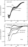
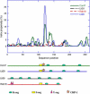
Dehydrins constitute a class of intrinsically disordered proteins that are expressed under conditions of water-related stress. Characteristic of the dehydrins are some highly conserved stretches of seven to 17 residues that are repetitively scattered in their sequences, the K-, S-, Y-, and Lys-rich segments. In this study, we investigate the putative role of these segments in promoting structure. The analysis is based on comparative analysis of four full-length dehydrins from Arabidopsis (Arabidopsis thaliana; Cor47, Lti29, Lti30, and Rab18) and isolated peptide mimics of the K-, Y-, and Lys-rich segments. In physiological buffer, the circular dichroism spectra of the full-length dehydrins reveal overall disordered structures with a variable content of poly-Pro helices, a type of elongated secondary structure relying on bridging water molecules. Similar disordered structures are observed for the isolated peptides of the conserved segments. Interestingly, neither the full-length dehydrins nor their conserved segments are able to adopt specific structure in response to altered temperature, one of the factors that regulate their expression in vivo. There is also no structural response to the addition of metal ions, increased protein concentration, or the protein-stabilizing salt Na(2)SO(4). Taken together, these observations indicate that the dehydrins are not in equilibrium with high-energy folded structures. The result suggests that the dehydrins are highly evolved proteins, selected to maintain high configurational flexibility and to resist unspecific collapse and aggregation. The role of the conserved segments is thus not to promote tertiary structure, but to exert their biological function more locally upon interaction with specific biological targets, for example, by acting as beads on a string for specific recognition, interaction with membranes, or intermolecular scaffolding. In this perspective, it is notable that the Lys-rich segment in Cor47 and Lti29 shows sequence similarity with the animal chaperone HSP90.
Figures


 .
.
Similar articles
-
Stress-induced accumulation and tissue-specific localization of dehydrins in Arabidopsis thaliana.Plant Mol Biol. 2001 Feb;45(3):263-79. doi: 10.1023/a:1006469128280. Plant Mol Biol. 2001. PMID: 11292073
-
Membrane-Induced Folding of the Plant Stress Dehydrin Lti30.Plant Physiol. 2016 Jun;171(2):932-43. doi: 10.1104/pp.15.01531. Epub 2016 Apr 26. Plant Physiol. 2016. PMID: 27208263 Free PMC article.
-
F-segments of Arabidopsis dehydrins show cryoprotective activities for lactate dehydrogenase depending on the hydrophobic residues.Phytochemistry. 2020 May;173:112300. doi: 10.1016/j.phytochem.2020.112300. Epub 2020 Feb 19. Phytochemistry. 2020. PMID: 32087435
-
The Disordered Dehydrin and Its Role in Plant Protection: A Biochemical Perspective.Biomolecules. 2022 Feb 11;12(2):294. doi: 10.3390/biom12020294. Biomolecules. 2022. PMID: 35204794 Free PMC article. Review.
-
Plant dehydrins--tissue location, structure and function.Cell Mol Biol Lett. 2006;11(4):536-56. doi: 10.2478/s11658-006-0044-0. Epub 2006 Sep 14. Cell Mol Biol Lett. 2006. PMID: 16983453 Free PMC article. Review.
Cited by
-
Multifarious roles of intrinsic disorder in proteins illustrate its broad impact on plant biology.Plant Cell. 2013 Jan;25(1):38-55. doi: 10.1105/tpc.112.106062. Epub 2013 Jan 29. Plant Cell. 2013. PMID: 23362206 Free PMC article. Review.
-
Sweeping away protein aggregation with entropic bristles: intrinsically disordered protein fusions enhance soluble expression.Biochemistry. 2012 Sep 18;51(37):7250-62. doi: 10.1021/bi300653m. Epub 2012 Sep 5. Biochemistry. 2012. PMID: 22924672 Free PMC article.
-
GRAS transcription factors emerging regulator in plants growth, development, and multiple stresses.Mol Biol Rep. 2022 Oct;49(10):9673-9685. doi: 10.1007/s11033-022-07425-x. Epub 2022 Jun 17. Mol Biol Rep. 2022. PMID: 35713799 Review.
-
Functional characterization of an acidic SK(3) dehydrin isolated from an Opuntia streptacantha cDNA library.Planta. 2012 Mar;235(3):565-78. doi: 10.1007/s00425-011-1531-8. Epub 2011 Oct 8. Planta. 2012. PMID: 21984262
-
Structure and function of a mitochondrial late embryogenesis abundant protein are revealed by desiccation.Plant Cell. 2007 May;19(5):1580-9. doi: 10.1105/tpc.107.050104. Epub 2007 May 25. Plant Cell. 2007. PMID: 17526751 Free PMC article.
References
-
- Alsheikh MK, Heyen BJ, Randall SK (2003) Ion binding properties of the dehydrin ERD14 are dependent upon phosphorylation. J Biol Chem 278: 40882–40889 - PubMed
-
- Bochicchio B, Tamburro AM (2002) Polyproline II structure in proteins: identification by chiroptical spectroscopies, stability, and functions. Chirality 14: 782–792 - PubMed
-
- Boudet J, Buitink J, Hoekstra FA, Rogniaux H, Larre C, Satour P, Leprince O (2006) Comparative analysis of the heat-stable proteome of radicles of Medicago truncatula seeds during germination identifies late embryogenesis abundant proteins associated with desiccation tolerance. Plant Physiol 140: 1418–1436 - PMC - PubMed
-
- Bourhis JM, Johansson K, Receveur-Brechot V, Oldfield CJ, Dunker KA, Canard B, Longhi S (2004) The C-terminal domain of measles virus nucleoprotein belongs to the class of intrinsically disordered proteins that fold upon binding to their physiological partner. Virus Res 99: 157–167 - PubMed
Publication types
MeSH terms
Substances
Associated data
- Actions
- Actions
- Actions
- Actions
LinkOut - more resources
Full Text Sources
Molecular Biology Databases

