. 2006 Apr;168(4):1119-33.
doi: 10.2353/ajpath.2006.050507.

The critical roles of serum/glucocorticoid-regulated kinase 3 (SGK3) in the hair follicle morphogenesis and homeostasis: the allelic difference provides novel insights into hair follicle biology

Affiliations

The critical roles of serum/glucocorticoid-regulated kinase 3 (SGK3) in the hair follicle morphogenesis and homeostasis: the allelic difference provides novel insights into hair follicle biology

Taro Okada et al. Am J Pathol. 2006 Apr.

Abstract

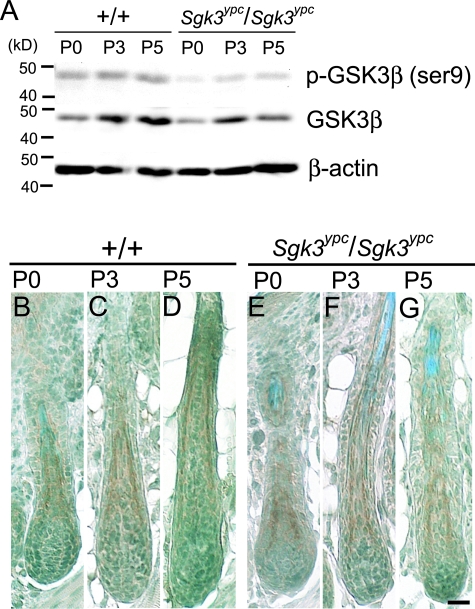
Mutation in the serum/glucocorticoid regulated kinase 3 (Sgk3, also known as Sgkl or Cisk) gene causes both defective hair follicle development and altered hair cycle in mice. We examined Sgk3-mutant YPC mice (YPC-Sgk3(ypc)/Sgk3(ypc)) and found expression of SGK3 protein with altered function. In the hair follicles of YPC mice, the aberrant differentiation and poor proliferation of hair matrix keratinocytes during the period of postnatal hair follicle development resulted in a complete lack of hair medulla and weak hair. Surprisingly, the length of postnatal hair follicle development and anagen term was shown to be dramatically shortened. Also, phosphorylation of GSK3beta at Ser9 and the nuclear accumulation of beta-catenin were reduced in the developing YPC hair follicle, suggesting that phosphorylation of GSK3beta and WNT-beta-catenin pathway takes part in the SGK3-dependent regulation of hair follicle development. Moreover, the above-mentioned features, especially the hair-cycling pattern, differ from those in other Sgk3-null mutant strains, suggesting that the various patterns of dysfunction in the SGK3 protein may result in phenotypic variation. Our results indicate that SGK3 is a very important and characteristic molecule that plays a critical role in both hair follicle morphogenesis and hair cycling.


Figures

Figure 1-6804
Figure 1-6804
Severe hair coat and hair structural defect in YPC mice (Sgk3ypc/Sgk3ypc). A: Gross appearance of ICR and YPC mice at the age of P70. The YPC mouse looks almost bald, but short, weak, and curved hairs appear on the skin. B and C: Development of WT (+/+, left) and mutant (Sgk3ypc/Sgk3ypc, right) of the same littermates obtained from the F1 male and female between ICR and YPC. At P7 (B), the mutant littermates show a thin hair coat compared with the WT, and it is more prominent at P9 (C). D–G: Gross appearance of the WT (+/+, left) and mutant (Sgk3ypc/Sgk3ypc, right) of the same littermates of C57BL/6J background. At P5 (D), the WT and mutant look the same, but the black coloring is less in the mutant than the WT. At P7 (E), the mutant shows a bald-like appearance (however, very short hairs appeared), whereas the WT bore short hairs at that age. The mutant shows the poor-coated phenotype while the WT littermate grew as normal B6 mouse at P21 (F) and P84 (G). H and I: Hairs plucked from +/+ (H) and Sgk3ypc/Sgk3ypc (I) mice on B6 background at P84, which were shown in G. Note that each type of hairs in Sgk3ypc/Sgk3ypc is curly and shorter and thinner than that in WT. Distinction of awl and zigzag hair in Sgk3ypc/Sgk3ypc was done based on the length of each hair. Scale bar, 1 mm.
Figure 2-6804
Figure 2-6804
Expression of Sgk3 mRNA in the ICR mouse hair follicle. A: The expression of the Sgk3 mRNA in different stages of the hair cycle with RT-PCR using RNA samples obtained from whole dorsal skin of P0 to P28 ICR mice. B–D: A digoxigenin-labeled Sgk3 cRNA probe was hybridized with the skin section of ICR mice at various ages. At the early stage of postnatal hair follicle morphogenesis, Sgk3 mRNA was restrictedly expressed in IRS (B, P3), and at late morphogenesis, the IRS-specific expression of Sgk3 mRNA is still observed (C, P14). At early catagen, Sgk3 mRNA is still expressed in the remaining IRS (D, P18), but the expression disappears thereafter, along with the involution of IRS through catagen progression. Scale bar, 60 μm.
Figure 3-6804
Figure 3-6804
Expression of SGK3 protein in ICR (WT, +/+) and Sgk3-mutant YPC mice (YPC-Sgk3ypc/Sgk3ypc) hair follicle. A: Western blot against protein samples extracted from whole skin of WT and YPC mice at P0, P3, and P5. The top bands are detected against anti-SGK3 antibody, and the bottom bands are against anti-β-actin antibody. SGK3 was equally expressed in both strains at every age examined. No size difference between the WT and YPC was detected in the experiment, and no extra bands were observed in any of the samples examined. B–G: Immunohistochemical detection of SGK3 protein on the paraffin sections of the WT (left) and YPC (right) mice at P0 (B and C), P3 (D and E), and P5 (F and G) skin. Positive signals were observed in the cytoplasm of the hair follicle keratinocytes, especially in hair bulb, ORS, IRS, cuticle/cortex and bulge, or sebaceous glands. Some differences between the WT and YPC, for example, the expression in bulb keratinocytes were observed at P3 and P5. Scale bar, 50 μm.
Figure 4-6804
Figure 4-6804
Drastically altered hair-cycling pattern in Sgk3-mutant YPC mouse. A: Comparison of the hair-cycling pattern between ICR (WT, +/+) and YPC (YPC-Sgk3ypc/Sgk3ypc) mice. The hair follicle length, which corresponds to the length from the base of the hair follicle bulb to the surface of the epidermis, represents the hair follicle cycling pattern. Each bar represents the SE. B: Percentage of hair follicles in different hair cycle stages in the YPC mouse from P5 to P14. Orange is anagen or morphogenesis, green is catagen, and blue is telogen. Bars represent SD. C–N: Hair follicle histology of the WT (left) and YPC mice (right) at different ages. At P0, no significant differences were observed between the WT and YPC mice (C and D). However, at P5, the YPC hair follicles are apparently shorter than the WT hair follicles (E and F). At P7 and P11, the WT hair follicles are in anagen (G and I), but, at P7, the YPC hair follicles begin to regress and become shorter than those at P5 (H). Then, they completely regress within the dermis at P11 (J). At P14, the WT hair follicles are in late anagen (K), and the YPC hair follicles grow again (L). At P18, the WT hair follicles are in late catagen (M), while the YPC hair follicles are still growing (N). O–R: Detailed histology of the YPC hair follicle in regression, quiescence, and regeneration phase. At P7, pyknotic keratinocytes are observed in the YPC hair follicle (O, arrowhead), which is positive for TUNEL staining (P, arrowhead). At P11, completely regressed hair follicle with club hair (arrow) is observed (Q), and, at P12, newly formed hair follicle bulb (arrow) and club hair (arrowhead) are observed (R). Scale bars: 100 μm (N); 40 μm (O, R).
Figure 5-6804
Figure 5-6804
Abnormal hair follicle structure in the YPC mouse. A–H: Immunohistochemistry for acidic hair keratin (AE13, A–D) and trichohyalin (AE15, E–H) for the WT and YPC hair follicles. Acidic hair keratin and trichohyalin are expressed in the cuticle and cortex and the IRS and medulla, respectively. In the WT hair follicle, AE13-positive cuticle and cortex are clearly observed at P3 (A) and P5 (B), and the IRS, but not medulla, is positive for AE15 at P3 (E) and both are positive at P5 (F). In the YPC hair follicle, AE13-positive cuticle and cortex are observed in the center of the hair follicle at P3 (C) and P5 (D), and the IRS but not medulla is positive for AE15 at both P3 (G) and P5 (H). I–L: Ultrastructual features of the WT and YPC hair follicles. All hair follicle layers, including the medulla, were clearly observed in the WT hair follicle at P3 (I) and P5 (K). In the YPC hair follicle, irregular sizes of the cells in each sub layer and a larger size of trichohyalin granules are found in the IRS, and an irregular arrangement and partial lack of hair shaft cuticle, and a complete lack of medulla are found in hair shaft at P3 (J) and P5 (L). Scale bars: 60 μm (H); 6 μm (L).
Figure 6-6804
Figure 6-6804
Proliferating activity in the WT and YPC hair follicles. A–D: Immunohistochemistry for phospho-histone H3 (p-histone H3). Positive cells correspond to the cells in the G2-M phase of cell cycle. A: WT mice, P3; B: YPC mice, P3; C: WT mice, P5; D: YPC mice, P5. Positive signals are found at the hair bulb, ORS, and a part of epidermal keratinocytes. E: The percentage of p-histone H3-positive keratinocytes in the bulb and ORS. Bars indicate mean ± SE. *P < 0.01, significantly different between WT and YPC mice (Student’s t-test). Scale bar, 50 μm.
Figure 7-6804
Figure 7-6804
Ser9 phosphorylation of GSK3β in ICR (WT, +/+) and Sgk3-mutant YPC mice (YPC-Sgk3ypc/Sgk3ypc) hair follicle. A: Western blot against protein samples extracted from whole skin of the WT and YPC mice at P0, P3, and P5. The top bands are detected with anti-phospho-GSK3β antibody, the middle bands are with anti-GSK3β antibody, and the bottom bands are with anti-β-actin antibody. At P0 and P3, the amount of total GSK3β protein in YPC is the same as that in the WT; however, Ser9-phosphorylated GSK3β is apparently less than the WT. At P5, both total GSK3β and phospho-GSK3β (Ser9) in YPC are less than those in the WT. B–G: Immunohistochemistry against phospho-GSK3β (Ser9) on the paraffin sections of WT (B, D, F) and YPC (C, E, G) dorsal skin at P0, P3, and P5. In both strains, Ser9-phosphorylated GSK3β is detected in the IRS of hair follicle at every age examined. No difference in the spatial distribution of phospho-GSK3β (Ser9) was observed between the two strains. Scale bar, 60 μm.
Figure 8-6804
Figure 8-6804
A–D: Immunofluorescence for β-catenin of the WT hair follicles at P3 (A) and P5 (C) and of the YPC hair follicles at P3 (B) and P5 (D). Green and red signals are β-catenin and propidium iodide, respectively. Nuclear β-catenin is seen as a yellow signal (arrowheads). E–H: Immunohistochemistry for phospho-smad1/5/8 of the WT hair follicle at P3 (E) and P5 (G) and of the YPC hair follicle at P3 (F) and P5 (H). I–L: Immunohistochemistry for GATA3 of the WT hair follicle at P3 (I) and P5 (K) and of the YPC hair follicle at P3 (J) and P5 (L). Insets in J and L show higher magnification of the positive cells. Scale bars, 60 μm.

Similar articles

Cited by

References

    1. Fuchs E, Merrill BJ, Jamora C, DasGupta R. At the roots of a never-ending cycle. Dev Cell. 2001;1:13–25. - PubMed
    1. Stenn KS, Paus R. Controls of hair follicle cycling. Physiol Rev. 2001;81:449–494. - PubMed
    1. Niemann C, Watt FM. Designer skin: lineage commitment in postnatal epidermis. Trends Cell Biol. 2002;12:185–192. - PubMed
    1. Paus R, Cotsarelis G. The biology of hair follicles. N Engl J Med. 1999;341:491–497. - PubMed
    1. Muller-Rover S, Handjiski B, van der Veen C, Eichmuller S, Foitzik K, McKay IA, Stenn KS, Paus R. A comprehensive guide for the accurate classification of murine hair follicles in distinct hair cycle stages. J Invest Dermatol. 2001;117:3–15. - PubMed

Publication types

MeSH terms