. 2006 Apr;168(4):1169-78; quiz 1404-5.
doi: 10.2353/ajpath.2006.050875.

Kupffer cell-dependent hepatitis occurs during influenza infection

Affiliations

Kupffer cell-dependent hepatitis occurs during influenza infection

Noelle K Polakos et al. Am J Pathol. 2006 Apr.

Abstract

Respiratory infections, including influenza in humans, are often accompanied by a hepatitis that is usually mild and self-limiting. The mechanism of this kind of liver damage is not well understood. In the present study, we show that influenza-associated hepatitis occurs due to the formation of inflammatory foci that include apoptotic hepatocytes, antigen-specific CD8(+) T cells, and Kupffer cells. Serum aminotransaminase levels were elevated, and both the histological and serum enzyme markers of hepatitis were increased in secondary influenza infection, consistent with a primary role for antigen-specific T cells in the pathogenesis. No virus could be detected in the liver, making this a pure example of "collateral damage" of the liver. Notably, removal of the Kupffer cells prevented the hepatitis. Such hepatic collateral damage may be a general consequence of expanding CD8(+) T-cell populations during many extrahepatic viral infections, yielding important implications for liver pathobiology.


Figures

Figure 1
Figure 1
Elevation of serum liver transaminase levels and absence of intrahepatic virus. A and B: Fifteen healthy male volunteers aged 18 to 45 years were intranasally infected with 107 TCID50 influenza A/Kawasaki on day 0. Plotted are serum alanine aminotransferase, ALT (A), and aspartate aminotransferase, AST (B), levels from those subjects exhibiting elevations above the upper limit of normal (dotted line) during the course of infection. Those individuals without enzyme elevations are not shown. C and D: Blood serum was also obtained from mice infected with influenza A/HKx31 and analyzed for ALT (C) or AST (D). ○, individual mice; ▪, means of three or more mice for each time point. E: RNA was isolated from liver homogenates or from a day-4 lung (LG) and subjected to quantitative real-time RT-PCR for influenza virus RNA. Copy numbers from uninfected and infected mice were indistinguishable. Detection limit is 2.6 copies of viral genome.
Figure 2
Figure 2
Kinetics and magnitude of hepatitis and virus-specific CD8+ immune response during primary and secondary influenza infections. A: Hepatitic foci are plotted after primary infection (black circle) of naïve B6 mice with 105 EID50 influenza A/HK/x31 or secondary infection (gray square) of HK/x31-immune B6 mice with 105 EID50 influenza A/PR/8. Hepatitis was scored as number of foci per square centimeter of H&E-stained liver sections, counted by two blinded individuals. B: Numbers of influenza-specific Db/NP tetramer+ CD8+ T cells in the lung airway (BAL) are plotted after primary influenza infection (black circle) of naïve B6 mice or secondary infection (gray square) of HK/x31-immune B6 mice, as in A. Mean numbers of NP+CD8+ T cells in the BAL were calculated by multiplying flow cytometric percentages by cell counts for pooled BAL samples and then correcting to represent the expected value from a single mouse. C: Influenza-specific Db/NP tetramer+ CD8+ T cells in the liver at the peak of the immune response primary infection (day 8). Percentage of lymphocytes that are CD8+ NP-tetramer+ is shown next to region. D: During the peak of secondary infection, a higher proportion of influenza-specific Db/NP tetramer+ CD8+ T cells localize to the liver. Percentage of lymphocytes that are CD8+ NP-tetramer+ on day 7 is shown next to the region.
Figure 3
Figure 3
Histological evidence of hepatitis in peptide-treated and influenza-infected mice. A–F: Formalin-fixed liver sections stained with hematoxylin and eosin (A, B, and D) or anti-F4/80 (C, E, and F); arrows indicate inflammatory foci. A: C57BL/6 mice received 5 × 10⁶ adoptively transferred OT-I CD8+ T cells followed by activation with intraperitoneal injection of 25 nmol/L SIINFEKL peptide in saline on days 0, 1, and 2; sections taken on day 5 (Original magnification, ×20). B: High magnification (×400) focus after T-cell activation by peptide; arrow indicates Councilman body. C: C57BL/6 livers harvested on day 8 after primary intranasal infection with 105 EID50 influenza A/HKx31 (Original magnification, ×20). D: High magnification focus after primary influenza infection (×400). E: Hepatic sections 6 days after secondary influenza A/PR/8 infection (Original magnification, ×20). F: Liver section 30 days after primary influenza infection (Original magnification, ×20).
Figure 4
Figure 4
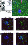
Antigen-specific CD8+ T cells are a necessary component of hepatitic foci. A: Naïve Thy1.1+ CD8+ OT-I T cells (5 × 10⁶) were transferred into C57BL/6 (Thy1.2+) mice that were subsequently infected with 103 EID50 of influenza WSN-OVAI. On day 9 after infection, intrahepatic lymphocytes were stained with antibodies against CD8, Thy1.1, and Thy1.2 for flow cytometric analysis; plot is gated on live lymphocytes. Numbers indicate the percentage of CD8s (in parentheses, percentage of total lymphocytes) staining with indicated markers. B: Identification of virus-specific CD8+ T cells in the foci of infected mice. Frozen sections of livers from A were stained with anti-Thy1.1 showing donor OT-I cells (red), anti-Thy1.2 showing host lymphocytes (blue), and nuclear counterstain Sytox Green. C–F: Immunohistochemical analysis of individual foci during influenza infection in frozen liver sections stained with combinations of anti-CD3-FITC (green), TUNEL reagent (red), and F4/80-APC (blue), as indicated (Original magnification, ×400). Cellular makeup of foci includes numerous CD8+ T cells and Kupffer cells.
Figure 5
Figure 5
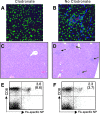
Kupffer cells are required for influenza-induced collateral liver damage. On day 2 of secondary infection with influenza A/PR8, mice received 200 μl of clodronate liposomes intraperitoneally. On day 5, livers were sectioned and stained. A and B: Frozen sections were stained with anti-F4/80 (Kupffer cells, blue), TUNEL (red), and nuclear counterstain Sytox Green (×400). Kupffer cells were absent after administration of clodronate (A) compared with PBS controls B. C and D: H&E-stained sections (×40) from clodronate-treated (C) and PBS control mice (D) show that focus formation was abrogated by Kupffer cell depletion. E and F: Flow cytometric analysis of virus NP-specific CD8+ T cells after clodronate (E) or PBS (F) injection. Numbers indicate percentage of virus-specific CD8s in livers; lower numbers in parentheses indicate percentage in lung airways.
Figure 6
Figure 6
Neither NK nor CD4+ T cells are required for the induction of CD8-dependent collateral liver damage. A: NK-deficient mice were infected with 105 EID50 influenza A/HK/x31, and day-8 liver sections were H&E-stained, revealing numerous foci and Councilman bodies (×400). Mean serum transaminase levels are indicated (in parentheses, control group transaminases). B: Focus typical of H&E-stained liver sections from MHC class II-deficient mice, infected 9 days prior with 105 EID50 influenza A/HK/x31 (×400). Mean serum transaminase levels are indicated (in parentheses, control group transaminases). C: TCR Cα−/− mice (with no endogenous T-cell response) received 2 × 10⁷ DO11.10 lymphocytes (CD4+ T cells specific for OVA323–339) intravenously and were subsequently infected with 500 pfu of influenza WSN-OVA323–339. Day-9 livers were harvested, and sections were H&E-stained. These mice contained influenza-activated CD4+ T cells in the absence of CD8+ T cells, and correspondingly, hepatitis was not visible histologically (×100).

Comment in

References

    1. Wirth S. [“Accompanying hepatitis” in viral infections]. Dtsch Med Wochenschr. 1995;120:461. - PubMed
    1. Yuen KY, Chan PK, Peiris M, Tsang DN, Que TL, Shortridge KF, Cheung PT, To WK, Ho ET, Sung R, Cheng AF. Clinical features and rapid viral diagnosis of human disease associated with avian influenza A H5N1 virus. Lancet. 1998;351:467–471. - PubMed
    1. Chan PK. Outbreak of avian influenza A(H5N1) virus infection in Hong Kong in 1997. Clin Infect Dis. 2002;34(Suppl 2):S58–S64. - PubMed
    1. Huang L, Soldevila G, Leeker M, Flavell R, Crispe IN. The liver eliminates T cells undergoing antigen-triggered apoptosis in vivo. Immunity. 1994;1:741–749. - PubMed
    1. Russell JQ, Morrissette GJ, Weidner M, Vyas C, Aleman-Hoey D, Budd RC. Liver damage preferentially results from CD8(+) T cells triggered by high affinity peptide antigens. J Exp Med. 1998;188:1147–1157. - PMC - PubMed

Publication types

MeSH terms

Substances