. 2006 Apr;168(4):1179-88.
doi: 10.2353/ajpath.2006.051081.

Therapeutic effect of vasoactive intestinal peptide on experimental autoimmune encephalomyelitis: down-regulation of inflammatory and autoimmune responses

Affiliations

Therapeutic effect of vasoactive intestinal peptide on experimental autoimmune encephalomyelitis: down-regulation of inflammatory and autoimmune responses

Elena Gonzalez-Rey et al. Am J Pathol. 2006 Apr.

Abstract

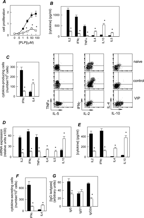
Multiple sclerosis (MS) is a disabling inflammatory, autoimmune demyelinating disease of the central nervous system. Despite intensive investigation, the mechanisms of disease pathogenesis remain unclear, and curative therapies are unavailable for MS. The current study describes a possible new strategy for the treatment of MS, based on the administration of the vasoactive intestinal peptide (VIP), a well-known immunosuppressive neuropeptide. Treatment with VIP significantly reduced incidence and severity of experimental autoimmune encephalomyelitis (EAE), in a MS-related rodent model system. VIP suppressed EAE neuropathology by reducing central nervous system inflammation, including the regulation of a wide spectrum of inflammatory mediators, and by selectively blocking encephalitogenic T-cell reactivity. Importantly, VIP treatment was therapeutically effective in established EAE and prevented the recurrence of the disease. Consequently, VIP represents a novel multistep therapeutic approach for the future treatment of human MS.


Figures

Figure 1
Figure 1
VIP treatment reduces EAE severity and incidence. A and B: VIP inhibits the progression of actively induced EAE and reduces the severity and relapses of RR-EAE. Active EAE was induced in C57BL/6 mice by immunization with MOG35-55, and RR-EAE was induced in SJL/J mice by immunization with PLP139-151. Immunized mice were treated intraperitoneally for 3 days with PBS (control) or with VIP (2 nmol/day, arrows) starting on day 5 or after the onset of clinical signs (days 10, onset; 16, acute phase; or 20, relapsing phase) as described in Materials and Methods. The data represent the mean clinical score for each group. Numbers in parentheses represent disease incidence of each group (percent mice with disease throughout the entire period). C: VIP effect on EAE is dose-dependent. SJL/J mice with RR-EAE were treated with different doses of VIP starting at the onset of clinical signs. The data represent the mean clinical score for each group. Numbers in parentheses represent disease incidence of each group (percent mice with disease throughout the entire period). *P < 0.001 versus control from day of onset (n = 12 to 22 mice/group).
Figure 2
Figure 2
VIP reduces the histopathology in the CNS of mice with EAE. SJL/J mice were induced with RR-EAE and treated with PBS (control, ▪) or with VIP at the onset of disease (□) as in Figure 1. Naïve animals without any treatment were used as negative controls. A–C: VIP treatment decreases demyelination, oligodendrocyte cell death, and inflammatory infiltration in the CNS. A: Transverse sections of several regions of the spinal cord (n = 6) randomly selected at the peak of clinical disease were stained with Luxol Fast Blue/periodic acid-Schiff (for demyelination) or with H&E (for inflammatory infiltration). A representative histological section from a control EAE mouse shows areas of demyelination (arrows) that correspond to areas of leukocytic infiltration (not shown). A comparable region of spinal cord from a VIP-treated mouse shows no histopathological signs, similar to naïve animals. Mean pathological scores of demyelination and lymphocyte infiltration were determined as described in Materials and Methods. B: Immunohistological evaluation of CNS infiltrates. Numbers of CD3+, CD4+, Gr-1+, and Mac-1+ cells in infiltrates per 104 square pixels from spinal cord sections were determined (n = 6). Immunohistological samples for CD4+ T cells are shown. C: Infiltrating mononuclear cells were isolated from brain at the peak of disease, and the numbers of CD4+ and CD8+ T cells, microglia (F4/80+CD45low), macrophages (MΦ, F4/80+CD45high), and dendritic cells (DCs, CD11c+) were determined by flow cytometry (n = 5). Numbers of infiltrating cells in naïve sham animals were lower than 104 cells, less microglia that were 2.4 × 10⁵ cells. D: The numbers of white matter oligodendrocytes in the lumbar dorsal cord were determined by immunohistochemistry for CNPase. Axonal damage was determined by Western blot analysis for abnormally dephosphorylated neurofilament H (NF-H) in whole spinal cord homogenates. Naïve animals showed 17.2 ± 2.3 CNPase+ cells/mm2. *P < 0.001 versus control.
Figure 3
Figure 3
VIP reduces the inflammation in the CNS of mice with EAE. SJL/J mice were induced with RR-EAE and treated with PBS (control, ▪) or with VIP at the onset of disease (□) as in Figure 1. A: VIP treatment decreases the expression of inflammatory mediators in the CNS. Total RNA was purified from spinal cords harvested at the peak of clinical disease and the expression of inflammatory cytokines, chemokines, chemokine receptors, and iNOS was determined by RPA. Data represent the mean ± SD of five mice per group. Representative RPA blots for each set of genes are shown. Samples from naïve animals were used as control for basal gene expression. B: BMNCs were pooled from five mice per group at the peak of disease and stimulated in vitro with PLP139-151 and splenocytes (as antigen-presenting cells, APCs). Cytokine and nitric oxide (NO) levels in supernatants were determined. Cells obtained from naïve animals or from EAE mice cultured with an irrelevant Ag (OVA, 10 μg/ml) or with medium alone did not produce any cytokines: <30 pg IP-10/ml, <20 pg TNF-α/ml, <15 pg IL-1β/ml, <50 pg RANTES/ml, <0.1 ng MIP-2/ml. Data represent the mean ± SD of five mice per group. *P < 0.001 versus control.
Figure 4
Figure 4
VIP decreases the Th1 cell autoreactive response in EAE. A–C: VIP treatment decreases Th1-mediated response in the periphery. SJL/J mice suffering from RR-EAE were treated with PBS (control, ▪) or with VIP at the onset of disease (□). DLN cells isolated at the peak of clinical disease were stimulated with different concentrations of PLP139-151, and the cell proliferative response (A) and the cytokine levels in supernatants (B) were determined. The number of PLP-specific T cells producing IFN-γ or IL-4 was determined by ELISPOT (C, left) and the expression of intracellular cytokines was determined by flow cytometry in gated CD4+ cells (C, right). DLN cells from naïve animals or from EAE mice cultured with an irrelevant Ag (OVA, 10 μg/ml) or with medium alone did not proliferate (A450 < 0.110) or produce any cytokines: <0.1 ng IFN-γ/ml, <30 pg IL-4/ml, <20 pg TNF-α/ml, <0.1 ng IL-10/ml, <0.1 ng/ml IL-2, and <50 pg IL-5/ml. Data represent the mean ± SD of eight mice per group. D–F: VIP regulates the Th1/Th2 balance in the CNS. Spinal cord and brain were isolated from the different experimental groups (n = 8) at the peak of clinical disease. D: Total RNA from spinal cords was isolated and the levels of gene expression for different Th1/Th2 cytokines were determined by RPA. BMNCs were stimulated with PLP139-151 and spleen APCs. E: Cytokine levels in supernatants were determined by enzyme-linked immunosorbent assay. Cells cultured with medium alone did not induce any cytokines. F: Number of PLP-specific T cells producing IFN-γ or IL-4 was determined by ELISPOT. Data are the mean ± SD of eight mice per group. G: VIP treatment regulates PLP-specific IgG levels. Sera were collected and the levels of PLP-specific IgG, IgG1, and IgG2a were determined by enzyme-linked immunosorbent assay. Serum obtained from naïve animals showed undetectable levels of PLP-specific IgG. Data are represented as the mean ± SD using arbitrary units, as analyzed in three separate experiments (eight mice per group per experiment). *P < 0.001 versus control.

References

    1. Steinman L. Assessment of animal models for MS and demyelinating disease in the design of rational therapy. Neuron. 1999;24:511–514. - PubMed
    1. Bauer J, Rauschka H, Lassmann H. Inflammation in the nervous system: the human perspective. Glia. 2001;36:235–243. - PubMed
    1. Owens T. The enigma of multiple sclerosis: inflammation and neurodegeneration cause heterogeneous dysfunction and damage. Curr Opin Neurol. 2003;16:259–265. - PubMed
    1. Pozo D, Delgado M. The many faces of vasoactive intestinal peptide in neuroimmunology: a cytokine rather a neuropeptide? FASEB J. 2004;18:1325–1334. - PubMed
    1. Delgado M, Pozo D, Ganea D. The significance of the vasoactive intestinal peptide in immunomodulation. Pharmacol Rev. 2004;56:249–290. - PubMed

Publication types

MeSH terms

Substances