. 2006 Apr;168(4):1200-9.
doi: 10.2353/ajpath.2006.050332.

A tumor necrosis factor receptor 1-dependent conversation between central nervous system-specific T cells and the central nervous system is required for inflammatory infiltration of the spinal cord

Affiliations

A tumor necrosis factor receptor 1-dependent conversation between central nervous system-specific T cells and the central nervous system is required for inflammatory infiltration of the spinal cord

Mary Ann Gimenez et al. Am J Pathol. 2006 Apr.

Abstract

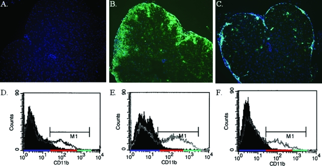
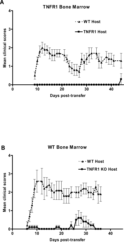
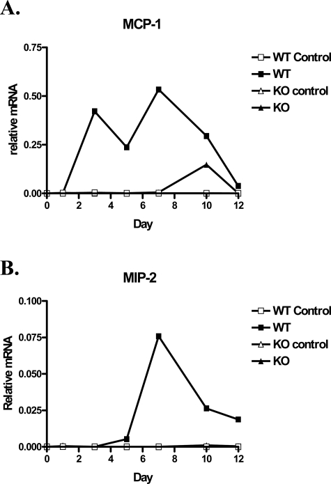
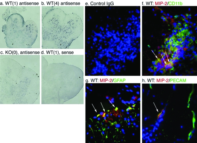
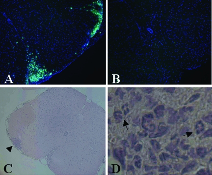
We examined the role of tumor necrosis factor receptor 1 (TNFR1) in inflammation initiated by the adoptive transfer of central nervous system (CNS)-specific Th1 cells in experimental autoimmune encephalomyelitis, a murine model of multiple sclerosis. This adoptive transfer paradigm eliminates the confounding effects of bacterial adjuvants in the analysis of inflammation. We found that although T cells could reach the meninges and perivascular space in the absence of TNFR1, recruitment of other inflammatory cells from the blood was dramatically reduced. The reduction in the recruitment of CD11b(hi) cells correlated with a dramatic reduction in the production of the chemokines CCL2 (MCP-1) and CXLC2 (MIP-2) in TNFR1-deficient hosts. Bone marrow chimera experiments demonstrated that TNF can be effectively supplied by either the hematopoietic system or the CNS, but the essential TNFR1-responsive cells reside in the CNS. Previous work has demonstrated that microglia produce CCL2, and here we demonstrate that astrocytes and endothelial cells produced CXCL2 in the early stages of inflammation. Therefore, productive inflammation results from a conversation, or mutually responding signals, between the initiating T cells and cells in the parenchyma of the spinal cord.


Figures

Figure 1
Figure 1
Recruitment of CD11b+ cells to the CNS is attenuated in TNFR1-null mice. Th1-skewed Thy1.1 WT CD4+ MOG-specific T cells (5 × 10⁶) were injected intravenously into sublethally irradiated (450 R) 6- to 8-week-old Thy1.2 B6 WT and TNFR1-null recipients. Frozen sections of lumbar-sacral spinal cord from naïve B6 (A); day 12 WT, clinical score = 2 (B); and day 12 TNFR1-null, clinical score = 0 (C) were immunostained for CD11b (green) and counterstained with DAPI (blue). Flow cytometry for CD11b was also performed on cells isolated from the brain and spinal cord of similar, naïve (M1 MFI = 140) (D); WT, clinical score = 2 (M1 MFI = 339) (E); and TNFR1, clinical score = 0 (M1 MFI = 187) (F), hosts day 12 after transfer. The solid histogram is staining with an isotype control antibody and represents CD11bneg cells highlighted by the blue underscore on the x axis. CD11blo cells are highlighted by the red underscore and CD11bhi cells highlighted by the green underscore. The MFIs were calculated for cells in the M1 gate. Original magnifications, ×65.
Figure 2
Figure 2
WT bone marrow does not reconstitute disease in TNFR1-null animals. EAE was passively induced into bone marrow chimeras. A: TNFR1-null bone marrow in lethally irradiated WT (n = 9, ▴), or TNFR1-null (n = 9, ▪) mice. B: TNFR1-null bone marrow in lethally irradiated WT (n = 9, ▴), or TNFR1-null (n = 9, ▪). Th1-skewed Thy1.1 WT CD4+ MOG-specific T cells (1 × 10⁷) were injected intravenously 6 to 8 weeks after bone marrow transplantation. Clinical signs of disease were monitored daily and graded on a scale of 0 to 5 as described previously.
Figure 3
Figure 3
Time course of relative mRNA levels of MCP-1 (A) and MIP-2 (B) in the spinal cord of WT and TNFR1-null mice. Th1-skewed Thy1.1 WT CD4+ MOG-specific T cells (5 × 10⁶) or PBS were injected intravenously into sublethally irradiated (450 R) 6- to 8-week-old Thy1.2 B6 WT and TNFR1-null recipients. On days 1, 3, 5, 7, 10, and 12 after transfer, total RNA was isolated from spinal cords and mRNA transcripts were analyzed by quantitative real-time RT-PCR. All samples were normalized to HPRT.
Figure 4
Figure 4
CXCL2 is produced by astrocytes and vascular endothelium of the CNS as well as CD11b+ cells from the inflammatory infiltrate early in the disease course. In situ hybridization analysis of spinal cord sections of irradiated WT (a, b, and d) and TNFR1-null (c) mice 6 days after transfer of 5 × 10⁶ Thy 1.1 cells using digoxigenin-labeled anti-sense (a–c) and sense (d) riboprobes. Clinical scores of animals are shown in parentheses. Immunohistochemical analyses of CXCL2 (red) expression within CD11b- (green) (f), GFAP- (green) (g), and PECAM-expressing cells (green) (h). Nuclei have been counterstained with DAPI. Arrowheads: Co-localization (yellow) of CXCL2- and CD11b- (f), GFAP- (g), and PECAM- (h) positive cells. Arrows: MIP-2-negative, PECAM-positive endothelium. e: Control IgG does not demonstrate any specific staining. Analyses performed on sections from six animals, n = three mice per group. Original magnifications, ×350.
Figure 5
Figure 5
Recruitment of peripheral GR1+ cells to the CNS is blocked in TNFR1-null mice. Th1-skewed Thy1.1 WT CD4+ MOG-specific T cells (5 × 10⁶) were injected intravenously into sublethally irradiated (450 R) 6- to 8-week-old Thy1.2 B6 WT and TNFR1-null recipients. Frozen sections of lumbar-sacral spinal cord from day 6 WT, CS = 1.5 (A), and day 6 TNFR1-null, CS = 0 (B) were immunostained for GR-1 (green) and counterstained with DAPI (blue). C and D: H&E section of lumbar-sacral spinal cord from day 7 WT, CS = 1, shows a focus of inflammatory infiltrate (arrowhead) invading the parenchyma, which contains many neutrophils (arrows). Original magnifications: ×65 (A–C); and ×260 (D).

References

    1. Goverman J, Brabb T. Rodent models of experimental allergic encephalomyelitis applied to the study of multiple sclerosis. Lab Anim Sci. 1996;46:482–492. - PubMed
    1. Lublin FD, Reingold SC. Defining the clinical course of multiple sclerosis: results of an international survey. National Multiple Sclerosis Society (USA) Advisory Committee on Clinical Trials of New Agents in Multiple Sclerosis. Neurology. 1996;46:907–911. - PubMed
    1. Lucchinetti CF, Brueck W, Rodriguez M, Lassmann H. Multiple sclerosis: lessons from neuropathology. Semin Neurol. 1998;18:337–349. - PubMed
    1. Prineas JW. The Neuropathology of Multiple Sclerosis. Koetsier JC, editor. New York: Elsevier,; 1985:p 213.
    1. Raine CS. Biology of disease. Analysis of autoimmune demyelination: its impact upon multiple sclerosis. Lab Invest. 1984;50:608–635. - PubMed

Publication types

MeSH terms