. 2006 Apr;168(4):1227-40.
doi: 10.2353/ajpath.2006.050125.

Type I interferon production by tertiary lymphoid tissue developing in response to 2,6,10,14-tetramethyl-pentadecane (pristane)

Affiliations

Type I interferon production by tertiary lymphoid tissue developing in response to 2,6,10,14-tetramethyl-pentadecane (pristane)

Dina C Nacionales et al. Am J Pathol. 2006 Apr.

Abstract

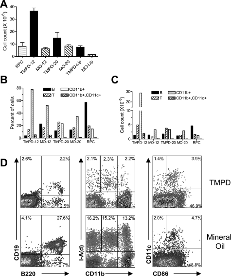
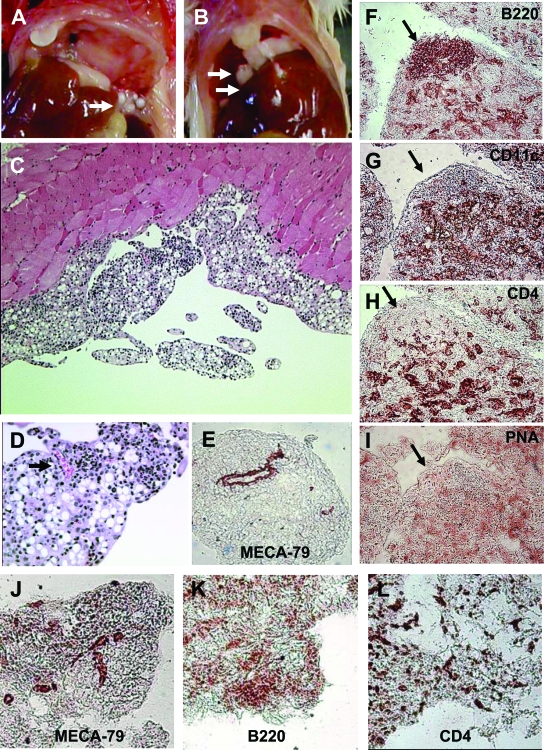
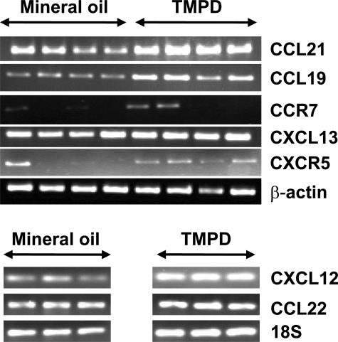
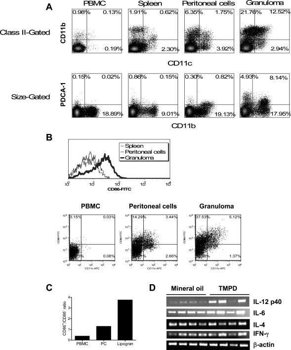
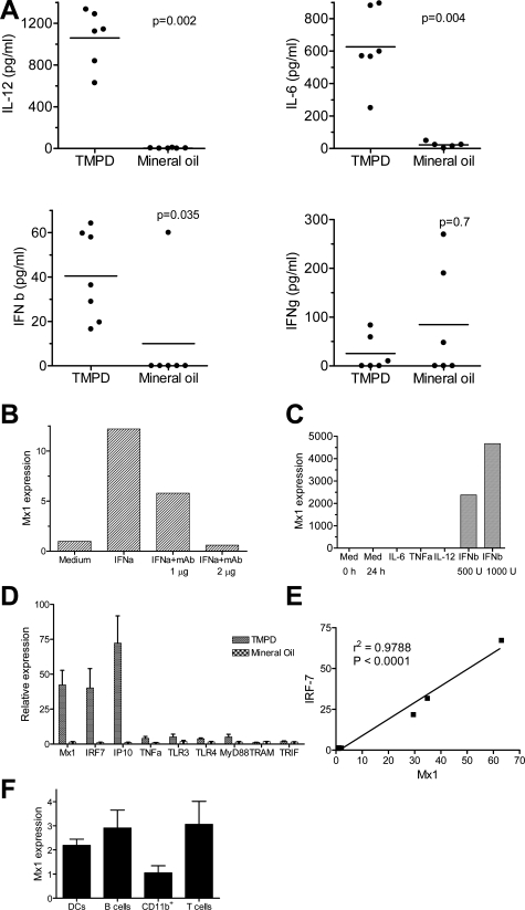
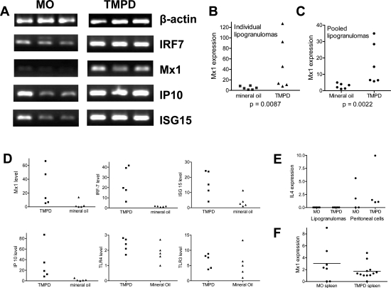
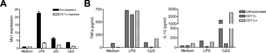
Lymphoid neogenesis is associated with antibody-mediated autoimmune diseases such as Sjogren's syndrome and rheumatoid arthritis. Although systemic lupus erythematosus is the prototypical B-cell-mediated autoimmune disease, the role of lymphoid neogenesis in its pathogenesis is unknown. Intraperitoneal injection of 2,6,10,14-tetramethyl-pentadecane (TMPD, pristane) or mineral oil causes lipogranuloma formation in mice, but only TMPD-treated mice develop lupus. We report that lipogranulomas are a form of lymphoid neogenesis. Immunoperoxidase staining of lipogranulomas revealed B cells, CD4(+) T cells, and dendritic cells and in some cases organization into T- and B-cell zones. Lipogranulomas also expressed the lymphoid chemokines CCL21, CCL19, CXCL13, CXCL12, and CCL22. Expression of the type I interferon (IFN-I)-inducible genes Mx1, IRF7, IP-10, and ISG-15 was greatly increased in TMPD- versus mineral oil-induced lipogranulomas. Dendritic cells from TMPD lipogranulomas underwent activation/maturation with high CD86 and interleukin-12 expression. Magnetic bead depletion of dendritic cells markedly diminished IFN-inducible gene (Mx1) expression. We conclude that TMPD-induced lupus is associated with the formation of ectopic lymphoid tissue containing activated dendritic cells producing IFN-I and interleukin-12. In view of the increased IFN-I production in systemic lupus erythematosus, these studies suggest that IFN-I from ectopic lymphoid tissue could play a role in the pathogenesis of experimental lupus in mice.


Figures

Figure 1
Figure 1
Flow cytometry of peritoneal exudate cells. BALB/c mice were treated intraperitoneally with TMPD or mineral oil (MO). Peritoneal exudate cells were harvested by lavage 12 or 20 weeks later (TMPD-12, MO-12, TMPD-20, and MO-20, respectively). Resident peritoneal cells (RPCs) from three untreated BALB/c mice were analyzed for comparison. Pooled lipogranulomas from individual mice were harvested at 20 weeks after TMPD or mineral oil injection (TMPD-Lip and MO-Lip, respectively) and treated with collagenase and dispase. A: Total peritoneal cell counts. Total cell counts were determined using a hemocytometer (mean and SE are shown). B and C: Cellular composition of peritoneal exudates by flow cytometry. Peritoneal cells were analyzed by flow cytometry using anti-B220 and CD19 (B cells), anti-CD3 (T cells), anti-CD11b (monocytes and some B cells), and CD11c (dendritic cells). The percent (B) and absolute numbers (C) of B and lymphocytes, CD11b+ cells, and CD11b+, CD11c+ monocyte-derived dendritic cells are shown. D: Representative examples of the staining for mineral oil- versus pristane-treated mice. Peritoneal cells were stained with anti-B220 and anti-CD19 mAbs (left), anti-CD11b and anti-I-Ad mAbs (middle), or anti-CD11c and anti-CD86 mAbs (right).
Figure 2
Figure 2
Pathology of lipogranulomas. A: Lipogranulomas adherent to the mesothelial surface of a mouse treated 12 weeks earlier with TMPD (arrow). B: Lipogranulomas adherent to the mesothelial surface of the diaphragm of a mouse treated 12 weeks earlier with mineral oil (arrows). C: H&E staining of lipogranulomas from a TMPD-treated mouse. Note numerous oil droplets and lymphocytic infiltration. D and E: Neovascularization of TMPD-induced lipogranulomas. D: H&E staining of a peritoneal lipogranuloma from a mouse treated 12 weeks earlier with TMPD showing a blood vessel filled with erythrocytes (arrow). E: Immunohistochemistry of frozen tissue (5-μm sections, immunoperoxidase technique) from a peritoneal lipogranuloma from a mouse treated 12 weeks earlier with TMPD and stained with MECA-79 antibody (anti-peripheral lymph node addressin, a marker for high endothelial venules). F–I: Immunohistochemistry of serial sections through a TMPD lipogranuloma. A snap-frozen lipogranuloma was sectioned at 5 μm and stained by the immunoperoxidase technique as follows: biotinylated anti-mouse CD45R/B220 (F), biotinylated anti-mouse CD11c (G), biotinylated anti-mouse CD4 (H), and biotinylated peanut agglutinin (I). Arrow indicates the location of a collection of B cells in the serial sections. J–L: Immunohistochemistry of mineral oil lipogranulomas. Snap-frozen lipogranulomas from mineral oil-treated mice were sectioned (5 μm) and stained by the immunoperoxidase technique using biotinylated MECA-70 (anti-PNAd) (J), B220 (B cells) (K), or CD4 (T cells) (L).
Figure 3
Figure 3
Expression of chemokines and chemokine receptors in lipogranulomas. RNA isolated from pooled lipogranulomas (12 weeks) from four TMPD-treated and four mineral oil-treated mice was used to synthesize cDNA. Expression of SLC (CCL21), ELC (CCL19), CCR7, BLC (CXCL13), and CXCR5 (top), as well as SDF-1 (CXCL12) and MDC (CCL22) (bottom) was analyzed by RT-PCR, normalized to β-actin (top) or 18S ribosomal RNA (bottom).
Figure 4
Figure 4
TMPD lipogranulomas contain activated dendritic cells. A: Accumulation of dendritic cells in the lipogranulomas. Peripheral blood mononuclear cells (PBMCs), spleen, peritoneal exudate cells, and pooled lipogranuloma cells from a representative BALB/c mouse 20 weeks after intraperitoneal TMPD injection were analyzed by flow cytometry. Top: Percentages of CD11c/CD11b+ monocytes/macrophages, CD11c+/CD11b+ monocyte-derived (?) dendritic cells, and CD11c+/CD11b dendritic cells in each location (PBMCs, spleen, peritoneal exudate, and lipogranulomas) are indicated. Bottom: Percentages of PDCA-1+ dendritic cells in each location (peripheral blood, spleen, peritoneal exudate, and lipogranulomas) are shown. B: Lipogranuloma dendritic cells express high levels of CD86. Expression of CD86 by CD11c+ cells in the peripheral blood, spleen, peritoneal exudate, and lipogranulomas from representative TMPD-treated mice was determined by flow cytometry. Top: Mean fluorescence intensity of CD86 in CD11c-gated cells from spleen, peritoneum, and lipogranulomas. Bottom: CD11c and CD86 staining of PBMCs, peritoneal cells, and isolated lipogranuloma cells. C: Dendritic cells in lipogranulomas preferentially express CD86. Shown is the ratio of CD86+ to CD86 dendritic cells (CD11c+) in peripheral blood mononuclear cells (PBMCs), peritoneal cells (PCs), and TMPD-treated mouse lipogranulomas. D: Increased expression of IL-12 p40 in TMPD lipogranulomas. RT-PCR was performed using cDNA from lipogranulomas from four mineral oil-treated mice and four TMPD-treated mice. Sequences of the IL-12 p40, IL-6, IL-4, IFN-γ, and β-actin primers used are shown in Table 1.
Figure 5
Figure 5
IFN-I-inducible gene expression in peritoneal exudate cells. A: Cytokine expression in peritoneal washings. Peritoneal lavage was performed in mice treated with TMPD or mineral oil and levels of IL-12, IL-6, IFN-β, and IFN-γ in the washings were measured by sandwich ELISAs. Levels were compared using the Mann-Whitney test. B: Stimulation of Mx1 gene expression by IFN-α. RAW 264.7 cells were treated for 24 hours with IFN-α (1000 U/ml) in the absence or presence of anti-IFN-α neutralizing antibodies (1 or 2 μg/ml). Mx1 expression (normalized to β-actin) was measured from cDNA by real-time PCR. C: Specificity of Mx1 expression for IFN-I. RAW 264.7 cells were treated with IFN-β (1000 or 500 U/ml), IL-6 (5 ng/ml), TNF-α (20 ng/ml), or IL-12 (10 or 20 ng/ml) or with medium (Med) alone. After 24 hours, cells were harvested and RNA extracted for cDNA synthesis. Mx1 expression was determined by real-time PCR, normalized to β-actin. D: Increased expression in TMPD- versus mineral oil-elicited peritoneal cells. RNA was extracted and cDNA was synthesized from freshly collected peritoneal exudate cells of mice treated with TMPD or mineral oil. Expression of mRNAs for the IFN-inducible genes Mx1, IRF-7, and IP-10 as well as Toll-like receptors (TLR3, TLR4) and adapter proteins MyD88, TRAM, and TRIF was measured by real-time PCR normalized to β-actin. E: Correlation of Mx1 and IRF-7 expression. Expression of Mx1 and IRF-7 was measured (real-time PCR) in RNA isolated from either unstimulated peritoneal cells or LPS-stimulated peritoneal cells from the same group of mice (seven measurements). Expression of the two IFN-inducible genes correlated highly (r2 = 0.9788, P < 0.0001). F: Mx1 expression by individual cell types. Dendritic cells (CD11c+), B cells (CD19+), monocytes/macrophages (CD11b+), and T cells (CD3+) were isolated from peritoneal exudates of TMPD-treated mice using magnetic beads and RNA was extracted. Mx1 expression by each cell type was determined by real-time PCR normalized to β-actin.
Figure 6
Figure 6
IFN-I-inducible gene expression in lipogranulomas. BALB/c mice were treated with TMPD or mineral oil and RNA was isolated from lipogranulomas adherent to the peritoneal lining or diaphragm at 12 weeks and used to synthesize cDNA. A: Expression of IFN-inducible genes. RT-PCR was performed for β-actin, IRF-7, Mx1, IP-10, and ISG-15 using lipogranuloma cDNA from mineral oil (MO)- or TMPD-treated mice. B: Mx1 expression, individual lipogranulomas. Expression of Mx1 normalized to β-actin was measured in individual lipogranulomas (n = 6) from mice treated with either TMPD or mineral oil by real-time PCR. Mx1 expression was significantly higher in TMPD versus mineral oil lipogranulomas (P = 0.0087, Mann-Whitney test). C: Mx1 expression, pooled lipogranulomas. Expression of Mx1 normalized to β-actin was measured in pooled lipogranulomas from six mice treated with TMPD and six mice treated with mineral oil. Mx1 expression (real-time PCR) was significantly higher in TMPD versus mineral oil lipogranulomas (P = 0.0022, Mann-Whitney test). D: Expression of other IFN-I-inducible genes, individual lipogranulomas. Gene expression in individual lipogranulomas from representative TMPD or mineral oil mice (five lipogranulomas per mouse) was measured by real-time PCR, normalized to β-actin. Expression of IFN-inducible genes Mx1, IRF-7, ISG-15, and IP-10 as well as TLR3 and TLR4 expression is shown. E: IL-4 expression in pooled lipogranulomas and peritoneal exudate. Expression of IL-4 normalized to β-actin (real-time PCR) was measured in pooled lipogranulomas and peritoneal exudate cells from four mice treated with TMPD and four mice treated with mineral oil (MO). F: Mx1 expression in the spleen. Mx1 expression normalized to β-actin (real-time PCR) was measured in spleen cells from mice treated with mineral oil (MO, n = 7) or TMPD (n = 12).
Figure 7
Figure 7
IFN-I and IL-12 production by CD11c+ peritoneal cells. A: Reduced TLR ligand stimulated Mx1 expression after depleting CD11c+ cells. Unfractionated peritoneal exudate cells and peritoneal exudate cells depleted of CD11c+ cells (magnetic beads) were treated with LPS (10 μg/ml), CpG ODN no. 1826 (10 μg/ml), or poly (I:C) (50 μg/ml) for 4 hours before extracting RNA. Mx1 expression was quantified by real-time PCR (normalized to β-actin). B: Reduced TLR ligand stimulated IL-12 production after depleting CD11c+ cells. CD11c+ cells from peritoneal exudate of a mouse treated 5 months previously with TMPD were depleted with magnetic beads, and TNF-α (left) and IL-12 (right) production in culture supernatants in response to LPS (10 μg/ml) or CpG DNA (10 μg/ml ODN no. 1826) were determined 24 hours later by ELISA.

Similar articles

Cited by

References

    1. Kratz A, Campos-Neto A, Hanson MS, Ruddle NH. Chronic inflammation caused by lymphotoxin is lymphoid neogenesis. J Exp Med. 1996;183:1461–1472. - PMC - PubMed
    1. Drayton DL, Ying X, Lee J, Lesslauer W, Ruddle NH. Ectopic LT alpha beta directs lymphoid organ neogenesis with concomitant expression of peripheral node addressin and a HEV-restricted sulfotransferase. J Exp Med. 2003;197:1153–1163. - PMC - PubMed
    1. Luther SA, Lopez T, Bai W, Hanahan D, Cyster JG. BLC expression in pancreatic islets causes B cell recruitment and lymphotoxin-dependent lymphoid neogenesis. Immunity. 2000;12:471–481. - PubMed
    1. Yoneyama H, Matsuno K, Zhang Y, Murai M, Itakura M, Ishikawa S, Hasegawa G, Naito M, Asakura H, Matsushima K. Regulation by chemokines of circulating dendritic cell precursors, and the formation of portal tract-associated lymphoid tissue, in a granulomatous liver disease. J Exp Med. 2001;193:35–49. - PMC - PubMed
    1. Hjelmstrom P, Fjell J, Nakagawa T, Sacca R, Cuff CA, Ruddle NH. Lymphoid tissue homing chemokines are expressed in chronic inflammation. Am J Pathol. 2000;156:1133–1138. - PMC - PubMed

Publication types

MeSH terms