. 2006 Apr;168(4):1241-8.
doi: 10.2353/ajpath.2006.050360.

Deficiency of C4 from donor or recipient mouse fails to prevent renal allograft rejection

Affiliations

Deficiency of C4 from donor or recipient mouse fails to prevent renal allograft rejection

Tao Lin et al. Am J Pathol. 2006 Apr.

Abstract

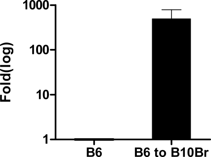
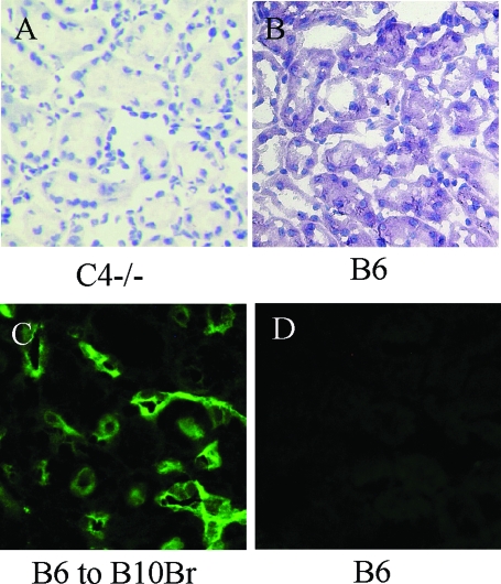
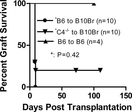
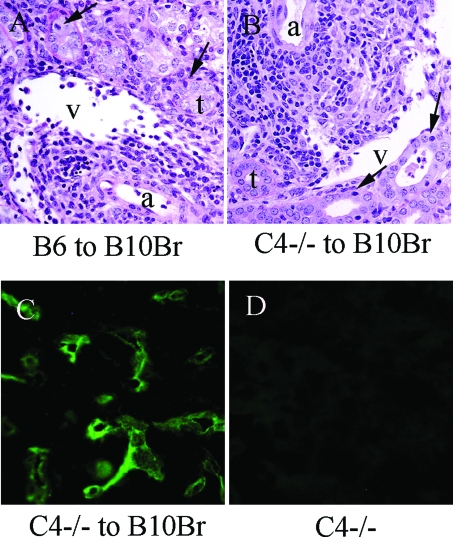
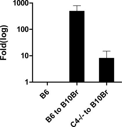
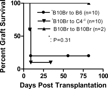
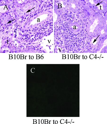
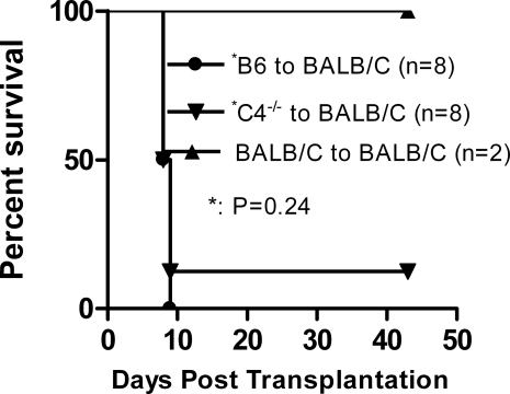
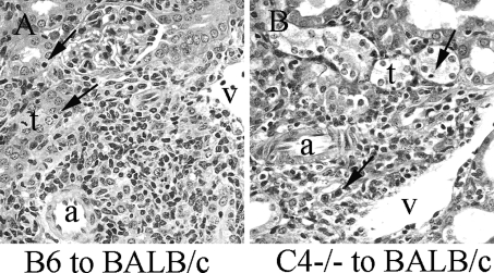
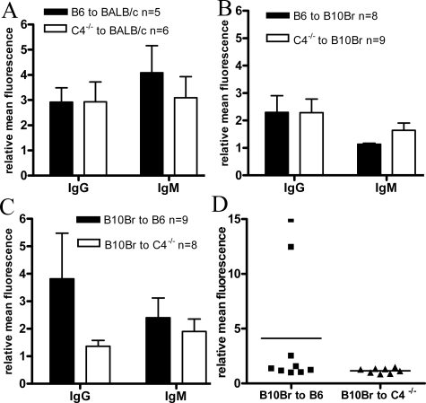
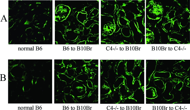
Complement effector products generated in the transplanted kidney are known to mediate transplant rejection, but which of the three main activation pathways of complement trigger this response is unclear. Here we assessed the role of the classical and lectin pathways by studying the common component C4 in mouse kidney transplant rejection. We transplanted wild-type or C4-null H-2(b) donor kidneys into H-2(k) or H-2(d) recipients, or vice-versa, to assess the roles of donor kidney and recipient expression of complement. Intragraft C4 gene expression rose substantially during rejection. However, we found no significant association between graft acceptance and the presence of C4 in either the donor kidney or recipient mouse. At the time of rejection, we found no significant differences in alloantibody response in the different groups. Tubular deposition of C3 to C9 occurred regardless of the absence or presence of C4 in either the donor or recipient mouse, indicating that C4 was dispensable for complement activation at this site. These data suggest that complement activation and renal allograft rejection are independent of the classical and lectin pathways in these models, implying that in the absence of these pathways the alternative pathway is the main trigger for complement-mediated rejection.


Figures

Figure 1
Figure 1
Complement C4 mRNA expression in normal B6 kidneys and rejecting B6 grafts. Real-time RT-PCR of C4 transcripts in rejecting B6 grafts. C4 mRNA levels are expressed as fold increase over normal B6 kidneys. Each analysis was performed on RNA isolated from three individual kidneys, and assays were run in duplicate.
Figure 2
Figure 2
Localization of C4 expression in wild-type donor kidney. A and B: In the wild-type normal B6 kidney, in situ hybridization of C4 mRNA showed positive staining in the cytoplasm of tubule cells. There was no staining found in C4−/− kidney tissue. C: Immunofluorescence staining for C4 in allografts showed staining predominantly of peritubular capillary walls. D: Background C4 staining in normal B6 kidney tissue. Original magnifications, ×250.
Figure 3
Figure 3
Effect of donor C4 status on graft survival: B10.Br recipients of wild-type kidney reject their transplants within 9 days. Eight of ten B10.Br recipients of C4−/− grafts rejected their transplants in 9 days; the other two recipients did not acutely reject but survived until killing at day 72 or day 110. P = 0.42 comparing C4−/− and wild-type grafts.
Figure 4
Figure 4
Histology and C4 staining in the rejecting B6 grafts. A: Typical rejecting kidney allograft (B6 to B10.Br) at day 8 showing heavy interstitial infiltration, venulitis, and tubulitis (→) (PAS stain). B: Typical rejecting C4−/− kidney graft at day 8 showing a similar pattern to B6 wild-type graft (PAS stain). C: Diffuse C4 deposition was detected in the peritubular capillary walls of C4−/− grafts, which was not present in normal nontransplanted C4−/− kidney (D). a, Arteriole; v, vein; t, tubule. Original magnifications, ×250.
Figure 5
Figure 5
Real-time RT-PCR of C4 transcripts in rejecting grafts. C4 mRNA levels are expressed as fold increase over normal B6 kidneys. Three samples were used in each analysis, and assays were run in duplicate.
Figure 6
Figure 6
Effect of recipient C4 on graft survival. In B10.Br to B6 group, two recipients were sacrificed at days 56 and 82; the others rejected their grafts at day 8 or day 9. C4−/− recipients rejected 9 of 10 B10.Br grafts before day 9. Another recipient was killed at day 34.
Figure 7
Figure 7
Histology and C4 staining in the rejecting B10.Br grafts. A and B: B10.Br allografts in wild-type and C4−/− B6 recipients at day 8 showing similar pattern of interstitial infiltration, venulitis, and tubulitis (→) (PAS stain). C: Immunofluorescence staining for C4 in B10.Br to C4−/− allografts showed background staining. Original magnifications, ×250.
Figure 8
Figure 8
Kidney transplant rejection in BALB/c recipients. Seven of eight BALB/c recipients of C4−/− grafts rejected their transplants within 9 days. The other one was killed at day 43. In B6 to BALB/c group, acute rejection occurred in every case.
Figure 9
Figure 9
Histology of rejecting B6 graft. A: Rejecting kidney allograft (B6 into BALB/c) at day 8 typically showing a heavy interstitial infiltration, venulitis, and tubulitis (→) (PAS stain). B: Typical rejecting C4−/− kidney graft at day 8 showing a similar pattern to B6 wild-type graft (PAS stain). Original magnifications, ×250.
Figure 10
Figure 10
Donor-specific alloantibody response. IgG and IgM alloantibodies were measured by flow cytometry in serum from recipients 8 days after transplantation. A–C: Detection of alloantibodies in recipients of different strain combination, P > 0.05 when comparing IgG or IgM in every pair of groups. D: Individual IgG responses in C4+/+ or C4−/− B6 recipients of B10.Br grafts.
Figure 11
Figure 11
Complement detection in rejecting renal allografts. A: Strong, diffuse C3d deposition was detected in the distribution of the tubular basement membrane and peritubular capillaries of rejecting grafts in all of the groups. B: Staining for C9 in rejecting grafts showed a similar pattern of staining as C3d. Original magnifications, ×250.

References

    1. Sacks SH, Chowdhury P, Zhou W. Role of the complement system in rejection. Curr Opin Immunol. 2003;15:487–492. - PubMed
    1. Baldwin WM, III, Larsen CP, Fairchild RL. Innate immune responses to transplants: a significant variable with cadaver donors. Immunity. 2001;14:369–376. - PubMed
    1. Korb LC, Ahearn JM. C1q binds directly and specifically to surface blebs of apoptotic human keratinocytes: complement deficiency and systemic lupus erythematosus revisited. J Immunol. 1997;158:4525–4528. - PubMed
    1. Gasque P. Complement: a unique innate immune sensor for danger signals. Mol Immunol. 2004;41:1089–1098. - PubMed
    1. Fujita T. Evolution of the lectin-complement pathway and its role in innate immunity. Nat Rev Immunol. 2002;2:346–353. - PubMed

Publication types