. 2006 Apr;168(4):1321-34.
doi: 10.2353/ajpath.2006.041220.

Peroxisomal multifunctional protein-2 deficiency causes motor deficits and glial lesions in the adult central nervous system

Affiliations

Peroxisomal multifunctional protein-2 deficiency causes motor deficits and glial lesions in the adult central nervous system

Steven Huyghe et al. Am J Pathol. 2006 Apr.

Abstract

In humans, mutations inactivating multifunctional protein-2 (MFP-2), and thus peroxisomal beta-oxidation, cause neuronal heterotopia and demyelination, which is clinically reflected by hypotonia, seizures, and death within the first year of life. In contrast, our recently generated MFP-2-deficient mice did not show neurodevelopmental abnormalities but exhibited aberrations in bile acid metabolism and one of three of them died early postnatally. In the postweaning period, all survivors developed progressive motor deficits, including abnormal cramping reflexes of the limbs and loss of mobility, with death at 6 months. Motor impairment was not accompanied by lesions of peripheral nerves or muscles. However, in the central nervous system MFP-2-deficient mice overexpressed catalase in glial cells, accumulated lipids in ependymal cells and in the molecular layer of the cerebellum, exhibited severe astrogliosis and reactive microglia predominantly within the gray matter of the brain and the spinal cord, whereas synaptic and myelin markers were not affected. This culminated in degenerative changes of astroglia cells but not in overt neuronal lesions. Neither the motor deficits nor the brain lesions were aggravated by increasing the branched-chain fatty acid concentration through dietary supplementation. These data indicate that MFP-2 deficiency in mice causes a neurological phenotype in adulthood that is manifested primarily by astroglial damage.


Figures
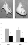
Figure 1
Figure 1
Development of motor deficits in MFP-2-deficient mice. Beginning at the age of 1 month, the MFP-2 knockout mice lose the characteristic leg-spreading reflex elicited by being suspended without contact to a supporting surface, as shown for a control mouse in A. Instead, mice progressively clasp both fore- and hindlegs (B), a phenomenon often observed in supraspinal CNS lesions. C: In parallel to the development of pathological reflexes, animals lose grip strength and motor coordination, cutting down their time on a rotarod instrument to <50% of that of control mice. Mean ± SEM of three control and three MFP2−/− 8-week-old mice is shown. *P = 0.05, **P < 0.005.
Figure 2
Figure 2
Lipid accumulations in the CNS. Oil Red O-positive lipid inclusions are present in the ependymal cells of the entire ventricular system (A–D) and in cerebellar Bergmann glia fibers (E–K) of 5-month-old mice. Within the lateral ventricles (overview in A and B, the labels D, L, M, V indicating the dorsal, lateral, medial, and ventral direction, respectively) the size of the inclusions strikingly correlates with the position of the ependymal cells. They remain small and dust-like in the medial wall (inset a in A) and progressively increase in the dorsal (b) and lateral wall (c). Lipid inclusions are likewise present in the lining of the third ventricle (C) and the central canal of the spinal cord (not shown). On the TEM level, the inclusions appear as round, membrane-bound structures filled with coarse, highly contrasted material in a translucent matrix (D). Note that the choroid plexus epithelium is not affected by lipid storage, which abruptly stops at the junction to the ependymal layer (arrows in insets d and e). In the cerebellar cortex of 5-month-old MFP-2 knockout mice, Oil Red O-positive inclusions (black arrows in F, compare to aged-matched WT mice in E) are only present in the molecular zone (MoZ), but remain notably absent from the white matter (WM) and the (neuronal) internal granule cell layer (IGL). The lipid droplets line up in straight pearl chain patterns extending to the surface (small arrows in G) and also concentrate on cell bodies (large arrows) between Purkinje cells (PC), both features indicative of Bergmann glial cells. This is further confirmed by a co-stain of Oil Red O and GFAP (H). Although no lipid droplets are detectable even with the more sensitive dark field optics in adult WT cerebella (I), at 4 weeks postnatally lipid droplets accumulate between (but not in) Purkinje cell bodies, but only at low levels in the molecular zone, and sometimes meningeal cells (Men) (J). The characteristic pearl chain pattern is established at 8 weeks (K), but intensifies further later on (compare to F). Scale bars: 200 μm (A–C); 5 μm (D); 100 μm (E, F); 20 μm (G, H); 50 μm (I–K).
Figure 3
Figure 3
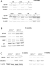
Time course of expression of catalase as well as neuronal and glial markers in MFP-2-deficient mice. A: A significant up-regulation of catalase protein could be seen by Western blotting in brain stem (BS), cerebellum (cere), diencephalon (Di), hippocampus (hippo), cortex, olfactory bulb (OB), and spinal cord (SC), with a concomitant increase of GFAP expression. In whole brain homogenates (shown for 5 months in B) the expression of oligodendroglial MBP and neuronal synaptophysin (SY) remained unchanged, indicating the absence of major white matter and neuronal defects. C: It is striking thereby that catalase up-regulation is a very early event, clearly being present at ∼3 weeks postnatally, a time at which GFAP expression levels were still fully normal. At 8 weeks a slight increase in GFAP expression but clear-cut increase of catalase were noticed (C). Incubation times and concentration of the primary and secondary antibodies were the same for all blots but developing time with the alkaline-phosphatase substrate was different.
Figure 4
Figure 4
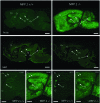
MFP-2 deficiency causes up-regulation of catalase and gliosis in the gray matter. Five-month-old MFP-2-deficient brains are characterized by a significant up-regulation of catalase in all brain regions, whereby only in white matter tracts like fimbria or corpus callosum immunoreactivity is not visibly increased beyond WT level. This increase is paralleled by reactive astrogliosis (GFAP staining) in a similar distribution, again sparing white matter tracts. Co, cortex; Cc, corpus callosum; Fi, fimbria hippocampi; Hi, hippocampus; Cb, cerebellum. Scale bars, 2 mm.
Figure 5
Figure 5
MFP-2 deficiency causes microglia activation in the gray matter, but no apparent white matter defects. The up-regulation of catalase and the astrogliosis seen in the gray matter of MFP-2-deficient mice is paralleled by an intense microglial reaction, both evidenced by a drastically increased stainability for F4/80 and binding of RCA-1, both again sparing white matter tracts like fimbria (Fi) or corpus callosum (Cc). Note that no change is visible in the expression of the myelin marker MBP. Co, cortex; Cc, corpus callosum; Fi, fimbria hippocampi; Hi, hippocampus; Cb, cerebellum. Scale bars, 2 mm.
Figure 6
Figure 6
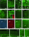
Increased reactivity for catalase and glial activation and degeneration. Increased catalase immunoreactivity in Bergmann glial fibers is visible as early as 3 weeks postnatally (shown at 5 weeks in A and B) and becomes more evident thereafter. C and D show a stain with GFAP and catalase on adjacent sections at 8 weeks, E and F a comparison of WT and MFP-2−/− mice at 22 weeks (see insets in F for a double stain of catalase and GFAP in Bergmann glia at 22 weeks). A similar localization of catalase overexpression to astroglial cells is seen in the neocortex (shown at 22 weeks in I–K). Intriguingly, similar cells in MFP-2-deficient, but not WT brains stain positive for Fluoro-Jade B, a marker for neuronal and possibly astroglial degeneration and glial activation (G, H, and L). Whereas the localization of Fluoro-Jade reactivity to Bergmann glial fibers (G, H) is evident from their unique morphology, the direct vascular and surface relationship (processes with endfeet formation) serves as an additional criterion for neocortical astrocytes next to their morphology (arrow in L). Purkinje cells, as stained by anti-calbindin, are present in normal numbers (M, N) and do not stain for Fluoro-Jade (G, H). However, in the cerebellar, but not cerebral white matter a moderate number of Fluoro-Jade-positive axons is invariably present (O, P), in knockout, but not WT mice. Scale bars: 40 μm (A–H); 60 μm (I, J); 40 μm (K, L); 120 μm (M–P).

References

    1. Van Veldhoven PP, Casteels M, Mannaerts GP, Baes M. Further insights into peroxisomal lipid breakdown via α- and β-oxidation. Biochem Soc Trans. 2001;29:292–298. - PubMed
    1. Wanders RJA, Vreken P, Ferdinandusse S, Jansen GA, Waterham HR, Van Roermund CWT, van Grunsven EG. Peroxisomal fatty acid α- and β-oxidation in humans: enzymology, peroxisomal metabolite transporters and peroxisomal diseases. Biochem Soc Trans. 2001;29:250–267. - PubMed
    1. Sprecher H. Metabolism of highly unsaturated n-3 and n-6 fatty acids. Biochim Biophys Acta. 2000;1486:219–231. - PubMed
    1. Ferdinandusse S, Meissner T, Wanders RJA, Mayatepek E. Identification of the peroxisomal β-oxidation enzymes involved in the degradation of leukotrienes. Biochem Biophys Res Commun. 2002;293:269–273. - PubMed
    1. Powers JM. The pathology of peroxisomal disorders with pathogenetic considerations. J Neuropathol Exp Neurol. 1995;54:710–719. - PubMed

Publication types

MeSH terms

LinkOut - more resources