. 2006 Apr;168(4):1365-74.
doi: 10.2353/ajpath.2006.050861.

Prolactin potentiates transforming growth factor alpha induction of mammary neoplasia in transgenic mice

Affiliations

Prolactin potentiates transforming growth factor alpha induction of mammary neoplasia in transgenic mice

Lisa M Arendt et al. Am J Pathol. 2006 Apr.

Abstract

Prolactin influences mammary development and carcinogenesis through endocrine and autocrine/paracrine mechanisms. In virgin female mice, pro-lactin overexpression under control of a mammary selective nonhormonally responsive promoter, neu-related lipocalin, results in estrogen receptor alpha (ERalpha)-positive and ERalpha-negative adenocarcinomas. However, disease in vivo occurs in the context of dysregulation of multiple pathways. In this study, we investigated the ability of prolactin to modulate carcinogenesis when co-expressed with the potent oncogene transforming growth factor alpha (TGFalpha) in bitransgenic mice. Prolactin and TGFalpha cooperated to reduce dramatically the latency of mammary macrocyst development, the principal lesion type induced by TGFalpha. In combination, prolactin and TGFalpha also increased the incidence and reduced the latency of other preneoplastic lesions and increased cellular turnover in structurally normal alveoli and ducts compared with single transgenic females. Bitransgenic glands contained higher levels of phosphorylated ERK1/2 compared with single TGFalpha transgenic glands, suggesting that this kinase may be a point of signaling crosstalk. Furthermore, transgenic prolactin also reversed the decrease in ERalpha induced by neu-related lipocalin-TGFalpha. Our findings demonstrate that locally produced prolactin can strikingly potentiate the carcinogenic actions of another oncogene and modify ovarian hormone responsiveness, suggesting that prolactin signaling may be a potential therapeutic target.


Figures

Figure 1-6813
Figure 1-6813
Effect of transgene on tumor latency. Transgenic mice of all lines were monitored until tumors reaching 1.5 cm in diameter were detected (end stage). Nontransgenic littermates remained tumor free at ≥19 months of age. The latencies were compared by Kaplan-Meier analysis, and differences were detected using the Mantel-Haenszel test. NRL-PRL lines have a significantly longer tumor latency than NRL-TGFα females (P < 0.002) or NRL-TGFα/PRL bitransgenic lines (P < 0.0001). Both NRL-TGFα/PRL bitransgenic lines have a significantly shorter latency than TGFα females (P < 0.0001) and are not significantly different from each other.
Figure 2-6813
Figure 2-6813
Macrocysts, preneoplastic lesions, and ERα expression in mammary glands of virgin bitransgenic female mice. Macrocysts from NRL-PRL (1655-8) × TGFα female (A) and NRL-PRL (1647-13) × TGFα female (B) with areas of simple and papillary cystic epithelial lining. C: Focus of adenosis in mammary gland of NRL-PRL (1647-13) × TGFα female. D: Macrocyst from NRL-PRL (1647-13) × TGFα female with simple to dense glandular epithelium. E: Higher magnification of D showing cellular detail. Epithelial lining varies from simple to multilayered and in many areas forms solid foci. There is moderate variability in nuclear size and shape and occasional mitotic figures (arrow) and apoptotic bodies (arrowheads). F: Macrocyst with papillary lining with high ERα expression in TGFα female. G: Macrocyst with papillary lining that is ERα negative in NRL-PRL (1655-8) × TGFα female. H: BrdU-labeled epithelial cells in duct from NRL-PRL (1647-13) × TGFα female. I: Apoptotic bodies (arrowheads) in duct from NRL-PRL (1647-13) × TGFα female. J: Phospho-ERK 1/2 staining in duct from NRL-PRL (1647-13) × TGFα female. Original magnification in A, B, and C, ×25; in D, ×100; and in E, F, G, H, I, and J), ×400.
Figure 3-6813
Figure 3-6813
Stromal density, ERα expression, proliferation, and apoptosis rates in macrocyst lesions in NRL-TGFα and bitransgenic NRL-TGFα/PRL females. A: Stromal density surrounding macrocysts (HPF, high powered field; magnification, ×400). B: Percentage of ERα+ cells present in mammary macrocysts. Each symbol represents a single macrocyst. C and D: Proliferation (C) and apoptosis (D) indices in macrocysts. BrdU labeling, apoptotic indices, and stromal density were determined as described in Materials and Methods and expressed as means ± SD. Letters represent significant differences using Kruskal-Wallis test with Mann-Whitney post test (P < 0.05).
Figure 4-6813
Figure 4-6813
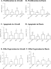
Proliferation rates, apoptosis indices, and ERα expression in morphologically normal alveoli and ducts. A and B: BrdU labeling in alveoli (A) and ducts (B). C and D: Apoptosis indices in alveoli (C) and ducts (D). E and F: ERα expression in alveoli (E) and ducts (F). Analysis was performed on mammary glands of 6-month-old virgin nontransgenic (NonTG), NRL-TGFα (TGFα), NRL-PRL 1647-13 (PRL), and bitransgenic NRL-TGFα/PRL 1647-13 (PRL × TGFα) females. Labeling was determined as described in Materials and Methods, and data are expressed as means ± SD. Different lowercase letters denote statistical differences among structures of the different lines determined by the Kruskal-Wallis test followed by Mann-Whitney post test (P < 0.05). NonTG and NRL-PRL proliferation and apoptosis rates were described previously.
Figure 5-6813
Figure 5-6813
Proliferation (Α), apoptosis (B), and ERα (C) expression in epithelial hyperplasias in 6-month-old NRL-TGFα and NRL- TGFα/PRL (line 1647-13) bitransgenic virgin females. NRL-PRL and nontransgenic (NonTG) females did not develop epithelial hyperplasias by this age. Labeling was determined as described in Materials and Methods, and data are expressed as means ± SD. Different lowercase letters denote statistical differences among structures of the different lines determined by the Kruskal-Wallis test followed by Mann-Whitney post test (P < 0.05).
Figure 6-6813
Figure 6-6813
Representative whole mounts from inguinal glands of the different genotypes at 13 weeks of age and end stage. A and B: Bitransgenic NRL-PRL (line 1647-13) × TGFα. C and D: TGFα. E and F: NRL-PRL (line 1647-13). G and H: nontransgenic mice (NonTG). The first column represents inguinal mammary whole mounts from 13-week-old females (A, C, E, and G). The second column represents inguinal mammary whole mounts collected at end stage, after the mice developed an adenocarcinoma or macrocyst reaching 1.5 cm in diameter (B, D, F, and H). Mammary whole mounts from NonTG female cage mates were also collected at this time. Mammary glands were collected and prepared as described in Materials and Methods. The large dark oval in each mammary gland is a lymph node.
Figure 7-6813
Figure 7-6813
PRL raises levels of phosphorylated ERK1/2 above TGFα alone. A: Mammary glands from 3-month-old NRL-TGFα and bitransgenic NRL-PRL (line 1647-13) × TGFα virgin females were disrupted with a polytron, and lysates were examined for phosphorylated ERK1/2, total ERK1/2, and keratin 8 by Western analysis. B: Signals in A were quantitated densitometrically and expressed as phosphorylated ERK1/2/total ERK1/2/keratin 8 signal (n = 3, means ± SD). Different lowercase letters denote statistically significant differences after one-way analysis of variance (P < 0.02). C: EGFR levels in the mono- and bitransgenic glands examined by Western analysis as in A, corrected for keratin 8 expression. (n = 3, means ± SD).
Figure 8-6813
Figure 8-6813
PRL and TGFα together prolong ERK1/2 phosphorylation. A: Western analysis of levels of phosphorylated ERK1/2 in PRL-deficient MCF-7 cells after stimulation with PRL and or/TGFα. Cells were serum-starved for 24 hours before incubation with vehicle, 4 nmol/L PRL, and/or 30 ng/ml TGFα for the times shown. Cellular lysates (30 μg protein) were analyzed for phosphorylated ERK1/2 and total ERK1/2 by Western analysis. B: Fold change in phosphorylated ERK1/2 levels after 120 minutes incubation with ligand compared with vehicle-treated controls from three independent experiments (means ± SD). Different lowercase letters denote statistically significant differences after one-way analysis of variance (P < 0.05).

References

    1. Clevenger CV, Furth PA, Hankinson SE, Schuler LA. Role of prolactin in mammary carcinoma. Endocr Rev. 2003;24:1–27. - PMC - PubMed
    1. Reynolds C, Montone KT, Powell CM, Tomaszewski JE, Clevenger CV. Expression of prolactin and its receptor in human breast carcinoma. Endocrinology. 1997;138:5555–5560. - PubMed
    1. Mertani HC, Garcia-Caballero T, Lambert A, Gérard F, Palayer C, Boutin JM, Vonderhaar BK, Waters MJ, Lobie PE, Morel G. Cellular expression of growth hormone and prolactin receptors in human breast disorders. Int J Cancer. 1998;79:202–211. - PubMed
    1. Touraine P, Martini JF, Zafrani B, Durand JC, Labaille F, Malet C, Nicolas A, Trivin C, Postel-Vinay MC, Kuttenn F, Kelly PA. Increased expression of prolactin receptor gene assessed by quantitative polymerase chain reaction in human breast tumors versus normal breast tissues. J Clin Endocrinol Metab. 1998;83:667–674. - PubMed
    1. Vonderhaar BK. Prolactin in human breast cancer development. Ethier SP, editor. Humana Press; Totawa, NJ: Endocrine Oncology. 2000:pp 101–120.

Publication types

MeSH terms

Substances