

Functional segregation of cortical language areas by sentence repetition
- PMID: 16565949
- PMCID: PMC6871319
- DOI: 10.1002/hbm.20250
Functional segregation of cortical language areas by sentence repetition
Abstract
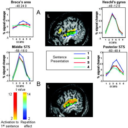
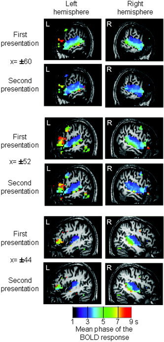
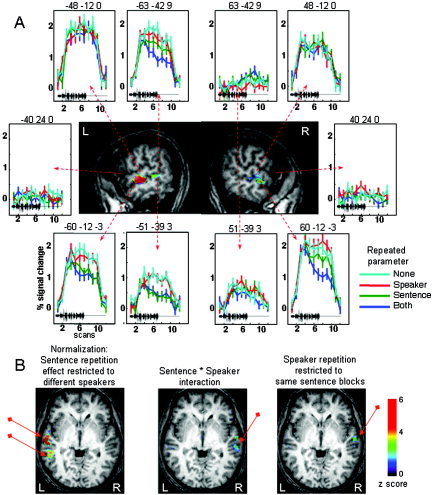
The functional organization of the perisylvian language network was examined using a functional MRI (fMRI) adaptation paradigm with spoken sentences. In Experiment 1, a given sentence was presented every 14.4 s and repeated two, three, or four times in a row. The study of the temporal properties of the BOLD response revealed a temporal gradient along the dorsal-ventral and rostral-caudal directions: From Heschl's gyrus, where the fastest responses were recorded, responses became increasingly slower toward the posterior part of the superior temporal gyrus and toward the temporal poles and the left inferior frontal gyrus, where the slowest responses were observed. Repetition induced a decrease in amplitude and a speeding up of the BOLD response in the superior temporal sulcus (STS), while the most superior temporal regions were not affected. In Experiment 2, small blocks of six sentences were presented in which either the speaker voice or the linguistic content of the sentence, or both, were repeated. Data analyses revealed a clear asymmetry: While two clusters in the left superior temporal sulcus showed identical repetition suppression whether the sentences were produced by the same speaker or different speakers, the homologous right regions were sensitive to sentence repetition only when the speaker voice remained constant. Thus, hemispheric left regions encode linguistic content while homologous right regions encode more details about extralinguistic features like speaker voice. The results demonstrate the feasibility of using sentence-level adaptation to probe the functional organization of cortical language areas.
Figures
References
-
- Andoh J, Artiges E, Pallier C, Riviere D, Mangin JF, Cachia A, Plaze M, Paillere‐Martinot ML, Martinot JL (2006): Modulation of language areas with functional MR image‐guided magnetic stimulation. Neuroimage 29: 619–627. - PubMed
-
- Belin P, Zatorre RJ (2003): Adaptation to speaker's voice in right anterior temporal lobe. Neuroreport 14: 2105–2109. - PubMed
-
- Belin P, Fecteau S, Bedard C (2004): Thinking the voice: neural correlates of voice perception. Trends Cogn Sci 8: 129–135. - PubMed
-
- Binder JR, Frost JA, Hammeke TA, Bellgowan PS, Springer JA, Kaufman JN, Possing ET (2000): Human temporal lobe activation by speech and non speech sounds. Cereb Cortex 10: 512–528. - PubMed
-
- Boemio A, Fromm S, Braun A, Poeppel D (2005): Hierarchical and asymmetric temporal sensitivity in human auditory cortices. Nat Neurosci 8: 389–395. - PubMed
Publication types
MeSH terms
LinkOut - more resources
Full Text Sources
