Comparative Study
. 2006 May 20;496(3):335-48.
doi: 10.1002/cne.20917.

Projections of the second cervical dorsal root ganglion to the cochlear nucleus in rats

Affiliations
Comparative Study

Projections of the second cervical dorsal root ganglion to the cochlear nucleus in rats

Xiping Zhan et al. J Comp Neurol. .

Abstract

Physiological, anatomical, and clinical data have demonstrated interactions between somatosensory and auditory brainstem structures. Spinal nerve projections influence auditory responses, although the nature of the pathway(s) is not known. To address this issue, we injected biotinylated dextran amine into the cochlear nucleus or dorsal root ganglion (DRG) at the second cervical segment (C2). Cochlear nucleus injections retrogradely labeled small ganglion cells in C2 DRG. C2 DRG injections produced anterograde labeling in the external cuneate nucleus, cuneate nucleus, nucleus X, central cervical nucleus, dorsal horn of upper cervical spinal segments, and cochlear nucleus. The terminal field in the cochlear nucleus was concentrated in the subpeduncular corner and lamina of the granule cell domain, where endings of various size and shapes appeared. Examination under an electron microscope revealed that the C2 DRG terminals contained numerous round synaptic vesicles and formed asymmetric synapses, implying depolarizing influences on the target cell. Labeled endings synapsed with the stalk of the primary dendrite of unipolar brush cells, distal dendrites of presumptive granule cells, and endings containing pleomorphic synaptic vesicles. These primary somatosensory projections contribute to circuits that are hypothesized to mediate integrative functions of hearing.


Figures

Figure 1
Figure 1
Retrograde labeling in C2 DRG. (1) Photomontage (left) and drawing (right) of injection site. The center of the injection site of BDA is located in the AVCN and extends in both directions yet is confined to the CN. It encroaches on the SPC posteriorly. Scale bar equals 200 µm. (B) Whole-mount preparation showing retrogradely labeled ganglion cells (asterisks) in C2 DRG. The relatively small somatic size suggests that these ganglion cells belong to the type B category. Scale bar equals 25 µm. (C) Raster plot of spikes in response to 200 msec tones of various frequencies at 70 dB SPL. The evoked spike rate is well above the spontaneous rate when the tone is within the unit’s response area. (D) On the basis of the maximal evoked spike discharge and minimal first spike latency, this unit was assigned a best frequency of 3.9 kHz. Dotted lines indicate 2 standard deviations above and below the spontaneous discharge rate.
Figure 2
Figure 2
Anterograde labeling from the C2 DRG to the brainstem. (A) Photomontage of the injection site in the C2 DRG. The whole-mount preparation also illustrates three rootlets that project into the brainstem (DR1, DR2, DR3). Note the labeled axons that are clearly shown in the peripheral nerve (C2). Scale bar equals 0.5 mm. (B) Photomicrograph of BDA labeled fibers and terminals in the ipsilateral cuneate nucleus. (C) Photomicrograph of labeled fibers and terminals in Nucleus X (X) and the external cuneate nucleus (ECu). The terminal field is squeezed between the inferior cerebellar peduncle (ICP) and the dorsal aspect of the spinal trigeminal tract (Sp5). (D) Photomicrograph of labeled fibers and swellings (arrows) in the subpeduncular corner of the cochlear nucleus GCD. (E) Photomicrograph of labeled fibers and swellings (arrows) in the lamina of the GCD. Scale bar for B–E equals 10 µm.
Figure 3
Figure 3
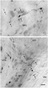
Photomicrographs of labeled fibers and swellings (arrows) in the subpeduncular corner (SPC) of the GCD. The SPC lies beneath the inferior cerebellar peduncle (ICP) and between the dorsal-medial extent of the ventral cochlear nucleus (VCN) laterally and the vestibular nerve root (VNr) medially. Most of the swellings are small (2–3 µm) but some appear as larger rosettes (4–8 µm). Scale bar equals 10 µm.
Figure 4
Figure 4
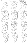
Drawings of sections through the cochlear nucleus illustrating distribution of labeled axons and terminals in the GCD (shaded gray). Sections are spaced 120 µm apart. Typically, projections from C2 DRG were concentrated in the subpeduncular corner (spc), medial sheet (ms), superficial lamina (sl) and lamina (lam). Abbreviations: ANr, auditory nerve root; AVCN, anteroventral cochlear nucleus; DCN, dorsal cochlear nucleus; Floc, flocculus of the cerebellum; ICP, inferior cerebellar peduncle; Sp5, spinal tract of the trigeminal nerve, VNr, vestibular nerve root. Scale bar equals 0.5 mm.
Figure 5
Figure 5
Electron micrograph of BDA-labeled endings in the subpeduncular corner. The dark DAB reaction product is clearly evident. This particular ending appeared as a string of en passant swellings when viewed in the light microscope but was considerably more complicated with respect to its shape and contacts when viewed with the electron microscope. Scale bar equals 1 µm.
Figure 6
Figure 6
Electron micrographs of labeled endings that synapse on dendrites. (A) This synapse (arrows) is asymmetric and upon a thin dendrite (d) of unknown origin but having features of a granule cell. (B) A larger ending formed a synapse on each of an array of dendrites (d1–d5). This relationship resembled that of a mossy fiber with dendrites of a granule cell “claw.” Asymmetric synapses are indicated by arrows. Scale bar equals 0.5 µm.
Figure 7
Figure 7
Electron micrograph of labeled endings that synapse on dendrites that contain ribosomes (arrowheads). Dendrites with ribosomes (polyribosomes) had been attributed to unipolar brush cells (UBC) in previous reports (Jaarsma et al., 1996). The synapses (arrows) are of the asymmetric variety. Scale bar equals 0.5 µm.
Figure 8
Figure 8
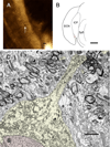
C2 DRG projection terminates on a unipolar brush cell (UBC). (A) Light photomicrograph of BDA-labeled terminal in the subpeduncular corner (arrow). Scale bar equals 10 µm. (B) Drawing of cochlear nucleus illustrating location of labeled terminal (asterisk). Abbreviations are provided in the legend for figure 4. Scale bar equals 0.5 mm. (C) Electron micrograph of labeled ending (asterisk) forming a synapse on the dendrite of a unipolar brush cell (UBC). This dendrite (yellow) is directly connected to the cell body, where the nucleus (pale red) exhibits a deep invagination of its envelope. Ribosomes are evident in the cell body and the dendrite. Dendrites (yellow) that branch off the main stalk as determined from serial section analysis are also shown. The morphology of the dendrites is a key feature of the UBC. Scale bar equals 2 µm.
Figure 9
Figure 9
Electron micrographs of endings forming asymmetric synapses (arrows) on structures containing pleomorphic synaptic vesicles. These target structures are called axon terminals (at) because of the vesicular content and continuity with axons (ax). Many of these postsynaptic terminals also contained vesicles with dense cores. Scale bar equals 0.5 µm.

References

    1. Abel MD, Levine RA. Muscle contractions and auditory perception in tinnitus patients and nonclinical subjects. Cranio. 2004;22:181–191. - PubMed
    1. Abrahams VC, Richmond FJ, Keane J. Projections from C2 and C3 nerves supplying muscles and skin of the cat neck: a study using transganglionic transport of horseradish peroxidase. J Comp Neurol. 1984;230:142–154. - PubMed
    1. Bae YC, Park KS, Bae JY, Paik SK, Ahn DK, Moritani M, Yoshida A, Shigenaga Y. GABA and glycine in synaptic microcircuits associated with physiologically characterized primary afferents of cat trigeminal principal nucleus. Exp Brain Res. 2005;162:449–457. - PubMed
    1. Baxter DA, Bittner GD. Intracellular recordings from crustacean motor axons during presynaptic inhibition. Brain Res. 1981;233:422–428. - PubMed
    1. Chen K, Waller HJ, Godfrey TG, Godfrey DA. Glutamergic transmission of neuronal responses to carbachol in rat dorsal cochlear nucleus slices. Neurosci. 1999;90:1043–1049. - PubMed

Publication types

MeSH terms