. 2006 Apr 4;45(13):4210-9.
doi: 10.1021/bi052382x.

Aberrant metabolites in mouse models of congenital blinding diseases: formation and storage of retinyl esters

Affiliations

Aberrant metabolites in mouse models of congenital blinding diseases: formation and storage of retinyl esters

Akiko Maeda et al. Biochemistry. .

Abstract

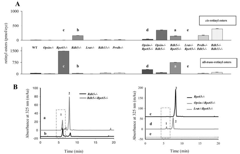
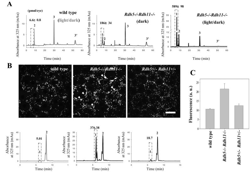
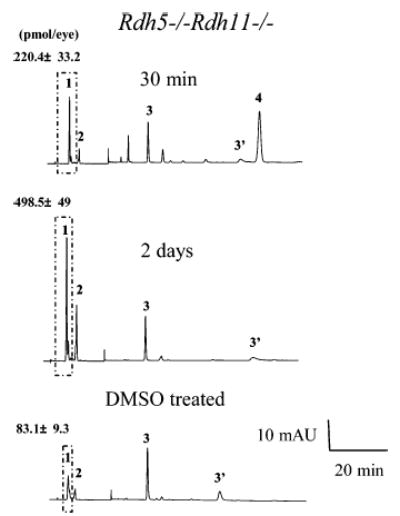
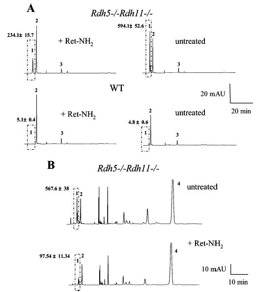
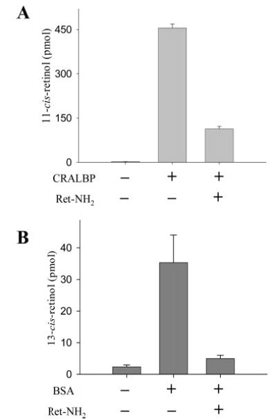
Regeneration of the visual chromophore, 11-cis-retinal, is a critical step in restoring photoreceptors to their dark-adapted conditions. This regeneration process, called the retinoid cycle, takes place in the photoreceptor outer segments and the retinal pigment epithelium (RPE). Disabling mutations in nearly all of the retinoid cycle genes are linked to human conditions that cause congenital or progressive defects in vision. Several mouse models with disrupted genes related to this cycle contain abnormal fatty acid retinyl ester levels in the RPE. To investigate the mechanisms of retinyl ester accumulation, we generated single or double knockout mice lacking retinoid cycle genes. All-trans-retinyl esters accumulated in mice lacking RPE65, but they are reduced in double knockout mice also lacking opsin, suggesting a connection between visual pigment regeneration and the retinoid cycle. Only Rdh5-deficient mice accumulate cis-retinyl esters, regardless of the simultaneous disruption of RPE65, opsin, and prRDH. 13-cis-Retinoids are produced at higher levels when the flow of retinoid through the cycle was increased, and these esters are stored in specific structures called retinosomes. Most importantly, retinylamine, a specific and effective inhibitor of the 11-cis-retinol formation, also inhibits the production of 13-cis-retinyl esters. The data presented here support the idea that 13-cis-retinyl esters are formed through an aberrant enzymatic isomerization process.


Figures

Figure 1
Figure 1
Chemistry of the retinoid cycle reactions in the vertebrate retina. The retinoid cycle reactions were reviewed recently (). In the rod outer segments (ROS), light causes the isomerization of the rhodopsin chromophore, 11-cis-retinylidene, to all-trans-retinylidene, which is hydrolyzed and released from opsin. All-trans-retinal is then reduced in a reaction catalyzed by all-trans-retinal-specific RDH(s) including prRDH. All-trans-retinol diffuses to retinal pigment epithelium (RPE) where it is esterified by LRAT to fatty acid all-trans-retinyl esters. All-trans-retinyl esters or its derivative is isomerized to 11-cis-retinol in a reaction that involves an abundant RPE protein, termed RPE65. 11-cis-Retinol is then oxidized by 11-cis-RDH (RDH5, RDH11) and other dehydrogenases to 11-cis-retinal, completing the cycle. 11-cis-Retinal diffuses across the extracellular space, is taken up by the ROS, and recombines with opsin to regenerate rhodopsin. In aberrant reactions, all-trans-retinyl esters or its derivative is isomerized to 13-cis-retinol. 13-cis-Retinol can also be esterified by LRAT to form 13-cis-retinyl esters, stored in retinosomes.
Figure 2
Figure 2
Retinyl esters in the eyes of mice of different genetic backgrounds. (A) All-trans- and cis-retinyl esters in mice of different genetic backgrounds. Retinoids were extracted from 6-week-old eyes and separated on normal-phase HPLC as described in Experimental Procedures. (B) Chromatographic separation of all-trans- and cis-retinyl esters from (a) Rpe65−/−, (b) opsin−/−Rdh65−/−, (c) Lrat−/−Rpe65−/−, (d) Rdh5−/−Rpe65−/−, and (e) Rdh5−/− mice. Retinoids were extracted from 6-week-old eyes and separated on normal-phase HPLC. The box represents cis-retinyl esters (>90% 13-cis-retinyl esters) also indicated as peak 1. Peak 2 represents all-trans-retinyl esters. The mice were reared under dim red light.
Figure 3
Figure 3
Chromatographic separation of nonpolar retinoids from opsin−/−Rdh5−/− mice. Retinoids were extracted from 6-week-old eyes and separated on normal-phase HPLC. The box represents cis-retinyl esters (>90% 13-cis-retinyl esters). A representative chromatogram is shown, and the average data from three mice are indicated with standard deviation above the ester peaks (mean ± SD).
Figure 4
Figure 4
Immunoblotting of eyecup extracts from wild-type and Rdh5−/−Rdh11−/− mice probed with anti-rhodopsin (1D4), anti-RPE65, or anti-LRAT antibodies. The eyecup extracts were prepared from mice by homogenizing with the sodium dodecyl sulfate–polyacrylamide gel electrophoresis (SDS–PAGE) sample buffer. The proteins (30 μg) were analyzed by 12.5% SDS–PAGE. The eyecup extract from Rdh5−/−Rdh11−/− mice showed no significant difference in the levels of rhodopsin, RPE65, or LRAT compared with wild-type mice.
Figure 5
Figure 5
Montage of cross sections of the retinas of 2-month-old Lrat−/−Rpe65−/− mice analyzed by transmission electron microscopy. Panel A shows the cross section of the RPE and the photoreceptor cells. Panels B and C show higher magnification sections of the RPE and ROS (B, C) and the IPL (D, E). (F–H) Montage of cross sections of the retinas of 2-month-old opsin−/−Rpe65−/− mice analyzed by transmission electron microscopy. Panel F shows the cross section of the RPE and the photoreceptor cells. Panels G and H show higher magnification sections of the RPE and ROS, repectively. (I–K) Montage of cross sections of the retinas of 2-month-old Rdh5−/−Rpe65−/−mice analyzed by transmission electron microscopy. Panel I shows the cross section of the RPE and photoreceptor cells. Panels J and K show a higher magnification of the RPE and ROS. The sections were prepared as described in Experimental Procedures. The scale bar represents 1 or 10 μm as indicated.
Figure 6
Figure 6
Light-dependent formation and storage of retinyl esters in eyes from Rdh5−/−Rdh11−/− mice. (A) Effect of dark or light rearing on the accumulation of 13-cis-retinyl esters in eyes from Rdh5−/−Rdh11−/− mice and wild-type controls. The dark-reared mice were exposed to no other light than dim red illumination. Peaks 1, 2, and 3/3′ represent cis-retinyl esters, all-trans-retinyl esters, and 11-cis-retinal oximes, respectively. The box represents cis-retinyl esters (>90% 13-cis-retinyl esters). A representative chromatogram is shown, and the average data from three mice are indicated with standard deviation above the ester peaks (mean ± SD). (B) Imaging of retinyl esters by two-photon microscopy (top row) and quantification of retinyl esters by HPLC (bottom row). Left column: Wild-type mice contained all-trans-retinyl esters in retinosomes (RESTs). Middle column: Rdh5−/−Rdh11−/− mice stored all-trans- and 13-cis-retinyl esters in retinosomes (white arrow). Retinosome fluorescence in Rdh5−/−Rdh11−/− mice is more intense than in wild-type mice (arrowheads). Right column: Distribution and quantity of all-trans-retinyl esters in eyes from Rdh5+/−Rdh+/− mice were similar to those of wild type. The box represents cis-retinyl esters (>90% 13-cis-retinyl esters). A representative chromatogram is shown, and the average data from three mice are indicated with standard deviation above the ester peaks (mean ± SD). (C) Quantification of fluorescence intensity measured by two-photon microscopy. Fluorescence intensity is higher in Rdh5−/−Rdh11−/− compared to wild-type and Rdh5+/−Rdh+/−mice (n = 3). The mean ± SD was indicated.
Figure 7
Figure 7
Enzymatic production of 13-cis-retinyl esters by Rdh5−/−Rdh11−/− mice. Mice were dark adapted for 48 h, followed by intravenous injection of 40 nmol of all-trans-retinol in 1 μL of DMSO with a 30 gauge needle under anesthesia. Retinoid analysis was performed both 30 min and 2 days after injection. All experiments were carried out in the dark. A representative chromatogram is shown, and the average data from three mice are indicated with standard deviation above the ester peaks (mean ± SD). Retinoids were extracted from 6-week-old eyes and separated on normal-phase HPLC. The box represents cis-retinyl esters (>90% 13-cis-retinyl esters). Peaks 1, 2, 3/3′, and 4 represent cis-retinyl esters, all-trans-retinyl esters, 11-cis-retinal oximes, and all-trans-retinol, respectively.
Figure 8
Figure 8
Effects of Ret-NH2 on 13-cis-retinyl ester production. (A) Effects of Ret-NH2 on 13-cis-retinyl ester production induced by light. Mice were dark adapted for 48 h, gavaged with 1 mg of Ret-NH2, and then exposed to intense light for 3 min at 500 cd·m−2, 24 h postgavage. A representative chromatogram is shown, and the average data from three mice are indicated with standard deviation above the ester peaks (mean ± SD). The box represents cis-retinyl esters (>90% 13-cis-retinyl esters). (B) Effects of Ret-NH2 on 13-cis-retinyl ester production from injected all-trans-retinol. Mice were dark adapted for 48 h and gavaged with 1 mg of Ret-NH2. After 24 h, 400 nmol of all-trans-retinol in 1 μL of DMSO was intravenously injected. A representative chromatogram is shown, and the average data from three mice are indicated with standard deviation above the ester peaks (mean ± SD). The box represents cis-retinyl esters (>90% 13-cis-retinyl esters). Peaks 1, 2, 3/3′, and 4 represent cis-retinyl esters, all-trans-retinyl esters, 11-cis-retinal oximes, and all-trans-retinol, respectively.
Figure 9
Figure 9
Influence of retinoid-binding proteins and Ret-NH2 on the formation of 11-cis-retinol and 13-cis-retinol. The isomerization reaction was performed in 10 mM BTP buffer, pH 7.5, containing 1 mM ATP and 10% BSA. UV-treated bovine RPE microsomes were used as a source of enzyme (150 μg of protein). For inhibition assays, all-trans-Ret-NH2 was used at a concentration of 3 mM. The reaction was initiated by adding all-trans-retinol in DMF and a solution of CRALBP or BSA to the final concentration of 10 mM or 10%, respectively. The reaction mixture was incubated in 37 °C for 1 h.

Similar articles

Cited by

References

    1. Filipek S, Stenkamp RE, Teller DC, Palczewski K. G protein-coupled receptor rhodopsin: A prospectus. Annu Rev Physiol. 2003;65:851–879. - PMC - PubMed
    1. Ridge KD, Abdulaev NG, Sousa M, Palczewski K. Phototransduction: crystal clear. Trends Biochem Sci. 2003;28:479–487. - PubMed
    1. Okada T, Ernst OP, Palczewski K, Hofmann KP. Activation of rhodopsin: new insights from structural and biochemical studies. Trends Biochem Sci. 2001;26:318–324. - PubMed
    1. Palczewski, K. (2006) G Protein-coupled receptor rhodopsin, Annu. Rev. Biochem. (in press). - PMC - PubMed
    1. Rando, R. R. (1991) Membrane phospholipids as an energy source in the operation of the visual cycle, Biochemistry 30, 595–602. - PubMed

Publication types

LinkOut - more resources