. 2006 Mar 27:7:9.
doi: 10.1186/1471-2091-7-9.

Identification and characterization of human polyserase-3, a novel protein with tandem serine-protease domains in the same polypeptide chain

Affiliations

Identification and characterization of human polyserase-3, a novel protein with tandem serine-protease domains in the same polypeptide chain

Santiago Cal et al. BMC Biochem. .

Abstract

Background: We have previously described the identification and characterization of polyserase-1 and polyserase-2, two human serine proteases containing three different catalytic domains within the same polypeptide chain. Polyserase-1 shows a complex organization and it is synthesized as a membrane-bound protein which can generate three independent serine protease domains as a consequence of post-translational processing events. The two first domains are enzymatically active. By contrast, polyserase-2 is an extracellular glycosylated protein whose three protease domains remain embedded in the same chain, and only the first domain possesses catalytic activity.

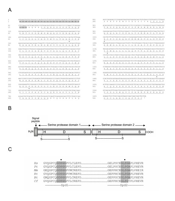
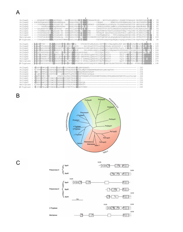
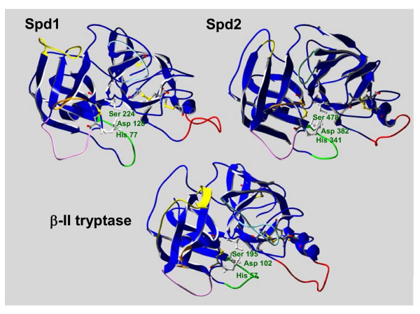
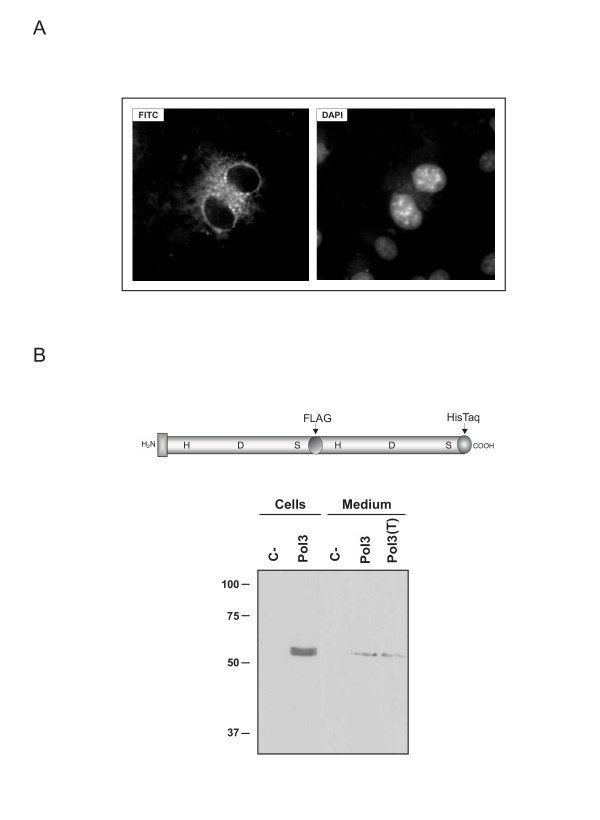
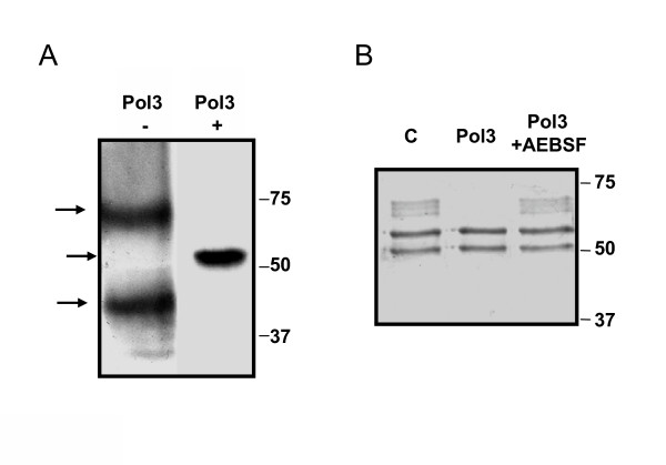
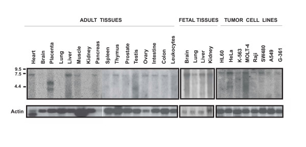
Results: Following our interest in the study of the human degradome, we have cloned a human liver cDNA encoding polyserase-3, a new protease with tandem serine protease domains in the same polypeptide chain. Comparative analysis of polyserase-3 with the two human polyserases described to date, revealed that this novel polyprotein is more closely related to polyserase-2 than to polyserase-1. Thus, polyserase-3 is a secreted protein such as polyserase-2, but lacks additional domains like the type II transmembrane motif and the low-density lipoprotein receptor module present in the membrane-anchored polyserase-1. Moreover, analysis of post-translational mechanisms operating in polyserase-3 maturation showed that its two protease domains remain as integral parts of the same polypeptide chain. This situation is similar to that observed in polyserase-2, but distinct from polyserase-1 whose protease domains are proteolytically released from the original chain to generate independent units. Immunolocalization studies indicated that polyserase-3 is secreted as a non-glycosylated protein, thus being also distinct from polyserase-2, which is a heavily glycosylated protein. Enzymatic assays indicated that recombinant polyserase-3 degrades the alpha-chain of fibrinogen as well as pro-urokinase-type plasminogen activator (pro-uPA). Northern blot analysis showed that polyserase-3 exhibits a unique expression pattern among human polyserases, being predominantly detected in testis, liver, heart and ovary, as well as in several tumor cell lines.

Conclusion: These findings contribute to define the growing group of human polyserine proteases composed at present by three different proteins. All of them share a complex structural design with several catalytic units in a single polypeptide but also show specific features in terms of enzymatic properties, expression patterns and post-translational maturation mechanisms.


Figures

Figure 1
Figure 1
Deduced amino acid sequence and domain organization of human polyserase-3 cDNA. A, the deduced amino acid sequence is shown in single-code letter. The signal peptide is shaded in gray. The two serine-protease domains are underlined. The residues His, Asp and Ser, corresponding to the catalytic triad of the serine protease domains, are indicated with a black dot. B, schematic representation of the domain organization of polyserase-3. The two predicted disulfide bonds around the putative activation site of both serine protease domains are indicated. C, Amino acid sequence alignment around the catalytic serine residues of human polyserase-3 serine protease domains with equivalent sequences deduced from other species. Spd, serine protease domain.Hs, Homo sapiens; Pt, Pan troglodytes, Mm, Mus musculus; Rn, Rattus norvegicus; Bt, Bos taurus; Cf, Canis familiaris.
Figure 2
Figure 2
Relationship between polyserase-3 and other serine proteases. A, sequence comparison of the serine protease domains of polyserase-3 with other related proteins. Active site amino acids are marked with a black dot. Pol3Spd, pol2Spd and pol1Spd indicate the different serine protease domains of each polyserase. B, phylogenetic tree of different human serine proteases related to polyserase-3. The analysis was performed using the serine protease domain of each enzyme and the Phylogenic Interface Environment program supplied by the Human Genome Mapping Project. C, organization of the human polyserase-3 gene and comparison with other serine protease genes. Relative positions of each exon are indicated by boxes. In the case of matriptase, only the exon/intron organization of its serine protease domain is represented. H, D and S refer to positions of the codons that encode the catalytic triad amino acids in each protease gene.
Figure 3
Figure 3
Homology models of the catalytic domains of polyserase-3 and β-II tryptase. The structural modeling of Spd-1 and Spd-2 reveals the high degree of similarity with the serine protease β-II tryptase. The twelve loops (six in each serine protease module) potentially involved in the dimerization of polyserase-3 as well as the 6 loops involved in β-II tryptase tetramerization are represented using the color code previously described by Sommerhoff et al. 1999 (33). The molecules are oriented towards the active site and the three residues that compose the catalytic triad of each serine protease are indicated. The backbone and side chains of the disulfide bonds are represented in CPK color scheme.
Figure 4
Figure 4
Immunocytochemical detection and analysis of recombinant polyserase-3 expression in human cell lines. A, image captures by fluorescence microscopy of 293-EBNA cells transfected with pCEP-pol3 vector and incubated with an anti-FLAG antibody, followed by incubation with a secondary fluorescein-conjugated antibody (FITC). This signal was not detected in cells transfected with an empty vector. The observed cytoplasmic fluorescence indicates that polyserase-3 is not a membrane-anchored protease. DNA in the cell nucleus was visualized with DAPI. B, representation of the recombinant polyserase-3 containing the indicated epitopes, and Western blot analysis of 293-EBNA cells and conditioned medium using an anti-FLAG antibody. Equivalent results were obtained using an anti His-Tag antibody (not shown). C- indicates cells or conditioned medium from cells transfected with the empty vector, and T indicates sample treated with 1 μg/mL tunicamycin. The concentration of the SDS-PAGE gel was 12%, and the molecular mass markers in kDa are indicated on the left.
Figure 5
Figure 5
Production and activity assays of recombinant Spd-1, Spd-2, and polyserase-3. A, 5 μL of bacterial extract transformed with the plasmids pGEX-Spd1 (lane3), pGEX-Spd2 (lane 5) and pGEX-pol3 (lane 9), and the corresponding purified fusion proteins (Spd1, lane 4; Spd2, lane 6, and polyserase3, lane 10). Lane 2, bacterial extract transformed with an empty pGEX-2TK. Lanes 7 and 8 correspond to induction and purification of ADAM23 disintegrin domain, respectively. Lane 1, molecular size markers, whose sizes in kDa are indicated on the left. B, Western blot analysis of the purified fusion proteins using an anti GST antibody. C, degradation of fibrinogen by the indicated fusion proteins. C, control, corresponds to incubation of fibrinogen without any recombinant protein. D, Inhibition analysis of polyserase-3 after preincubation with AEBSF (0.1 mM), EDTA (2 mM) and E-64 (10 μM). E, digestion of pro-uPA by purified polyserase-3. C indicates incubation of pro-uPA alone. F, incubation of different extracellular proteins with purified polyserase-3.
Figure 6
Figure 6
Polyserase-3 may form active dimers. A, Western blot analysis of His-tagged polyserase-3 in the absence (-) or presence (+) of 2% 2-mercaptoethanol, using an anti-HisTag antibody. Molecular size markers are indicated on the right, and detected bands are indicated with arrows on the left. B, degradation of fibrinogen by His-tagged polyserase 3 in the absence or presence of the serine protease inhibitor AEBSF.
Figure 7
Figure 7
Analysis of polyserase-3 expression in human tissues and tumor cell lines. Approximately 2 μg of polyadenylated RNA from the indicated tissues or tumor cell lines was hybridized with a probe specific for polyserase-3. The position of the RNA markers is shown. The filters were subsequently hybridized with an actin probe to ascertain the differences in RNA loading among the different tissues.

Similar articles

Cited by

  • Substrate specificity of human metallocarboxypeptidase D: Comparison of the two active carboxypeptidase domains.
    Garcia-Pardo J, Tanco S, Díaz L, Dasgupta S, Fernandez-Recio J, Lorenzo J, Aviles FX, Fricker LD. Garcia-Pardo J, et al. PLoS One. 2017 Nov 13;12(11):e0187778. doi: 10.1371/journal.pone.0187778. eCollection 2017. PLoS One. 2017. PMID: 29131831 Free PMC article.
  • Oral anticoagulation and VKORC1 polymorphism in patients with a mechanical heart prosthesis: a 6-year follow-up.
    Giansante C, Fiotti N, Altamura N, Pitacco P, Consoloni L, Scardi S, Mazzone C, Grassi G, Pandullo C, Di Lenarda A. Giansante C, et al. J Thromb Thrombolysis. 2012 Nov;34(4):506-12. doi: 10.1007/s11239-012-0740-8. J Thromb Thrombolysis. 2012. PMID: 22592842 Clinical Trial.
  • Identification of protease serine S1 family member 53 as a mitochondrial protein in murine islet beta cells.
    Mizusawa N, Harada N, Iwata T, Ohigashi I, Itakura M, Yoshimoto K. Mizusawa N, et al. Islets. 2022 Jan 1;14(1):1-13. doi: 10.1080/19382014.2021.1982325. Epub 2021 Oct 12. Islets. 2022. PMID: 34636707 Free PMC article.
  • Genome-wide association analysis identifies three psoriasis susceptibility loci.
    Stuart PE, Nair RP, Ellinghaus E, Ding J, Tejasvi T, Gudjonsson JE, Li Y, Weidinger S, Eberlein B, Gieger C, Wichmann HE, Kunz M, Ike R, Krueger GG, Bowcock AM, Mrowietz U, Lim HW, Voorhees JJ, Abecasis GR, Weichenthal M, Franke A, Rahman P, Gladman DD, Elder JT. Stuart PE, et al. Nat Genet. 2010 Nov;42(11):1000-4. doi: 10.1038/ng.693. Epub 2010 Oct 17. Nat Genet. 2010. PMID: 20953189 Free PMC article.
  • Identification of 15 new psoriasis susceptibility loci highlights the role of innate immunity.
    Tsoi LC, Spain SL, Knight J, Ellinghaus E, Stuart PE, Capon F, Ding J, Li Y, Tejasvi T, Gudjonsson JE, Kang HM, Allen MH, McManus R, Novelli G, Samuelsson L, Schalkwijk J, Ståhle M, Burden AD, Smith CH, Cork MJ, Estivill X, Bowcock AM, Krueger GG, Weger W, Worthington J, Tazi-Ahnini R, Nestle FO, Hayday A, Hoffmann P, Winkelmann J, Wijmenga C, Langford C, Edkins S, Andrews R, Blackburn H, Strange A, Band G, Pearson RD, Vukcevic D, Spencer CC, Deloukas P, Mrowietz U, Schreiber S, Weidinger S, Koks S, Kingo K, Esko T, Metspalu A, Lim HW, Voorhees JJ, Weichenthal M, Wichmann HE, Chandran V, Rosen CF, Rahman P, Gladman DD, Griffiths CE, Reis A, Kere J; Collaborative Association Study of Psoriasis (CASP); Genetic Analysis of Psoriasis Consortium; Psoriasis Association Genetics Extension; Wellcome Trust Case Control Consortium 2; Nair RP, Franke A, Barker JN, Abecasis GR, Elder JT, Trembath RC. Tsoi LC, et al. Nat Genet. 2012 Dec;44(12):1341-8. doi: 10.1038/ng.2467. Epub 2012 Nov 11. Nat Genet. 2012. PMID: 23143594 Free PMC article.

References

    1. Liu L, Wilson T, Hastings JW. Molecular evolution of dinoflagellate luciferases, enzymes with three catalytic domains in a single polypeptide. Proc Natl Acad Sci U S A. 2004;101:16555–16560. doi: 10.1073/pnas.0407597101. - DOI - PMC - PubMed
    1. Eberhardt RY, Gilbert HJ, Hazlewood GP. Primary sequence and enzymic properties of two modular endoglucanases, Cel5A and Cel45A, from the anaerobic fungus Piromyces equi. Microbiology. 2000;146 ( Pt 8):1999–2008. - PubMed
    1. Cal S, Quesada V, Llamazares M, Diaz-Perales A, Garabaya C, Lopez-Otin C. Human polyserase-2, a novel enzyme with three tandem serine protease domains in a single polypeptide chain. J Biol Chem. 2005;280:1953–1961. doi: 10.1074/jbc.M409139200. - DOI - PubMed
    1. Li L, Hong R, Hastings JW. Three functional luciferase domains in a single polypeptide chain. Proc Natl Acad Sci U S A. 1997;94:8954–8958. doi: 10.1073/pnas.94.17.8954. - DOI - PMC - PubMed
    1. Ouwendijk J, Peters WJ, van de Vorstenbosch RA, Ginsel LA, Naim HY, Fransen JA. Routing and processing of lactase-phlorizin hydrolase in transfected Caco-2 cells. J Biol Chem. 1998;273:6650–6655. doi: 10.1074/jbc.273.12.6650. - DOI - PubMed

Publication types

LinkOut - more resources