Comparative Study
. 2006 Mar;61(3):245-55.
doi: 10.1093/gerona/61.3.245.

Molecular regulation of apoptosis in fast plantaris muscles of aged rats

Affiliations
Comparative Study

Molecular regulation of apoptosis in fast plantaris muscles of aged rats

Emidio E Pistilli et al. J Gerontol A Biol Sci Med Sci. 2006 Mar.

Abstract

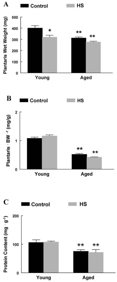
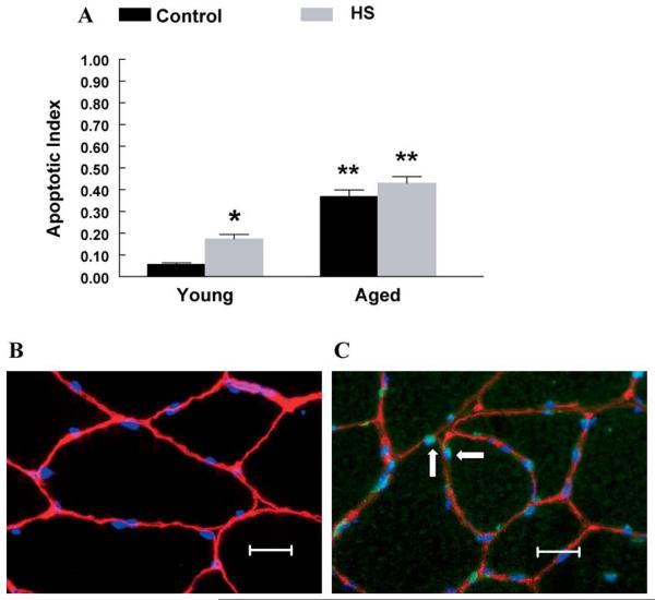
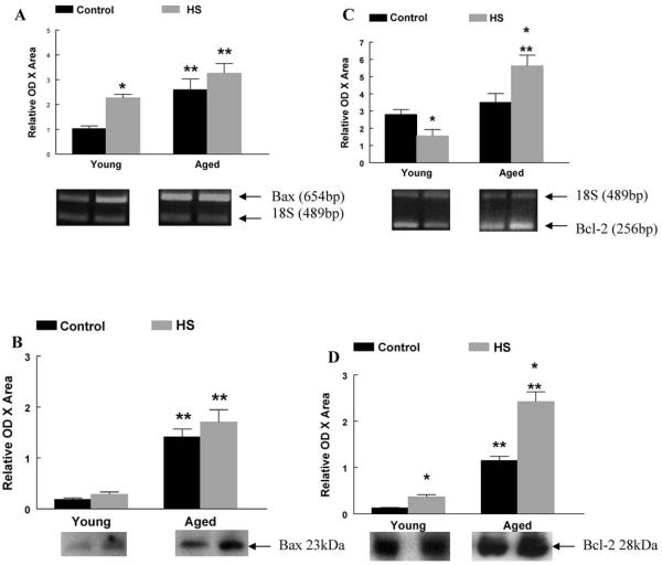
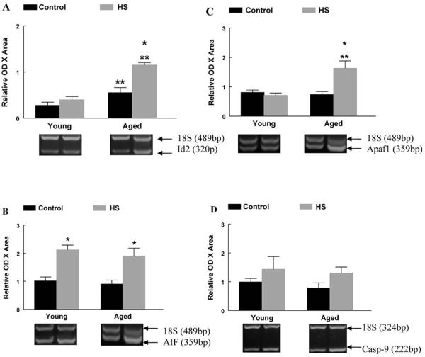
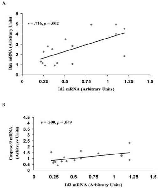
This study tested the hypothesis that aging exacerbates apoptotic signaling in rat fast plantaris muscle during muscle unloading. Plantaris muscle mass was 22% lower in aged animals and the apoptotic index was 600% higher, when compared to those in young adult animals. Following 14 days of hind-limb unloading, absolute plantaris muscle mass was 20% lower in young adult animals with a corresponding 200% higher elevation of the apoptotic index. Unloading had no affect on muscle weight or apoptotic index of aged plantaris muscles. The changes in pro-apoptotic messenger RNA (mRNA) for apoptotic protease activating factor-1 (Apaf-1), Bax, and inhibitor of differentiation protein-2 (Id2) were exacerbated with aging. Bax and Bcl-2 protein levels were also altered differently in aged muscle, compared to young. Significant positive correlations were observed between the changes in Id2 and Bax mRNA, and Id2 and caspase-9 mRNA. These data suggest that a pro-apoptotic environment may contribute to aging-associated atrophy in fast skeletal muscle, but apoptotic signaling differs by age.


Figures

Figure 1
Figure 1
A, Absolute plantaris muscle wet weight. Plantaris muscles from young and aged animals expressed as milligrams of tissue. *Significant effect of hind-limb suspension (HS; p < .05). **Significant effect of aging ( p < .05). Data are presented as means ± standard error (SE). B, Normalized plantaris muscle weight (mg · g −1). Plantaris muscles from young and aged animals were normalized to the animal body weight in grams. **Significant effect of aging ( p < .05). Data are presented as means ± SE. C, Plantaris muscle protein content (mg · g −1). Plantaris protein content was determined in young and aged plantaris muscles following HS. **Significant effect of aging ( p < .05). Data are presented as means ± SE.
Figure 2
Figure 2
A, Apoptotic index (optical density [OD] 405 · mg protein−1). The extent of DNA fragmentation was assayed using a cell death enzyme-linked immunosorbent assay (ELISA) kit. *Significant effect of hind-limb suspension (HS; p < .05). **Significant effect of aging ( p < .05). Data are presented as means ± standard error (SE). B, Fluorescent terminal deoxynucleotidyl transferase biotin-dUTP nick end labeling (TUNEL) stain in young control muscle. TUNEL staining was used to determine the extent of nuclear apoptosis in gastrocnemius muscles from young animals. Muscle fiber borders were visualized using a rat laminin antibody, and nonapoptotic nuclei were visualized using 4′,6-diamidino-2-phenylindole (DAPI). The image was obtained at an objective magnification of 40×. Bar, 10 μm. C, Fluorescent TUNEL stain in aged control muscle. TUNEL staining was used to determine the extent of nuclear apoptosis in gastrocnemius muscles from old animals. Muscle fiber borders were visualized using a rat laminin antibody. All nuclei were stained with DAPI. Arrows highlight TUNEL-positive nuclei. Nonapoptotic nuclei were TUNEL negative. The image was obtained at an objective magnification of 40×. Bar, 10 μm.
Figure 3
Figure 3
A, Bax messenger RNA (mRNA). The mRNA content for pro-apoptotic Bax was determined by reverse transcription–polymerase chain reaction (RT–PCR), with PCR products normalized to the 18S gene. Representative agarose gel images following electrophoresis are displayed for each group. *Significant effect of hind-limb suspension (HS; p < .05). **Significant effect of aging ( p < .05). Data are presented as means ± standard error (SE). B, Bax protein content. The protein content of pro-apoptotic Bax was determined by western immunoblot. Representative blots following antibody incubation are displayed. **Significant effect of aging ( p < .05). Data are presented as means ± SE. C, Bcl-2 mRNA. The mRNA content for anti-apoptotic Bcl-2 was determined by RT–PCR, with PCR products normalized to the 18S gene. Representative agarose gel images following electrophoresis are displayed for each group. *Significant effect of HS ( p < .05). **Significant effect of aging ( p < .05). Data are presented as means ± SE. D, Bcl-2 protein content. The protein content of anti-apoptotic Bcl-2 was determined by western immunoblot. Representative blots following antibody incubation are displayed. *Significant effect of HS ( p < .05). **Significant effect of aging ( p < .05). Data are presented as means ± SE.
Figure 4
Figure 4
A, Fluorescent image of Bax protein in young control muscle. The extent of cytoplasmic Bax protein was determined using fluorescence microscopy. A faint Bax stain was visualized in gastrocnemius muscles from young animals. Muscle fiber borders were visualized using a rat laminin antibody, and nonapoptotic nuclei were visualized using 4′,6-diamidino-2-phenylindole (DAPI). The image was obtained at an objective magnification of 40×. Bar, 10 μm. B, Fluorescent image of Bax protein in aged control muscle. The extent of cytoplasmic Bax protein was determined using fluorescence microscopy. Bax protein was visualized throughout the cytoplasm of aged muscle fibers. Muscle fiber borders were visualized using a rat laminin antibody, and nonapoptotic nuclei were visualized using DAPI. The image was obtained at an objective magnification of 40×. Bar, 10 μm.
Figure 5
Figure 5
A, Inhibitor of differentiation protein-2 (Id2) messenger RNA (mRNA). The mRNA content for Id2 was determined by reverse transcription–polymerase chain reaction (RT–PCR), with PCR products normalized to the 18S gene. Representative agarose gel images following electrophoresis are displayed for each group. *Significant effect of hind-limb suspension (HS; p < .05). **Significant effect of aging ( p < .05). Data are presented as means ± standard error (SE). B, Apoptosis-inducing factor (AIF) mRNA. The mRNA content for AIF was determined by RT–PCR, with PCR products normalized to the 18S gene. Representative agarose gel images following electrophoresis are displayed for each group. **Significant effect of aging ( p < .05). Data are presented as means ± SE. C, Apaf-1 mRNA. The mRNA content for Apaf-1 was determined by RT–PCR, with PCR products normalized to the 18S gene. Representative agarose gel images following electrophoresis are displayed for each group. *Significant effect of HS ( p < .05). **Significant effect of aging ( p < .05). Data are presented as means ± SE. D, Caspase-9 mRNA. The mRNA content for caspase-9 was determined by RT–PCR, with PCR products normalized to the 18S gene. Representative agarose gel images following electrophoresis are displayed for each group. Data are presented as means ± SE.
Figure 6
Figure 6
A, Correlational analysis of Bax and inhibitor of differentiation protein-2 (Id2) messenger RNA (mRNA). The relationship between the changes in Bax mRNA and Id2 mRNA were determined by calculating the Pearson correlation coefficient. B, Correlational analysis of caspase-9 and Id2 mRNA. The relationship between the changes in caspase-9 mRNA and Id2 mRNA were determined by calculating the Pearson correlation coefficient.

Similar articles

Cited by

References

    1. Alway SE, Coggan AR, Sproul MS, Abduljalil AM, Robitaille PM. Muscle torque in young and older untrained and endurance-trained men. J Gerontol. 1996;51A:B195–B201. - PubMed
    1. Daw CK, Starnes JW, White TP. Muscle atrophy and hypoplasia with aging: impact of training and food restriction. J Appl Physiol. 1988;64:2428–2432. - PubMed
    1. Degens H, Alway SE. Skeletal muscle function and hypertrophy are diminished at old age. Muscle Nerve. 2003;27:339–347. - PubMed
    1. Larsson L, Grimby G, Karlsson J. Muscle strength and speed of movement in relation to age and muscle morphology. J Appl Physiol. 1979;46:451–456. - PubMed
    1. Wickham CC, Cooper C, Margetts BM, Barker DJ. Muscle strength, activity, housing, and the risk of falls in elderly people. Age Ageing. 1989;18:47–51. - PubMed

Publication types

MeSH terms