Comparative Study
. 2006 Apr 17;203(4):919-32.
doi: 10.1084/jem.20052237. Epub 2006 Mar 27.

Secondary memory CD8+ T cells are more protective but slower to acquire a central-memory phenotype

Affiliations
Comparative Study

Secondary memory CD8+ T cells are more protective but slower to acquire a central-memory phenotype

Ali Jabbari et al. J Exp Med. .

Abstract

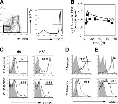
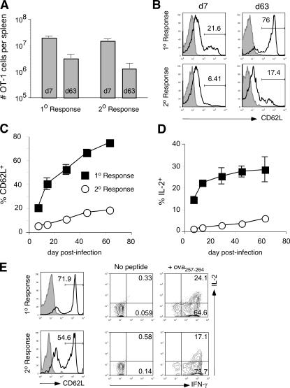
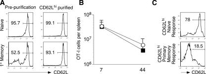
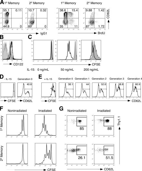
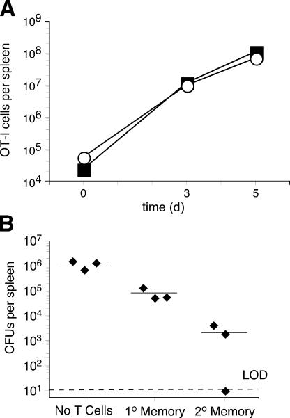
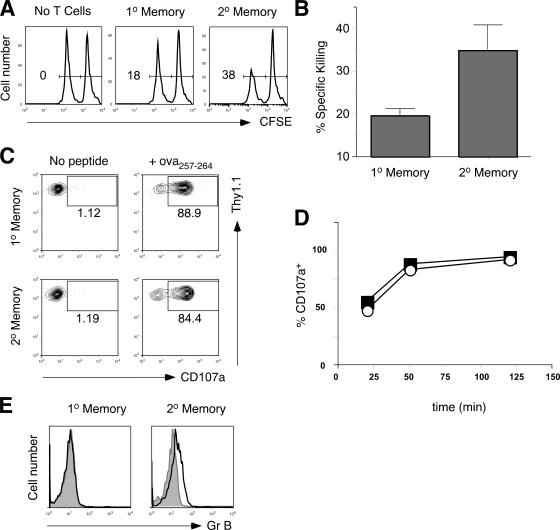
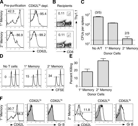
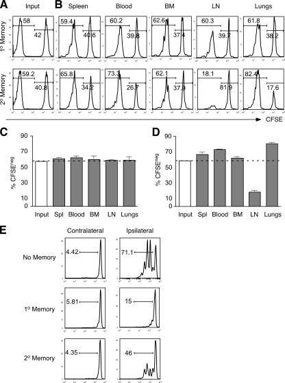
The formation of memory CD8 T cells is an important goal of vaccination. However, although widespread use of booster immunizations in humans generates secondary and tertiary CD8 T cell memory, experimental data are limited to primary CD8 T cell memory. Here, we show that, compared with primary memory CD8 T cells, secondary memory CD8 T cells exhibit substantially delayed conversion to a central-memory phenotype, as determined by CD62L expression and interleukin (IL)-2 production. This delayed conversion to a central-memory phenotype correlates with reduced basal proliferation and responsiveness to IL-15, although in vitro coculture with a high concentration of IL-15 is capable of inducing proliferation and CD62L upregulation. Functionally, secondary memory CD8 T cells are more protective in vivo on a per cell basis, and this may be explained by sustained lytic ability. Additionally, secondary memory CD8 T cells are more permissive than primary memory CD8 T cells for new T cell priming in lymph nodes, possibly suggesting a mechanism of replacement for memory T cells. Thus, primary and secondary memory CD8 T cells are functionally distinct, and the number of encounters with antigen influences memory CD8 T cell function.


Figures

Figure 1.
Figure 1.
Reduced CD62L expression of secondary compared with primary memory CD8 T cells in the same host. Approximately 2 × 10⁴ NP118-specific CD8 T cells from a LCMV-immune BALB/c, Thy1.2 mouse (day 129 after infection) were adoptively transferred into naive, Thy1.1 BALB/c mice. 1 d later, recipient mice were infected i.p. with LCMV- Armstrong. At various time points after infection, the number of NP118-specific CD8 T cells undergoing either a primary or secondary response was assessed in the spleen by MHC tetramers (left) and differential Thy1 expression (right). (A) Analysis of a representative mouse at day 8 after infection. Right panel gated on NP118/Ld tetramer+ cells. (B) Total number of NP118-specific CD8 T cells in the spleen undergoing either a primary (▪) or secondary (○) response over time. Data are mean ± SD for three mice/time point. (C) CD62L expression of NP118-specific cells undergoing either primary (top) or secondary (bottom) responses at day 8 (left) or day 72 (right) after infection. Open histograms, CD62L; shaded histograms, isotype control. (D) Adoptive transfer recipients were infected with 107 actA LM-NPs, and CD62L expression on primary (top) and secondary (bottom) NP118-specific memory CD8+ T cells was determined 39 d later. Shown are representative histograms of NP118/Ld tetramer gated cells. (E) Naive or LM-OVA–immune B6 mice were infected with 1.2 × 10⁷ actA LM-OVA, and CD62L expression on primary (top) and secondary (bottom) ova257-specific memory CD8+ T cells was determined 39 d later. Shown are representative histograms of ova257-specific gated cells. 1°, primary; 2°, secondary.
Figure 2.
Figure 2.
OT-I CD8 T cells undergoing a secondary response do not adopt central–memory-like characteristics until very late time points after infection. 104-purified naive or primary memory (day 126 after infection) OT-I/Thy1.1 cells were adoptively transferred into separate groups of naive, Thy1.2 B6 mice, and recipient mice were subsequently infected with 1.4 × 10⁷ actA LM-OVA to initiate primary and secondary responses, respectively. (A) Enumeration of OT-I cells (CD8+/Thy1.1+) in the spleen at days 7 and 63 after infection. Data are mean ± SD of three mice/group. (B) CD62L expression of OT-I cells in the spleen undergoing either primary (top) or secondary responses (bottom) at days 7 (left) or 63 (right) after infection. Representative histograms are gated on CD8+/Thy1.1+ splenocytes. Open histograms, CD62L; shaded histograms, isotype control. (C) CD62L expression by OT-I cells undergoing either primary (▪) or secondary (○) responses at various time points after infection. Data are mean ± SD of three mice/time point. (D) IL-2 production by primary and secondary memory OT-I cells detected by ova257-stimulated intracellular cytokine staining at various days after infection. Data are mean ± SD of three mice/time point. (E) Representative CD62L expression (left) and IL-2 production (middle and right) at 227 d after infection on primary (top) and secondary (bottom) memory OT-I T cells. 1°, primary; 2°, secondary.
Figure 3.
Figure 3.
TCM primary memory CD8 T cells exhibit delayed CD62L reacquisition after infection. Primary memory OT-I CD8 T cells were generated in vivo as described in Fig. 2. (A) CD62Lhi cells were purified from splenocytes of mice containing primary memory OT-I cells as well as from naive OT-I mice. (B) Approximately 5 × 10⁴ CD62Lhi-purified naive or primary memory CD8 T cells were then adoptively transferred into naive B6 mice, and these mice were infected 1 d later with actA LM-OVA. At days 7 and 44 after infection, the numbers of OT-I cells were assessed. (C) At day 44 after infection, the CD62L expression of cells that had undergone a primary (top) or secondary (bottom) response was assessed. 1°, primary.
Figure 4.
Figure 4.
Reduced IL-15R expression and reduced responsiveness to IL-15 correlate with decreased basal proliferation and delayed central–memory phenotype. Primary and secondary memory OT-I CD8 T cells were generated in vivo as described in Fig. 2. (A) Starting at day 69 after infection, mice received BrdU for the following 8 d. Representative profiles of primary and secondary memory OT-I cells for expression of surface CD62L and intracellular isotype control (IgG1, left two panels) or BrdU (right two panels). Contour plots are gated on CD8+Thy1.1+ cells. (B) Representative expression at 63 d after infection on primary memory OT-I cells of CD122 (open histogram), isotype control (vertical line-fill), or secondary memory OT-I cells (CD122, shaded histogram), or isotype control (diagonal line-fill). (C) Purified primary or secondary memory OT-I cells at 63 d after infection were labeled with CFSE and cocultured in the presence of 0, 50, or 200 ng/ml IL-15. After 3 d, dilution of CFSE as a measure of proliferation was assessed by flow cytometry. Open histograms, primary memory OT-I cells; shaded histograms, secondary memory OT-I cells. (D and E) Purified secondary memory OT-I cells at 75 d after infection were labeled with CFSE and cocultured for 3 d in the absence (D) or in the presence (E) of 200 ng/ml IL-15. Cells were subsequently stained for CD62L expression. Left-most panels, CFSE profile of cultured primary or secondary memory OT-I cells; right panels, CD62L profiles of CFSE peaks as gated in top panels. (F and G) CFSE-labeled Thy1.1-purified primary or secondary memory OT-I cells were adoptively transferred into nonirradiated B6 mice or B6 mice sublethally irradiated 24 h prior. 14 d later, spleens of mice were harvested and assessed for CFSE dilution (F) or CD62L expression (G). 1°, primary; 2°, secondary.
Figure 5.
Figure 5.
Robust expansion and enhanced protection in response to infection by secondary memory CD8 T cells. (A) 5 × 10⁵ purified primary or secondary memory CD8 T cells (day 66 after infection) were adoptively transferred into separate groups of naive, Thy1.2 B6 mice. 1 d later, mice were infected with 1.5 × 10⁷ actA LM-OVA. Shown are total numbers of OT-I cells/spleen at days 0, 3, and 5 after infection of recipients of primary (▪) or secondary (○) memory OT-I cells. Data represent mean ± SD from three mice/group/time point. (B) Approximately 5 × 10⁵ primary or secondary memory OT-I cells were adoptively transferred to naive, B6 recipients. 1 d later, mice were infected with 2.7 × 10⁵ virulent LM-OVA. The number of bacteria in the spleen was determined 3 d after challenge infection. LOD, limit of detection. 1°, primary; 2°, secondary.
Figure 6.
Figure 6.
Secondary memory CD8 T cells exhibit enhanced in vivo killing capacity, equivalent degranulation kinetics, and increased expression of granzyme B compared with primary memory cells. (A) Approximately 1.5 × 10⁶ primary (middle) or secondary memory (right) OT-I cells were adoptively transferred into naive B6 recipients. 1 d later, recipient mice and a group of naive B6 mice (left) were injected with target cells consisting of 2 × 10⁶ CFSEhi-labeled B6 splenocytes (no peptide) and 2 × 10⁶ CFSElo-labeled B6 splenocytes (coated with ova257 peptide). Representative histograms of CFSE-labeled cells recovered from each group at 2 h after target cell injection. Number in histograms is percentage of specific killing. (B) Cumulative data from three mice per group for the in vivo cytolysis assay. (C) Primary and secondary memory OT-I cells were assessed for degranulation (cell surface presence of CD107a) in the absence (left) or presence (right) of ova257 peptide at 50 min after stimulation. Contour plots are gated on CD8+Thy1.1+ cells. (D) Percentage of primary (▪) or secondary (○) memory OT-I T cells expressing surface CD107a at various time points after peptide stimulation. (E) Intracellular stain of primary (left) and secondary (right) memory OT-I cells obtained at 63 d after infection for granzyme B. Histograms are gated on CD8+Thy1.1+ cells. Open histograms, granzyme B; shaded histograms, isotype control. GrB, granzyme B. 1°, primary; 2°, secondary.
Figure 7.
Figure 7.
CD62Llo secondary memory CD8 T cells offer greater protection and exhibit more efficient cytolysis than CD62Llo, primary TEM. Primary and secondary memory OT-I/Thy1.1 CD8 T cells were generated in vivo as described in Fig. 2. Splenocytes were harvested and depleted of CD62Lhi-expressing cells. (A) CD62L expression on OT-I cells pre and after purification. (B) Approximately 9.2 × 10⁵ CD62Llo primary or secondary memory OT-I/Thy1.1 CD8 T cells were adoptively transferred into naive, Thy1.2-expressing B6 mice. 1 d later, spleens of recipient mice were harvested and stained for CD8 and Thy1.1. (C) 1 d after transfer, recipient mice were infected with 1.27 × 10⁶ virulent LM-OVA. 3 d after infection, the bacterial burden in the spleens of three to five infected mice per group was assessed as in Fig. 6. Two out of five naive mice died. One out of three recipients of secondary memory OT-I cells did not have detectable levels of LM-OVA. Line, limit of detection. (D) Recipients of CD62Llo primary or secondary memory CD8 T cells were injected with target cells consisting of 1.25 × 10⁶ CFSEhi-labeled B6 splenocytes (no peptide) and 1.25 × 10⁶ CFSElo-labeled B6 splenocytes (coated with ova257 peptide). Representative histograms of CFSE-labeled cells recovered from each group at 2 h after target cell injection. Number in histograms is percentage of specific killing. (E) Cumulative data from three mice per group for the in vivo cytolysis assay. (F and G) Unpurified primary (F) and secondary (G) memory OT-I cells were harvested from spleens and stained for CD62L and intracellular granzyme B directly ex vivo. Left panels are gated on OT-I cells, middle panels are gated on CD62Llo OT-I cells, and right panels are gated on CD62Lhi OT-I cells. 1°, primary; 2°, secondary.
Figure 8.
Figure 8.
Decreased capacity of secondary memory CD8 T cells to traffic to lymph nodes permits enhanced priming of naive CD8 T cells. (A) Primary memory OT-I cells generated in Thy1.2 B6 mice were labeled with CFSE and mixed with unlabeled primary (top) or secondary (bottom) memory OT-I cells (Thy1.1+). Histograms of the CFSE profiles are gated on CD8+Thy1.1+ cells. Approximately 3 × 10⁶ memory OT-I cells in these mixed populations were adoptively transferred into naive Thy1.2 B6 mice. (B) Representative histograms obtained 3 d after transfer of blood, spleens, bone marrow (BM), lymph nodes, and lungs were assessed for presence of unlabeled primary (top) or secondary (bottom) memory OT-I cells relative to CFSE-labeled primary memory OT-I cells. Representative histograms, gated on CD8+Thy1.1+ cells, are shown. (C) Percentage of unlabeled primary and (D) secondary memory OT-I cells (mean ± SD of three mice/group) in various tissues compared with input. (E) Primary and secondary memory OT-I T cells were generated in Thy1.2 B6 mice. At day 69 after infection, naive mice (top) or with preexisting primary (middle) or secondary (bottom) OT-I memory received 106 naive, CFSE-labeled OT-I cells. 1 d later, mice were injected with ∼1.2 × 10⁷ actA LM-OVA s.c. in the lower right flank. Proliferation of the CFSE-labeled naive OT-I cells was assessed in inguinal lymph nodes contralateral (left) and ipsilateral (right) to the injection site at 2 d after infection. Representative histograms, gated on CD8+Thy1.1+CFSE+OT-I cells, are shown. Numbers are the percentages of CFSE-labeled cells that had undergone cell division. 1°, primary; 2°, secondary.

Similar articles

Cited by

References

    1. Sprent, J., and C.D. Surh. 2002. T cell memory. Annu. Rev. Immunol. 20:551–579. - PubMed
    1. Woodland, D.L. 2004. Jump-starting the immune system: prime-boosting comes of antigene. Trends Immunol. 25:98–104. - PubMed
    1. Masopust, D., V. Vezys, A.L. Marzo, and L. Lefrancois. 2001. Preferential localization of effector memory cells in nonlymphoid tissue. Science. 291:2413–2417. - PubMed
    1. Harty, J.T., and V.P. Badovinac. 2002. Influence of effector molecules on the CD8(+) T cell response to infection. Curr. Opin. Immunol. 14:360–365. - PubMed
    1. Harty, J.T., A.R. Tvinnereim, and D.W. White. 2000. CD8+ T cell effector mechanisms in resistance to infection. Annu. Rev. Immunol. 18:275–308. - PubMed

Publication types

MeSH terms