Physiological features of the S- and M-cone photoreceptors of wild-type mice from single-cell recordings
- PMID: 16567464
- PMCID: PMC2151510
- DOI: 10.1085/jgp.200609490
Physiological features of the S- and M-cone photoreceptors of wild-type mice from single-cell recordings
Abstract
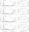
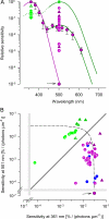
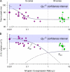
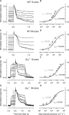
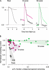
Cone cells constitute only 3% of the photoreceptors of the wild-type (WT) mouse. While mouse rods have been thoroughly investigated with suction pipette recordings of their outer segment membrane currents, to date no recordings from WT cones have been published, likely because of the rarity of cones and the fragility of their outer segments. Recently, we characterized the photoreceptors of Nrl(-/-) mice, using suction pipette recordings from their "inner segments" (perinuclear region), and found them to be cones. Here we report the use of this same method to record for the first time the responses of single cones of WT mice, and of mice lacking the alpha-subunit of the G-protein transducin (G(t)alpha(-/-)), a loss that renders them functionally rodless. Most cones were found to functionally co-express both S- (lambda(max) = 360 nm) and M- (lambda(max) = 508 nm) cone opsins and to be maximally sensitive at 360 nm ("S-cones"); nonetheless, all cones from the dorsal retina were found to be maximally sensitive at 508 nm ("M-cones"). The dim-flash response kinetics and absolute sensitivity of S- and M-cones were very similar and not dependent on which of the coexpressed cone opsins drove transduction; the time to peak of the dim-flash response was approximately 70 ms, and approximately 0.2% of the circulating current was suppressed per photoisomerization. Amplification in WT cones (A approximately 4 s(-2)) was found to be about twofold lower than in rods (A approximately 8 s(-2)). Mouse M-cones maintained their circulating current at very nearly the dark adapted level even when >90% of their M-opsin was bleached. S-cones were less tolerant to bleached S-opsin than M-cones to bleached M-opsin, but still far more tolerant than mouse rods to bleached rhodopsin, which exhibit persistent suppression of nearly 50% of their circulating current following a 20% bleach. Thus, the three types of mouse opsin appear distinctive in the degree to which their bleached, unregenerated opsins generate "dark light."
Figures


Similar articles
-
Photoreceptors of Nrl -/- mice coexpress functional S- and M-cone opsins having distinct inactivation mechanisms.J Gen Physiol. 2005 Mar;125(3):287-304. doi: 10.1085/jgp.200409208. J Gen Physiol. 2005. PMID: 15738050 Free PMC article.
-
Cone-like morphological, molecular, and electrophysiological features of the photoreceptors of the Nrl knockout mouse.Invest Ophthalmol Vis Sci. 2005 Jun;46(6):2156-67. doi: 10.1167/iovs.04-1427. Invest Ophthalmol Vis Sci. 2005. PMID: 15914637 Free PMC article.
-
A mouse M-opsin monochromat: retinal cone photoreceptors have increased M-opsin expression when S-opsin is knocked out.Vision Res. 2011 Feb 23;51(4):447-58. doi: 10.1016/j.visres.2010.12.017. Epub 2011 Jan 8. Vision Res. 2011. PMID: 21219924 Free PMC article.
-
Human retinal dark adaptation tracked in vivo with the electroretinogram: insights into processes underlying recovery of cone- and rod-mediated vision.J Physiol. 2022 Nov;600(21):4603-4621. doi: 10.1113/JP283105. Epub 2022 Jun 7. J Physiol. 2022. PMID: 35612091 Free PMC article. Review.
-
Why are rods more sensitive than cones?J Physiol. 2016 Oct 1;594(19):5415-26. doi: 10.1113/JP272556. Epub 2016 Jul 21. J Physiol. 2016. PMID: 27218707 Free PMC article. Review.
Cited by
-
Transcriptional control of cone photoreceptor diversity by a thyroid hormone receptor.Proc Natl Acad Sci U S A. 2022 Dec 6;119(49):e2209884119. doi: 10.1073/pnas.2209884119. Epub 2022 Dec 1. Proc Natl Acad Sci U S A. 2022. PMID: 36454759 Free PMC article.
-
Signaling properties of a short-wave cone visual pigment and its role in phototransduction.J Neurosci. 2007 Sep 19;27(38):10084-93. doi: 10.1523/JNEUROSCI.2211-07.2007. J Neurosci. 2007. PMID: 17881515 Free PMC article.
-
Implications of dimeric activation of PDE6 for rod phototransduction.Open Biol. 2018 Aug;8(8):180076. doi: 10.1098/rsob.180076. Open Biol. 2018. PMID: 30068567 Free PMC article.
-
Examining the Role of Cone-expressed RPE65 in Mouse Cone Function.Sci Rep. 2018 Sep 21;8(1):14201. doi: 10.1038/s41598-018-32667-w. Sci Rep. 2018. PMID: 30242264 Free PMC article.
-
Analysis of dim-light responses in rod and cone photoreceptors with altered calcium kinetics.J Math Biol. 2023 Oct 12;87(5):69. doi: 10.1007/s00285-023-02005-4. J Math Biol. 2023. PMID: 37823947 Free PMC article.
References
-
- Applebury, M.L., M.P. Antoch, L.C. Baxter, L.L. Chun, J.D. Falk, F. Farhangfar, K. Kage, M.G. Krzystolik, L.A. Lyass, and J.T. Robbins. 2000. The murine cone photoreceptor: a single cone type expresses both S and M opsins with retinal spatial patterning. Neuron. 27:513–523. - PubMed

