. 2006 Mar 27;172(7):1009-22.
doi: 10.1083/jcb.200511010.

Stu2p binds tubulin and undergoes an open-to-closed conformational change

Affiliations

Stu2p binds tubulin and undergoes an open-to-closed conformational change

Jawdat Al-Bassam et al. J Cell Biol. .

Abstract

Stu2p from budding yeast belongs to the conserved Dis1/XMAP215 family of microtubule-associated proteins (MAPs). The common feature of proteins in this family is the presence of HEAT repeat-containing TOG domains near the NH2 terminus. We have investigated the functions of the two TOG domains of Stu2p in vivo and in vitro. Our data suggest that Stu2p regulates microtubule dynamics through two separate activities. First, Stu2p binds to a single free tubulin heterodimer through its first TOG domain. A large conformational transition in homodimeric Stu2p from an open structure to a closed one accompanies the capture of a single free tubulin heterodimer. Second, Stu2p has the capacity to associate directly with microtubule ends, at least in part, through its second TOG domain. These two properties lead to the stabilization of microtubules in vivo, perhaps by the loading of tubulin dimers at microtubule ends. We suggest that this mechanism of microtubule regulation is a conserved feature of the Dis1/XMAP215 family of MAPs.


Figures
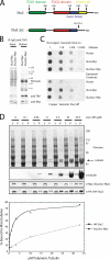
Figure 1.
Figure 1.
Coiled-coil domain of Stu2p is required for homodimerization, affinity for microtubules, and the function of Stu2p in vivo. (A) Scheme of Stu2p and the Stu2p mutant lacking the coiled-coil domain. Scheme shows the location of the first (green) and second TOG domain (red), basic linker domain (blue; previously termed the microtubule-binding domain), and the (predicted) coiled-coil domain (yellow). Boundaries of the TOG domains were determined from sequence alignments of Stu2p and Dis1/XMAP215 family members. Numbers indicate amino acid positions. (B) Immunoblot showing the results of a coimmunoprecipitation experiment. Stu2-myc or Stu2-myc lacking the coiled-coil domain (Stu2-Δcc–myc) was expressed in a yeast strain expressing Stu2-TAP. Stu2-TAP was immunoprecipitated from extracts, and the elutions were analyzed by SDS-PAGE and immunoblotting. (C) Stu2p function in vivo depends on the dimerization domain. Stu2-myc or Stu2–Δcoiled coil–myc (Stu2-Δcc–myc) was expressed in a yeast strain in which genomic Stu2p can be run down by adding copper. Strains were spotted in a dilution series on plates lacking or containing 300 μM CuCl2. (D) Deletion of the coiled-coil segment reduces cosedimentation of Stu2p with microtubules. (top) Immunoblot showing a microtubule cosedimentation assay with yeast extracts. Yeast extracts from a strain expressing HA-Stu2 and Stu2-myc lacking the dimerization domain (Stu2-Δcc–myc) were incubated with increasing amounts of taxol-stabilized microtubules. Subsequently, the reactions were spun to separate microtubule bound from unbound proteins. Supernatants (S) and pellets (P) were analyzed by immunoblotting using anti-HA, anti-myc, and antitubulin antibodies. (bottom) Graph shows the percentage of bound HA-Stu2 and Stu2-Δcc–myc plotted against the concentration of polymerized tubulin incorporated into microtubules. For clarity, a curve has been fit to the data points.
Figure 2.
Figure 2.
The first TOG domain of Stu2p is required in vivo to stabilize both cytoplasmic and nuclear microtubules. Stu2 or Stu2 lacking large parts of the first TOG domain (Stu2-ΔTOG1), both of which are COOH-terminally myc tagged, were expressed as exclusive Stu2 copies in yeast. The respective strains were arrested in early S phase with hydroxyurea and subsequently released from the hydroxyurea block, taking samples at 10-min time intervals for FACS and immunofluorescence analysis. Cytoplasmic microtubule lengths were determined from projected image stacks at time point zero; spindle lengths were determined from projected image stacks at the time points at which the peak of spindle elongation occurred. (A) Scheme of Stu2 and Stu2-ΔTOG1. See Fig. 1 A. (B) FACS profile of cultures before and during the hydroxyurea release. (C) Anti-Stu2p immunoblot of yeast extracts prepared from the cultures after the end of the hydroxyurea release. (D) Examples of antitubulin immunofluorescence of cytoplasmic microtubules at time point zero (bottom) and of microtubule spindles at the peak of spindle elongation (top). Tubulin, green; DNA, red. (right, bottom) Average cytoplasmic microtubule lengths (n = 590 for Stu2-myc and n = 366 for Stu2-ΔTOG1–myc). Error bars represent SEM. Bar, 2.5 μm. (right, top) Average microtubule spindle length (n = 195 for Stu2-myc and n = 178 for Stu2-ΔTOG1–myc). Error bars represent SD. Bar, 6 μm. Images have been processed using Photoshop, including some γ adjustments. (E) A yeast strain expressing an exclusive Stu2 copy of Stu2-ΔTOG1–myc shows enhanced sensitivity to the microtubule-destabilizing drug benomyl compared with a strain expressing intact Stu2-myc. Strains were spotted at the same cell densities onto YPD plates containing or lacking 10 μg/ml benomyl.
Figure 3.
Figure 3.
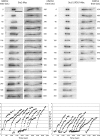
The fast phase of anaphase spindle elongation is compromised by the deletion of TOG1. (top) Still images of an elongating spindle, as followed by time-lapse microscopy, in cells expressing GFP-tubulin and either intact Stu2-myc (left) or Stu2-ΔTOG1–myc (right) as exclusive Stu2 copies. Bar, 3 μm. (bottom) Examples of traces of spindle lengths versus time. Individual traces have been shifted along the time axis to give clear spacing between them.
Figure 4.
Figure 4.
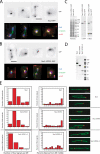
The first TOG domain of Stu2p is not required for the end association of Stu2p. (A and B) In vivo localization of Stu2-GFP and Stu2-ΔTOG1–GFP. (top) GFP localization was imaged directly from midlogarithmic yeast cultures expressing as exclusive Stu2 copies Stu2 (A) and Stu2-ΔTOG1 (B), which were both COOH-terminally GFP tagged. Bars, 5 μm. (bottom) Parts of the cultures were fixed and prepared for immunofluorescence imaging using anti-GFP and antitubulin (A, Stu2-GFP; B, Stu2-ΔTOG1–GFP). GFP, red; tubulin, green; DNA, blue. Images have been processed with Photoshop, including some γ adjustments. Bars (A), 2.2 μm; (B) 1.2 μm. (C) Anti-Stu2p and anti-GFP immunoblot of yeast extracts prepared from the yeast cultures used in A and B. (D) Coomassie-stained SDS-PAGE gel with equimolar amounts of the protein preparations used in E. (E) In vitro end-binding assay of Stu2p, Stu2p lacking the first TOG domain (Stu2-ΔTOG1), and Stu2p lacking both TOG domains (Stu2-ΔTOG1+2). Proteins were covalently labeled with Cy3 and incubated with taxol-stabilized microtubules partly labeled with Oregon green. Reactions were fixed, spun through a glycerol cushion, and resuspended before microscopic analysis. Fluorescence signals above 10% background level in the Cy3 channel were noted with their location along the microtubule. (left) Microtubule binding. The number of microtubules with a set number of Stu2 signals is displayed. (center) Microtubule end binding. Microtubules were divided in 10% intervals from the microtubule middle to the microtubule end, and the percentages of Stu2 signals found in each interval are displayed. (right) Three examples of microtubules with bound Stu2p fragments. Bar, 3 μm. Stu2 is in red, and microtubules are in green. For the purpose of this display only, images have been processed using Photoshop, including some γ adjustments.
Figure 5.
Figure 5.
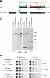
Duplicating TOG1 of Stu2p does not hyperstabilize microtubules. (A) Scheme of Stu2 and Stu2 with a duplicated first TOG domain (Stu2-2TOG1). See Fig. 1 A. (B) Immunoblot of yeast extracts prepared from cells expressing as exclusive Stu2 copies Stu2 or Stu2-2TOG1, which were both COOH-terminally myc tagged. Antibodies were anti-myc, antitubulin, an antibody raised against a COOH-terminal peptide of Stu2p, and an antibody raised against the NH2 terminus (covering TOG1) of Stu2p. (C) Benomyl sensitivity test. Stu2 or Stu2-2TOG1, both COOH-terminally myc tagged, was expressed as exclusive Stu2 copies in yeast cells. Strains were spotted at the same cell densities onto YPD plates containing or lacking 10, 12.5, or 15 μg/ml of the microtubule-destabilizing drug benomyl.
Figure 6.
Figure 6.
Stu2p and its TOG1 domain fragment bind an αβ-tubulin heterodimer. (A) Affinity chromatography using a recombinant first TOG domain of Stu2p (TOG1). 1.9 mg TOG1 or, as a control, 2.6 mg BSA were immobilized on NHS-activated columns (A, left; Coomassie-stained gel showing TOG1 and BSA before coupling to the column). Yeast extracts were prepared from a wild-type strain and passed through the column before washing and elution with high salt. Eluates were subjected to SDS-PAGE and the gel stained with Coomassie blue (A, right). The band present only in the elution of the column carrying TOG1 was identified by antitubulin immunoblotting (C) and mass spectrometry (not depicted) as yeast tubulin. (B) Affinity chromatography using a recombinant second TOG domain of Stu2p (TOG2). 2.2 mg TOG2 or, as a control, 2 mg BSA were immobilized on NHS-activated columns (left; Coomassie-stained gel showing TOG2 and BSA before coupling to the column). (C) Yeast extracts were prepared from a wild-type yeast strain and passed through the column carrying TOG1 before washing and elution with high salt. The eluates were subjected to SDS-PAGE, and the gel was stained with Coomassie (right) and antitubulin immunoblotting (left). (D) Size-exclusion chromatograms of recombinant TOG1 (blue) and unpolymerized αβ-tubulin dimer (red). Equimolar amounts of TOG1 and tubulin dimer were mixed and analyzed by gel filtration (green). SDS-PAGE of the peak fractions show that TOG1 coelutes with tubulins. The estimated stoichiometry is 1:1 for the TOG1/tubulin dimer from densitometry of the Coomassie blue–stained protein bands (Fig. S2 C, available at http://www.jcb.org/cgi/content/full/jcb.200511010/DC1). The elongated shape of TOG1 causes it to elute earlier than a globular protein of the same molecular weight (Table I). The unpolymerized αβ-tubulin chromatogram (red) and SDS-PAGE of fractions (bottom left) are also shown in E and F for comparison with other experiments. (E) Size exclusion chromatograms of recombinant TOG1–TOG2 (blue) and unpolymerized αβ-tubulin dimer (red). Equimolar amounts of TOG1–TOG2 and tubulin dimer were mixed and analyzed by gel filtration (green). SDS-PAGE of peak fractions shows that TOG1–TOG2 coelutes with tubulins. The estimated stoichiometry is 1:1 for the TOG1–TOG2/tubulin dimer (Fig. S2 C). Analytical ultracentrifugation shows that TOG1–TOG2 is a monomer with a molecular mass of 67 kD (Table II and Fig. S3), but its elongated shape causes it to elute earlier than a 67-kD globular protein (Table I). (F) Size-exclusion chromatograms of recombinant Stu2p (blue) and unpolymerized αβ-tubulin dimer (red). Equimolar amounts of Stu2p and tubulin dimer were mixed and analyzed by gel filtration (green). A complex forms with an apparent molecular mass slightly larger than that of free Stu2p, but excess, unbound tubulin is also detected. The Stu2p–tubulin complex contains one tubulin heterodimer per Stu2p homodimer (Fig. S2 C). Analytical ultracentrifugation shows that Stu2p is a dimer with a molecular mass of 200 kD (Table II and Fig. S3), but its elongated shape causes it to elute substantially earlier than expected for a protein of this size (Table I). (D–F) Arrows refer to the void volume of the size-exclusion column.
Figure 7.
Figure 7.
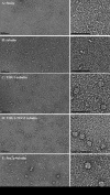
Stu2p, an elongated flexible homodimer, forms a compact complex with αβ-tubulin heterodimer. (A–E). Electron micrographs of negatively stained complexes obtained from the peak fractions illustrated in Fig. 6. The panels on the right are at twice the magnification of those on the left. Bars, 50 nm. (A) Stu2p homodimer. (B) Free αβ-tubulin heterodimer. (C) TOG1–tubulin complex. (D) TOG1–TOG2–tubulin complex. (E) Stu2p–tubulin complex. The uniform particles are slightly larger than those in the micrographs of TOG1–tubulin and TOG1–TOG2–tubulin.
Figure 8.
Figure 8.
Model for the mechanism of microtubule stabilization by Stu2p. (A) A model for Stu2p dimer–tubulin dimer complex assembly. Stu2p dimer in extended conformation consisting of two tandem arrangements of TOG1 (purple) and TOG2 (yellow) held together by a coiled coil. Stu2p becomes compact when it sequesters a single tubulin heterodimer (crosshatched dimer). (B) Model of the loading of tubulin dimer to a microtubule plus end from a Stu2p dimer complex. (I) Stu2p–tubulin complex associates with microtubule plus ends through its TOG2 domains. (II) Stu2p positions its sequestered tubulin at the end of a single protofilament on the microtubule plus end. (III) Stu2p dissociates from the assembled tubulin dimer. (IV) Stu2p released from the microtubule plus end returns to its flexible conformation. The loading process of tubulin (I–IV) can become a tubulin removal process (IV back to I) if the equilibrium is reversed in the absence of dimeric tubulin, leading to microtubule depolymerization.

References

    1. Brittle, A.L., and H. Ohkura. 2005. Mini spindles, the XMAP215 homologue, suppresses pausing of interphase microtubules in Drosophila. EMBO J. 24:1387–1396. - PMC - PubMed
    1. Carvalho, P., J.S. Tirnauer, and D. Pellman. 2003. Surfing on microtubule ends. Trends Cell Biol. 13:229–237. - PubMed
    1. Cassimeris, L., and J. Morabito. 2004. TOGp, the human homolog of XMAP215/Dis1, is required for centrosome integrity, spindle pole organization, and bipolar spindle assembly. Mol. Biol. Cell. 15:1580–1590. - PMC - PubMed
    1. Cassimeris, L., D. Gard, P.T. Tran, and H.P. Erickson. 2001. XMAP215 is a long thin molecule that does not increase microtubule stiffness. J. Cell Sci. 114:3025–3033. - PubMed
    1. Charrasse, S., M. Schroeder, C. Gauthier-Rouviere, F. Ango, L. Cassimeris, D.L. Gard, and C. Larroque. 1998. The TOGp protein is a new human microtubule-associated protein homologous to the Xenopus XMAP215. J. Cell Sci. 111:1371–1383. - PubMed

Publication types

MeSH terms