. 2006 Mar 27;172(7):1057-68.
doi: 10.1083/jcb.200508058.

CpG-induced tyrosine phosphorylation occurs via a TLR9-independent mechanism and is required for cytokine secretion

Affiliations

CpG-induced tyrosine phosphorylation occurs via a TLR9-independent mechanism and is required for cytokine secretion

Miguel A Sanjuan et al. J Cell Biol. .

Abstract

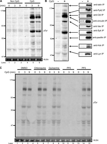
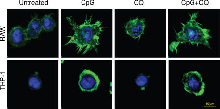
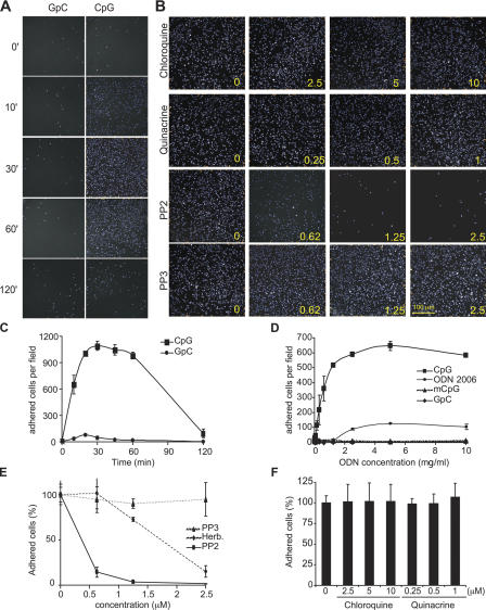
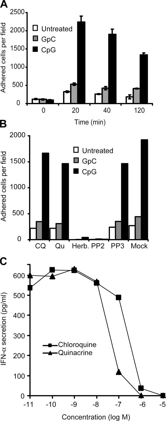
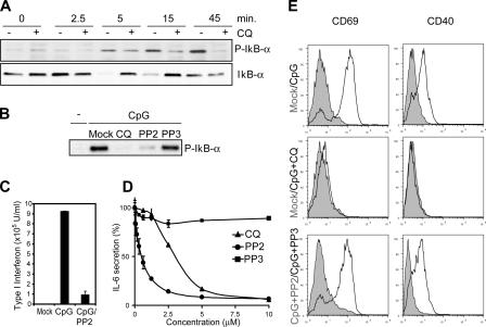
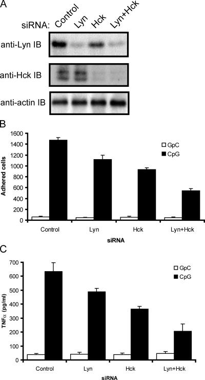
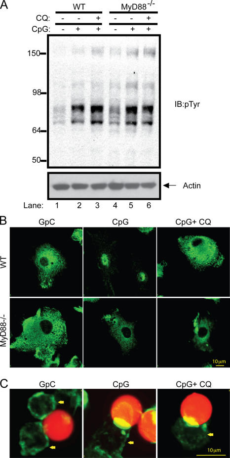
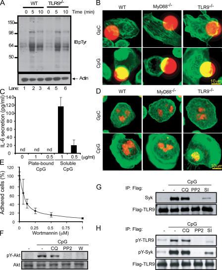
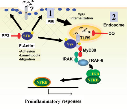
Toll-like receptors (TLRs) recognize molecular patterns preferentially expressed by pathogens. In endosomes, TLR9 is activated by unmethylated bacterial DNA, resulting in proinflammatory cytokine secretion via the adaptor protein MyD88. We demonstrate that CpG oligonucleotides activate a TLR9-independent pathway initiated by two Src family kinases, Hck and Lyn, which trigger a tyrosine phosphorylation-mediated signaling cascade. This cascade induces actin cytoskeleton reorganization, resulting in cell spreading, adhesion, and motility. CpG-induced actin polymerization originates at the plasma membrane, rather than in endosomes. Chloroquine, an inhibitor of CpG-triggered cytokine secretion, blocked TLR9/MyD88-dependent cytokine secretion as expected but failed to inhibit CpG-induced Src family kinase activation and its dependent cellular responses. Knock down of Src family kinase expression or the use of specific kinase inhibitors blocked MyD88-dependent signaling and cytokine secretion, providing evidence that tyrosine phosphorylation is both CpG induced and an upstream requirement for the engagement of TLR9. The Src family pathway intersects the TLR9-MyD88 pathway by promoting the tyrosine phosphorylation of TLR9 and the recruitment of Syk to this receptor.


Figures

Figure 1.
Figure 1.
SFKs initiate a chloroquine-insensitive, CpG-triggered tyrosine phosphorylation cascade. (A) THP-1 monocytes were stimulated with a control ODN, GpC (lanes 1–3), and CpG–ODN 2216 (lanes 4–6) at 1 μg/ml for the indicated times. 50-μg aliquots of lysate were subjected to SDS-PAGE followed by immunoblotting with an anti-pTyr antibody. Loading controls were immunoblotted with an anti-actin antibody. (B) Individual phosphoproteins were immunoprecipitated with the indicated antibodies from 100 μg of GpC- or CpG-stimulated (3 min) cell lysate and then immunoblotted with anti-pTyr antibody. (C) THP-1 cells were pretreated for 30 min with vehicle (lanes 1–3), 10 μM chloroquine (lanes 4–6), 1 μM quinacrine (lanes 7–9), 1 μM of the SFK inhibitor PP2 (lanes 10–13), and 1 μM of the control inhibitor PP3 (lanes 13–15). Lysates were prepared after CpG stimulation and immunoblotted with anti-pTyr antibody.
Figure 2.
Figure 2.
Chloroquine fails to block CpG-induced actin polymerization. Mouse macrophages (RAW) or human monocytes (THP-1) were pretreated for 15 min with 10 μM chloroquine (CQ) or vehicle control and then stimulated for 20 min with 1 μg/ml of the mouse-selective CpG–ODN 1668 or human CpG–ODN 2216, respectively. After fixation, cells were stained with FITC-phalloidin (green) and nuclei were visualized with TOPRO-3 (blue).
Figure 3.
Figure 3.
CpG-induced cell adhesion is insensitive to chloroquine but requires SFK activity. (A) Cells were stimulated with 1 μg/ml of a control GpC-ODN or with CpG–ODN 2216 for the indicated times (min). After fixation, cells were visualized as described in Materials and methods. (B) THP-1 cells were preincubated for 10 min with chloroquine, quinacrine, PP2, and PP3. The number at the lower right corner of each panel indicates the concentration for each inhibitor used (μM). Cells were then stimulated for 30 min with CpG, and adhesion was measured. Representative fields are presented. (C) THP-1 cells were stimulated as indicated in A, and images captured from 36 different fields from at least four different wells were quantified to obtain cell number per field and plotted with the SD. (D) THP-1 cells were stimulated for 20 min with increasing concentrations of CpG, control GpC, the B class oligo 2006 (ODN 2006), and murine CpG (mCpG), and adhesion was measured. (E) Adhesion was measured on THP-1 cells that were pretreated for 10 min with increasing concentrations of PP2, PP3, and herbimycin A and then stimulated with CpG for 30 min. Data are represented as percentage of adhered cells versus vehicle-pretreated cells. (F) THP-1 cells were preincubated for 10 min with increasing concentrations of chloroquine or quinacrine. Cells were then stimulated for 30 min with CpG, and the number of adhered cells from at least 36 different fields was quantified. Error bars indicate SD.
Figure 4.
Figure 4.
CpG-induced chloroquine-insensitive adhesion of human DCs. (A) Adhesion was measured in human monocyte–derived immature DCs, as described in Fig. 3. Cells were stimulated with CpG–ODN 2216, and the number of adhered cells was obtained at the indicated times. White bars represent background adhesion, gray bars indicate adhesion in the presence of a control GpC-ODN, and black bars indicate adhesion induced by CpG-ODN. Error bars indicate SD. (B) DCs were preincubated for 15 min with chloroquine (CQ; 10 μM), quinacrine (Qu; 1 μM), herbimycin A (5 μM), PP2 (1 μM), and PP3 (1 μM). Cells were then stimulated with GpC and CpG-ODN, and adhesion was quantified as previously described. (C) pDCs purified from human PBMCs were incubated with the indicated concentrations of chloroquine and quinacrine in the presence of CpG for 18 h. Culture supernatants were analyzed for secreted IFN-α by ELISA.
Figure 5.
Figure 5.
CpG-induced SFK activation is upstream of NF-κB activation. (A) Raw cells lysates were collected after CpG treatment (ODN 1585) in the presence or absence of 10 μM chloroquine (CQ). IκB-α was immunoprecipitated, and the product was resolved by SDS-PAGE and immunoblotted with a phospho–IκB-α antibody. The degradation of IκB-α was also detected using an IκB-α antibody. (B) Cells were pretreated with the SFK inhibitor PP2 and its control PP3. Lysates were analyzed as described in A. (C) Isolated pDCs from human blood were cultured for 18 h and then preincubated for 15 min with vehicle or PP2 (2 μM). Cells were then stimulated with 5 μM CpG. After 24 h of culture, supernatants were taken and analyzed for type I interferon levels. (D) Splenocytes from B6 mice were pretreated for 1 h with increasing concentrations of PP2, PP3, and chloroquine and then stimulated with CpG for 12 h. Supernatants were analyzed for IL-6 secretion by ELISA. Error bars indicate SD. (E) Splenocytes from D were analyzed for CpG-induced up-regulation of CD69 and CD40 by FACS. Unshaded versus shaded histogram for each panel represents the following: untreated versus CpG stimulated (top), and untreated versus CpG plus chloroquine treatment (middle). (bottom) B cells pretreated with PP2 (unshaded) versus PP3 (shaded) after CpG treatment.
Figure 6.
Figure 6.
Knockdown of SFK expression by siRNA significantly inhibits CpG-induced adhesion and cytokine production. (A) THP-1 cells were transfected with control-scrambled siRNA or siRNA pools for Lyn, Hck, or both kinases and then cultured for 48 h. Gene knockdown was visualized by immunoblotting lysates with anti-Lyn or anti-Hck antibodies. (B) Cells from A were stimulated with GpC control or CpG–ODN 2216 (1 μM) for 20 min and tested in an adhesion assay, as described in Fig. 3 A. (C) THP-1 cells from A were plated at 5 × 10⁵ cells/well in triplicate. Cells were stimulated with a GpC control or CpG–ODN 2216 for 4 h. TNFα in culture supernatants was measured by ELISA. Error bars indicate SD.
Figure 7.
Figure 7.
CpG activates SFKs at the plasma membrane and upstream of the MyD88 activation. (A) Thioglycollate-elicited peritoneal macrophages were obtained from wild-type and MyD88−/− mice and cultured for 24 h in medium containing 0.5% FBS. Cells were pretreated with 10 μM chloroquine (CQ) or vehicle for 15 min and stimulated with CpG–ODN 1585 for 5 min. Tyrosine phosphorylation after CpG treatment was visualized by immunoblotting with an anti-pTyr antibody. (B) Murine bone marrow–derived DCs from wild-type and MyD88−/− mice were pretreated with 10 μM chloroquine or vehicle for 15 min, and after CpG stimulation, cells were stained with an antibody to the p65 subunit of NF-κB. (C) CpG- or GpC-coated 10-μm red fluorescent polystyrene beads were added to RAW cells for 20 min. Cells were then fixed and stained with FITC-phalloidin. The yellow arrow indicates the location of cells.
Figure 8.
Figure 8.
CpG-induced SFK activation is a TLR9-independent event. (A) Thioglycollate-elicited peritoneal macrophages were obtained from wild-type (WT) and TLR9−/− mice. After 24 h of culture in medium (0.5% FCS), cells were stimulated with CpG–ODN 1585 for 5 and 10 min. Tyrosine phosphorylation after CpG treatment was visualized by immunoblotting with an anti-pTyr antibody. (B) Peritoneal macrophages obtained from wild-type, MyD88−/−, and TLR9−/− mice were cultured for 24 h, and CpG- or GpC-coated 15-μm red fluorescent polystyrene beads were added for 20 min. Cells were then fixed and stained with FITC-phalloidin. (C) High-binding 96-well plates were coated with 100 μl of a CpG solution for 15 h at 0.5 and 1 μg/ml, and unbounded ODN were removed by washing thoroughly. Peritoneal macrophages were plated at 2 × 10⁵ cells per well. A different group of cells was stimulated with soluble CpG at the indicated concentration. 15 h later, supernatants were collected and secreted IL-6 was measured by ELISA. (D) Freshly isolated peritoneal macrophages were allowed to attach for only 10 min to wells previously covered with CpG and GpC. After paraformaldehyde fixation, cells were stained with FITC-phalloidin (green) and nuclei were visualized with TOPRO-3 (red). (E) Adhesion was assayed in THP-1 cells pretreated with wortmannin at increasing concentrations, as previously described. (F) THP-1 cells were pretreated with 10 μM chloroquine (CQ), 1 μM PP2, and 1 μM wortmannin (W) for 20 min. After CpG stimulation for 5 min, the phosphorylation of Akt was analyzed by Western blot. (G) THP-1 cells expressing Flag-TLR9 were pretreated with 10 μM chloroquine and 1 μM PP2 for 20 min. After stimulating the cells for 5 min, samples were lysed and TLR9 was immunoprecipitated using an anti-Flag antibody. Flag-TLR9 and Syk were then detected by Western blot. (H) Cells were pretreated as before and also with Syk inhibitor (SI). Immunoprecipitated TLR9 was assayed with anti-pTyr (4G10).
Figure 9.
Figure 9.
A model for the two-step activation by CpG-DNA. We propose that the recognition of A class CpG-DNA is a two-step process that can begin at the plasma membrane (PM) by a TLR9-independent mechanism. We hypothesize the existence of a receptor, localized at the plasma membrane, which is able to distinguish CpG- versus GpC-DNA. CpG-DNA stimulation of this receptor would activate two SFKs, Hck and Lyn, that would initiate a tyrosine phosphorylation cascade that can be fully inhibited with the inhibitor PP2. SFK-mediated actin filament rearrangement is upstream of cell adhesion, spreading, and migration. One downstream effector of the SFK pathway, Syk, interacts with TLR9. The second CpG-induced activation event occurs at the endosome when internalized CpG-DNA binds TLR9 and initiates the MyD88-dependent cascade that is chloroquine sensitive.

Similar articles

Cited by

References

    1. Ahmad-Nejad, P., H. Hacker, M. Rutz, S. Bauer, R.M. Vabulas, and H. Wagner. 2002. Bacterial CpG-DNA and lipopolysaccharides activate Toll-like receptors at distinct cellular compartments. Eur. J. Immunol. 32:1958–1968. - PubMed
    1. Araiza-Casillas, R., F. Cardenas, Y. Morales, and M.H. Cardiel. 2004. Factors associated with chloroquine-induced retinopathy in rheumatic diseases. Lupus. 13:119–124. - PubMed
    1. Arbibe, L., J.P. Mira, N. Teusch, L. Kline, M. Guha, N. Mackman, P.J. Godowski, R.J. Ulevitch, and U.G. Knaus. 2000. Toll-like receptor 2-mediated NF-kappa B activation requires a Rac1-dependent pathway. Nat. Immunol. 1:533–540. - PubMed
    1. Baek, K.H., S.J. Ha, and Y.C. Sung. 2001. A novel function of phosphorothioate oligodeoxynucleotides as chemoattractants for primary macrophages. J. Immunol. 167:2847–2854. - PubMed
    1. Beaty, C.D., T.L. Franklin, Y. Uehara, and C.B. Wilson. 1994. Lipopolysaccharide-induced cytokine production in human monocytes: role of tyrosine phosphorylation in transmembrane signal transduction. Eur. J. Immunol. 24:1278–1284. - PubMed

MeSH terms

Substances