. 2006 Apr;55(4):901-10.
doi: 10.2337/diabetes.55.04.06.db05-0932.

Contribution of aldose reductase to diabetic hyperproliferation of vascular smooth muscle cells

Affiliations

Contribution of aldose reductase to diabetic hyperproliferation of vascular smooth muscle cells

Sanjay Srivastava et al. Diabetes. 2006 Apr.

Abstract

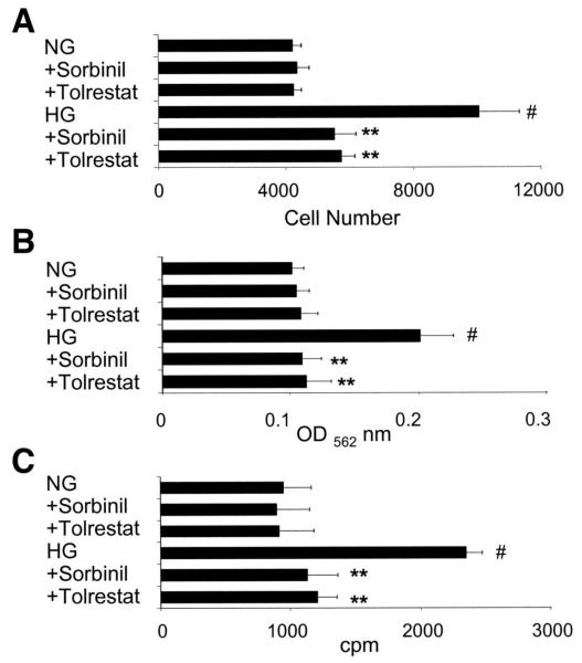
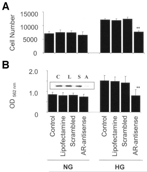
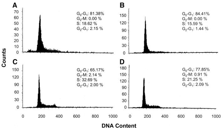
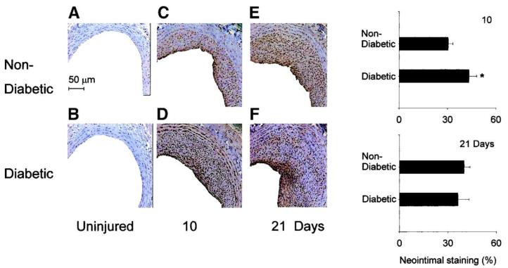
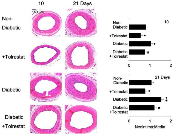
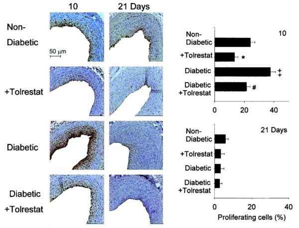
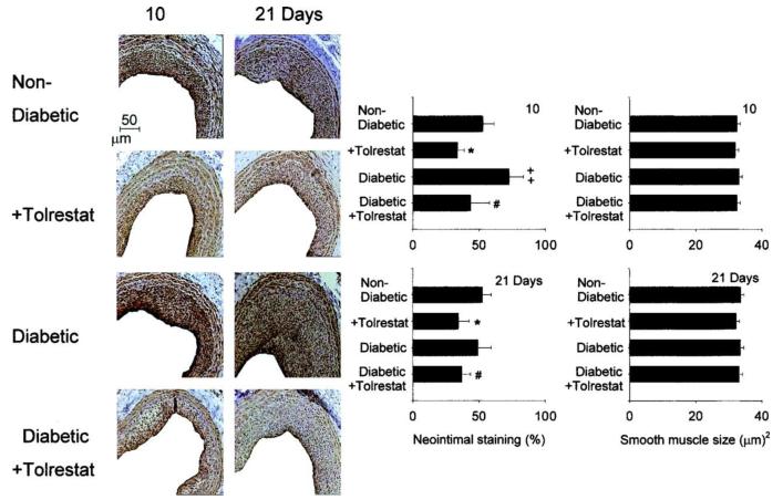
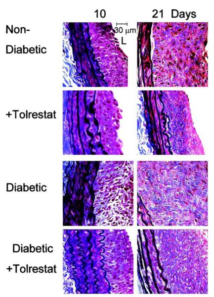
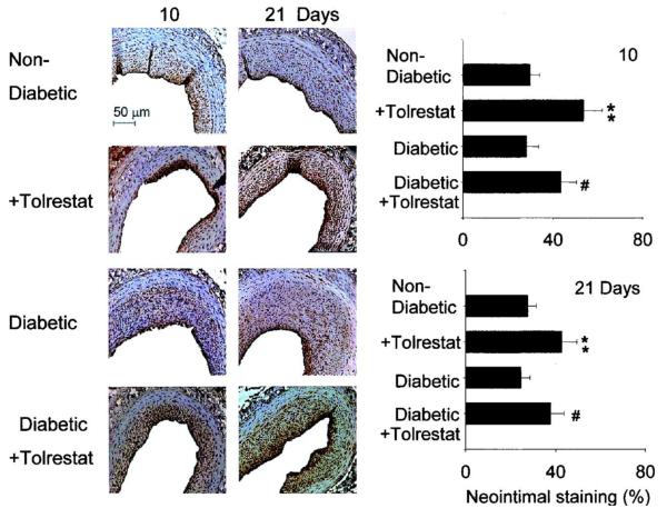
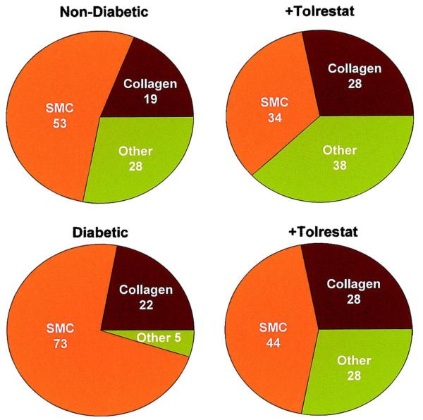
The objective of this study was to determine whether the polyol pathway enzyme aldose reductase mediates diabetes abnormalities in vascular smooth muscle cell (SMC) growth. Aldose reductase inhibitors (tolrestat or sorbinil) or antisense aldose reductase mRNA prevented hyperproliferation of cultured rat aortic SMCs induced by high glucose. Cell cycle progression in the presence of high glucose was blocked by tolrestat, which induced a G0-G1 phase growth arrest. In situ, diabetes increased SMC growth and intimal hyperplasia in balloon-injured carotid arteries of streptozotocin-treated rats, when examined 7 or 14 days after injury. Treatment with tolrestat (15 mg x kg(-1) x day(-1)) diminished intimal hyperplasia and decreased SMC content of the lesion by 25%. Although tolrestat treatment increased immunoreactivity of the lesion with antibodies raised against protein adducts of the lipid peroxidation product 4-hydroxy trans-2-nonenal, no compensatory increase in lesion fibrosis was observed. Collectively, these results suggest that inhibition of aldose reductase prevents glucose-induced stimulation of SMC growth in culture and in situ. Even though inhibition of aldose reductase increases vascular oxidative stress, this approach may be useful in preventing abnormal SMC growth in vessels of diabetic patients.


Figures

FIG. 1
FIG. 1
Inhibition of aldose reductase prevents high-glucose-induced VSMC growth. Growth-arrested VSMCs in 5.5 mmol/l glucose (NG) were either left untreated or stimulated with additional 19.5 mmol/l glucose (HG) in the absence and presence of 10 μmol/l sorbinil or tolrestat for 24 h. Cell growth was determined by counting the number of cells (A), MTT assay (OD562) (B), and the incorporation of [3H]thymidine added 6 h before the end of the experiment (C). Horizontal bars represent means ± SE (n = 4). **P < 0.001 vs. high-glucose cells without the inhibitor; #P < 0.001 vs. normal glucose.
FIG. 2
FIG. 2
Antisense ablation of aldose reductase prevents high-glucose-induced SMC growth. The VSMCs were either left untreated or treated with lipofectamine, aldose reductase (AR) antisense oligonucleotide, or scrambled oligonucleotides and cultured in normal (5.5 mmol/l) glucose (NG) or high (25 mmol/l) glucose (HG) for 24 h. Cell growth measured by counting the number of cells (A) and cell viability by MTT assay (OD562) (B). The bars represent means ± SE (n = 4). Inset of B shows a representative immunoblot of the control (C), lipofectamine-treated (L), scrambled oligonucleotide–treated (S), and aldose reductase antisense oligonucleotide-treated (A) SMCs probed with anti-aldose reductase antibodies. **P < 0.001 vs. cells transfected with the scrambled oligonucleotide.
FIG. 3
FIG. 3
Effect of aldose reductase inhibition on high-glucose-induced cell cycle progression. Quiescent VSMCs were incubated without or with sorbinil (10 μmol/l) in DMEM containing 0.1% FBS and 5.5 mmol/l glucose (A) with sorbinil (B) or 25 mmol/l glucose (C) with sorbinil (D) for 24 h. The cells were trypsinized, rinsed with PBS, and treated with 20 μg/ml RNase. DNA was stained with propidium iodide, and 1 × 10⁶ cells were analyzed by fluorescence-activated cell sorter analysis. Inset shows percent distribution of cells in G0, G1, S, G2, and M stages of the cell cycle for the representative dataset shown in the figure.
FIG. 4
FIG. 4
Expression of aldose reductase in the proliferating neointima in nondiabetic and diabetic rats. Cross sections of carotid arteries obtained from nondiabetic (control) and diabetic rats without injury (A and B) or 10 (C and D) and 21 (E and F) days after balloon injury were stained with anti-aldose reductase antibody. Immunoreactivity is evident as a dark brown stain, whereas nonreactive areas display only the background color (hematoxylin and eosin staining). The extent of staining was quantified using the MetaMorph imaging software. The bar graphs show means ± SE of neointimal staining (%) 10 and 21 days after injury. *P < 0.05 vs. control (lesions of nondiabetic rats).
FIG. 5
FIG. 5
Inhibition of aldose reductase diminishes neointima formation in balloon-injured carotid artery of nondiabetic and diabetic rats. Photomicrographs of cross sections of carotid arteries from nondiabetic and diabetic rats at 10 days (left) and 21 days (right) after balloon injury. Sections were stained with hematoxylin and eosin, and the neointima-to-media ratio was calculated by image analysis. The bar graph shows means ± SE of the neointima-to-media ratios. *P < 0.05 vs. control (nondiabetic lesion); +P < 0.05 and ++P < 0.01 vs. control; #P < 0.05 vs. untreated diabetic rats (diabetic).
FIG. 6
FIG. 6
Inhibition of aldose reductase prevents cell proliferation in balloon-injured carotid arteries of nondiabetic and diabetic rats. Cross sections of balloon-injured arteries were obtained from nondiabetic and diabetic rats 10 (left) and 21 (right) days after balloon injury and stained with anti-PCNA. Immunoreactivity is evident as a dark brown stain, whereas nonreactive areas display only the background color. The total number of cells was determined by counting the total number of propidium iodide-positive cells (data not shown), and the number of proliferating cells was determined by counting the number of PCNA-positive cells. The bar graphs show means ± SE. *P < 0.05, ++P < 0.01 vs. control; #P < 0.05 vs. untreated diabetic rats.
FIG. 7
FIG. 7
Inhibition of aldose reductase diminishes SMC proliferation in the neointima of the balloon-injured carotid arteries of nondiabetic and diabetic rats. Cross sections of balloon-injured arteries were obtained from nondiabetic and diabetic rats at 10 (left) and 21 (right) days after balloon injury and stained with anti-SMC α-actin antibody. Immunoreactivity is evident as a dark brown stain, whereas nonreactive areas display only the background color. Percent neointimal staining was determined by measuring positive immunoreactivity per unit area. SMCs in the lesion were quantitated by digital image analysis. The bar graphs show means ± SE. *P < 0.05, ++P < 0.01 vs. control; # P < 0.05 vs. sections obtained from untreated diabetic rats.
FIG. 8
FIG. 8
Inhibition of aldose reductase increases the abundance of collagen in the neointima of balloon-injured carotid artery of nondiabetic and diabetic rats. Cross sections of carotid arteries from nondiabetic and diabetic rats at 10 (left) and 21 (right) days after balloon injury, stained with Masson’s trichrome. Collagen was stained blue and elastin was stained black. All photomicrographs are in the same orientation. L, luminal surface.
FIG. 9
FIG. 9
Inhibition of aldose reductase increases the abundance of protein-HNE adducts in the neointima of the balloon-injured arteries of nondiabetic and diabetic rats. Cross sections of balloon-injured arteries were obtained from nondiabetic and diabetic rats 10 (left) and 21 (right) days after balloon injury and were stained with antibody directed against protein-HNE adducts. Immunoreactivity is evident as a dark brown stain; nonreactive areas display only the background color. The bar graphs show means ± SE of the percentage of neointima stained by the anti-protein-HNE antibody. **P < 0.05 vs. control; #P < 0.05 vs. untreated diabetic rats (diabetic).
FIG. 10
FIG. 10
Aldose reductase-dependent changes in lesion composition in diabetic and nondiabetic animals. Graph shows percent distribution of SMC, collagen, and other constituents in carotid arteries, 10 days after injury as determined by staining with anti-SMC α-actin and Masson’s trichrome stain (see Fig. 7 and Table 2).

References

    1. Beckman JA, Creager MA, Libby P. Diabetes and atherosclerosis: epidemiology, pathophysiology, and management. JAMA. 2002;287:2570–2581. - PubMed
    1. Kannel WB, McGee DL. Diabetes and cardiovascular risk factors: the Framingham study. Circulation. 1979;59:8–13. - PubMed
    1. Ceriello A, Motz E. Is oxidative stress the pathogenic mechanism underlying insulin resistance, diabetes, and cardiovascular disease? The common soil hypothesis revisited. Arterioscler Thromb Vasc Biol. 2004;24:816–823. - PubMed
    1. Stern MP. Diabetes and cardiovascular disease: the “common soil” hypothesis. Diabetes. 1995;44:369–374. - PubMed
    1. Barsness GW, Peterson ED, Ohman EM, Nelson CL, DeLong ER, Reves JG, Smith PK, Anderson RD, Jones RH, Mark DB, Califf RM. Relationship between diabetes mellitus and long-term survival after coronary bypass and angioplasty. Circulation. 1997;96:2551–2556. - PubMed

Publication types

MeSH terms