. 2006 Apr 4;103(14):5291-6.
doi: 10.1073/pnas.0509639103. Epub 2006 Mar 27.

Structural basis for histone N-terminal recognition by human peptidylarginine deiminase 4

Affiliations

Structural basis for histone N-terminal recognition by human peptidylarginine deiminase 4

Kyouhei Arita et al. Proc Natl Acad Sci U S A. .

Abstract

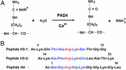
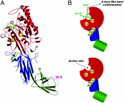
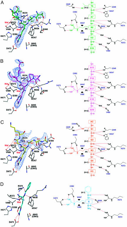
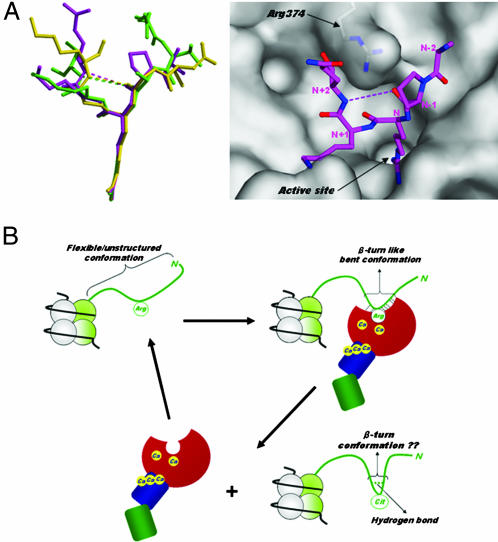
Histone arginine methylation is a posttranslational modification linked to the regulation of gene transcription. Unlike other posttranslational modifications, methylation has generally been regarded as stable, and enzymes that demethylate histone arginine residues have not been identified. However, it has recently been shown that human peptidylarginine deiminase 4 (PAD4), a Ca(2+)-dependent enzyme previously known to convert arginine residues to citrulline in histones, can also convert monomethylated arginine residues to citrulline both in vivo and in vitro. Citrullination of histone arginine residues by the enzyme antagonizes methylation by histone arginine methyltransferases and is thus a novel posttranslational modification that regulates the level of histone arginine methylation and gene activity. Here we present the crystal structures of a Ca(2+)-bound PAD4 mutant in complex with three histone N-terminal peptides, each consisting of 10 amino acid residues that include one target arginine residue for the enzyme (H3/Arg-8, H3/Arg-17, and H4/Arg-3). To each histone N-terminal peptide, the enzyme induces a beta-turn-like bent conformation composed of five successive residues at the molecular surface near the active site cleft. The remaining five residues are highly disordered. The enzyme recognizes each peptide through backbone atoms of the peptide with a possible consensus recognition motif. The sequence specificity of the peptide recognized by this enzyme is thought to be fairly broad. These observations provide structural insights into target protein recognition by histone modification enzymes and illustrate how PAD4 can target multiple arginine sites in the histone N-terminal tails.


Conflict of interest statement

Conflict of interest statement: No conflicts declared.

Figures

Fig. 1.
Fig. 1.
Citrullination (or deimination) of peptide substrates by PAD4. (A) Citrullination of arginine (R = H) and NG-monomethylarginine (R = CH3) residues by PAD4. Arginine residue is methylated to give NG-monomethylarginine, symmetric NG,NG-dimethylarginine, or asymmetric NG,NG-dimethylarginine by enzymes such as protein arginine methyltransferases (33). (B) Three histone N-terminal peptides, H3-1, H3-2, and H4. For each peptide, the target arginine residue is colored red and the five successive residues that form a β-turn-like bent conformation upon interaction with PAD4 are colored blue.
Fig. 2.
Fig. 2.
Structure of Ca2+-bound PAD4 (C645A) in complex with peptide H3-1. (A) Ribbon representation of the structure. Ca2+ ions and the histone peptide are shown as yellow balls and as a green stick model, respectively. The N-terminal subdomains 1 (residues 1–118) and 2 (residues 119–300) and the C-terminal domain (residues 301–663) are colored green, blue, and red, respectively. The nuclear localization signal (NLS) region in the subdomain 1 is shown as a dotted line. (B Upper) Schematic representation of the structure shown in A. The colors of the N-terminal domain (subdomains 1 and 2) and the C-terminal domain are the same as in A. Dotted lines show hydrogen bonds that form a consensus recognition motif at the molecular surface near the active site. (B Lower) For reference, the structure of the Ca2+-bound PAD4 (C645A) is shown.
Fig. 3.
Fig. 3.
Structures around the active sites of the Ca2+-bound PAD4 (C645A) in complex with peptides H3-1, H3-2, H4, and BA. (Left) Ball-and-stick representation of the structures. The protein moiety is colored gray, and the peptides, H3-1 (A), H3-2 (B), H4 (C), and BA (D), are colored green, magenta, yellow, and cyan, respectively. Superimposed are FoFc electron densities of the peptides, contoured at 2σ. (Right) Schematic diagrams of the structures in Left. Dotted lines and green half-circles show hydrogen bonds and hydrophobic interactions, respectively. The structure of the complex with BA was drawn by using the refined coordinates deposited in the Protein Data Bank (accession code 1WDA).
Fig. 4.
Fig. 4.
Histone N-terminal structures. (A Left) Structural comparison of PAD4-bound forms. Peptides H3-1, H3-2, and H4 are shown as ball-and-stick representations colored green, magenta, and yellow, respectively, as in Fig. 3 AC. (A Right) Top view of the peptide H3-2 structure shown in Left, together with a molecular surface representation near the active site cleft. The weak intrapeptide interactions between the backbone oxygen at (N − 1) position and the backbone nitrogen at (N + 2) position are shown as dotted lines. (B) Possible conformational change of the histone N-terminal tail in histone citrullination. The histone N-terminal tail protruding from the nucleosome core particle is shown as green ribbon. The structure of PAD4 is drawn in the same way as in Fig. 2B.
Fig. 5.
Fig. 5.
Stereogram showing the putative active site structure with a bound NG-monomethylarginine side chain. The van der Waals representation corresponds to moieties of the protein (gray) and the methyl group of the NG-monomethylarginine side chain (green).

Similar articles

Cited by

References

    1. Luger K., Mader A. W., Richmond R. K., Sargent D. F., Richmond T. J. Nature. 1997;389:251–260. - PubMed
    1. Felsenfeld G., Groudine M. Nature. 2003;421:448–453. - PubMed
    1. Khorasanizadeh S. Cell. 2004;116:259–272. - PubMed
    1. Cuthbert G. L., Daujat S., Snowden A. W., Erdjument-Bromage H., Hagiwara T., Yamada M., Schneider R., Gregory P. D., Tempst P., Bannister A. J., et al. Cell. 2004;118:545–553. - PubMed
    1. Wang Y., Wysocka J., Sayegh J., Lee Y. H., Perlin J. R., Leonelli L., Sonbuchner L. S., McDonald C. H., Cook R. G., Dou Y., et al. Science. 2004;306:279–283. - PubMed

Publication types