. 2006 Apr 4;103(14):5302-7.
doi: 10.1073/pnas.0507416103. Epub 2006 Mar 27.

Solution structure of the N-domain of Wilson disease protein: distinct nucleotide-binding environment and effects of disease mutations

Affiliations

Solution structure of the N-domain of Wilson disease protein: distinct nucleotide-binding environment and effects of disease mutations

Oleg Dmitriev et al. Proc Natl Acad Sci U S A. .

Abstract

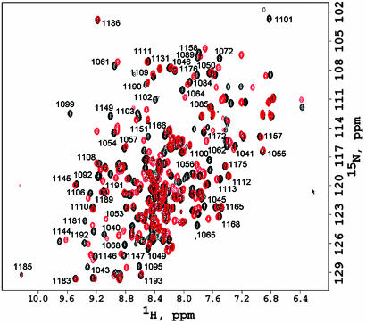
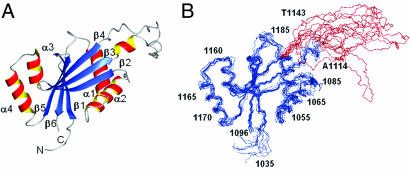
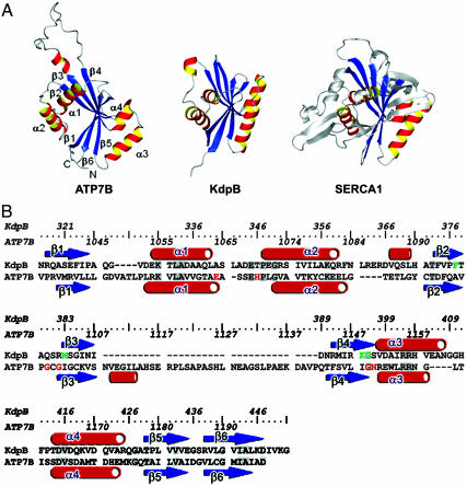
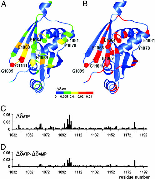
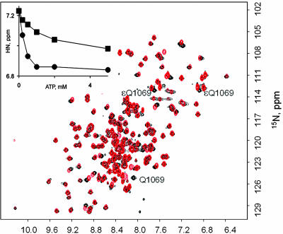
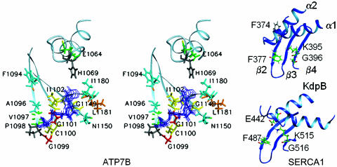
Wilson disease protein (ATP7B) is a copper-transporting P(1B)-type ATPase that regulates copper homeostasis and biosynthesis of copper-containing enzymes in human tissues. Inactivation of ATP7B or related ATP7A leads to severe neurodegenerative disorders, whereas their overexpression contributes to cancer cell resistance to chemotherapeutics. Copper-transporting ATPases differ from other P-type ATPases in their topology and the sequence of their nucleotide-binding domain (N-domain). To gain insight into the structural basis of ATP7B function, we have solved the structure of the ATP7B N-domain in the presence of ATP by using heteronuclear multidimensional NMR spectroscopy. The N-domain consists of a six-stranded beta-sheet with two adjacent alpha-helical hairpins and, unexpectedly, shows higher similarity to the bacterial K(+)-transporting ATPase KdpB than to the mammalian Ca(2+)-ATPase or Na(+),K(+)-ATPase. The common core structure of P-type ATPases is retained in the 3D fold of the N-domain; however, the nucleotide coordination environment of ATP7B within this fold is different. The residues H1069, G1099, G1101, I1102, G1149, and N1150 conserved in the P(1B)-ATPase subfamily contribute to ATP binding. Analysis of the frequent disease mutation H1069Q demonstrates that this mutation does not significantly affect the structure of the N-domain but prevents tight binding of ATP. The structure of the N-domain accounts for the disruptive effects of >30 known Wilson disease mutations. The unique features of the N-domain provide a structural basis for the development of specific inhibitors and regulators of ATP7B.


Conflict of interest statement

Conflict of interest statement: No conflicts declared.

Figures

Fig. 1.
Fig. 1.
1H,15N-HSQC spectra of the ATP7B N-domain recorded in the presence (black) and absence (red) of 5 mM ATP. Some of the backbone amide assignments made in the presence of ATP are shown. Residue numbers correspond to the full-length ATP7B.
Fig. 2.
Fig. 2.
Structure of the N-domain in the presence of ATP. (A) Ribbon diagram of the structure with the α-helices shown in red-yellow and β-sheet in blue. (B) The backbone traces of the 10 lowest energy structures of the N-domain illustrating folded regions (blue) and the flexible loop Ala-1114-Thr-1143 (red).
Fig. 3.
Fig. 3.
Comparison of the N-domains of ATP7B (P1B-ATPase), KdpB (P1A-ATPase), and SERCA1 Ca2+-ATPase (P2-ATPase). (A) The structures of KdpB (1SVJ) and SERCA1 (1T5S) N-domains were aligned to the ATP7B structure by using dali. rms deviation for the α-carbons of the aligned regions was 3.6 Å (ATP7B–KdpB alignment) and 4.2 Å (ATP7B–SERCA1 alignment). The common core is shown in red-yellow (α-helices) and blue (β-sheet). (B) Alignment of the secondary structure elements for ATP7B and KdpB. The residues predicted to be involved in ATP binding are shown in red (ATP7B) or green (KdpB). The identical residues are highlighted in gray.
Fig. 4.
Fig. 4.
Residues affected by nucleotide binding and the location of Wilson disease-causing mutations. (A) The structure of the ATP7B N-domain with a color map of backbone amide chemical shift changes induced by ATP binding. The secondary chemical shifts for each residue are expressed as Δδ = (((δHNNUC − δHNfree)/δHNNUC)2 + ((δNNUC − δNfree)/δNNUC)2)1/2, where δHNNUC and δNNUC are 1H and 15N chemical shifts of the backbone amide group in the presence of the nucleotide, and δHNfree and δNfree are chemical shifts measured without nucleotide present. Data for H1069 (gray) is not available. The residues invariant in the P1B-ATPases are shown as spheres. The disordered loop A1114-T1143 is not shown. (B) The location of disease causing mutations is shown in red. (C) Chemical shift changes (Δδ, defined above) at each amino acid residue upon binding of ATP. (D) Chemical shift changes at each residue observed on binding ATP minus the corresponding chemical shift changes on binding AMP.
Fig. 5.
Fig. 5.
Characterization of the H1069Q mutant. Overlay of the fingerprint spectra of the WT ATP7B N-domain (red) and the H1069Q mutant (black) are shown. Tentative Q1069 assignment is shown. (Inset) Chemical shift of the G1101 1HN signal as a function of added ATP in the wild type (•) and H1069Q mutant (■) of the ATP7B N-domain.
Fig. 6.
Fig. 6.
Organization of the nucleotide-binding site in the N-domains of ATP7B, SERCA1, and KdpB. (A) Stereo view of the ensemble of the ATP molecules docked into the ATP-binding site of ATP7B. The residues in proximity to ATP are colored according to the magnitude of the ATP-dependent secondary chemical shifts as in Fig. 4A. Data are not available for H1069 (shown in gray). (B) Fragments of the KdpB and SERCA1 N-domain structures with the residues involved in ATP binding.

References

    1. Puig S., Thiele D. J. Curr. Opin. Chem. Biol. 2002;6:171–180. - PubMed
    1. Daniel K. G., Harbach R. H., Guida W. C., Dou Q. P. Front. Biosci. 2004;9:2652–2662. - PubMed
    1. Ferenci P. Aliment. Pharmacol. Ther. 2004;19:157–165. - PubMed
    1. Gitlin J. D. Gastroenterology. 2003;125:1868–1877. - PubMed
    1. Shim H., Harris Z. L. J. Nutr. 2003;133:1527S–1531S. - PubMed

Publication types

MeSH terms

Associated data

LinkOut - more resources