. 2006 May;36(5):1241-53.
doi: 10.1002/eji.200535563.

The key regulators of adult T helper cell responses, STAT6 and T-bet, are established in early life in mice

Affiliations

The key regulators of adult T helper cell responses, STAT6 and T-bet, are established in early life in mice

Shawn Rose et al. Eur J Immunol. 2006 May.

Abstract

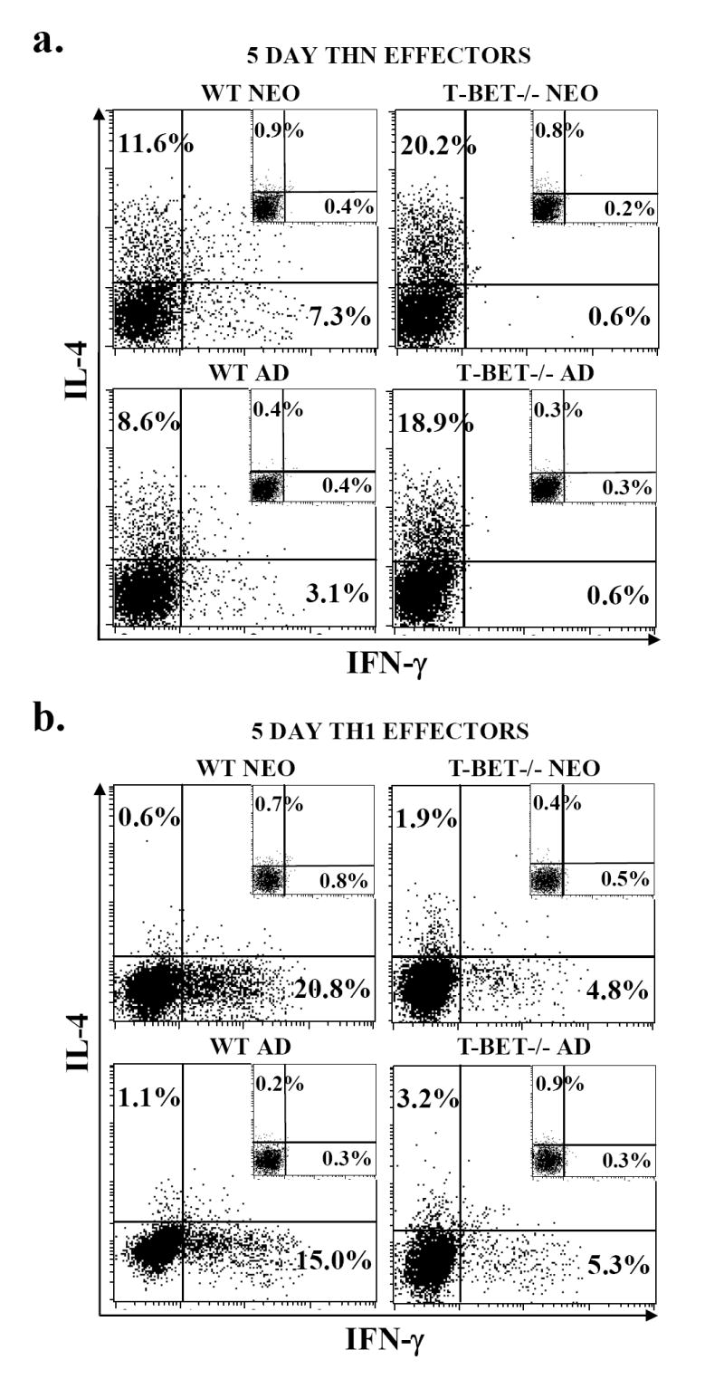
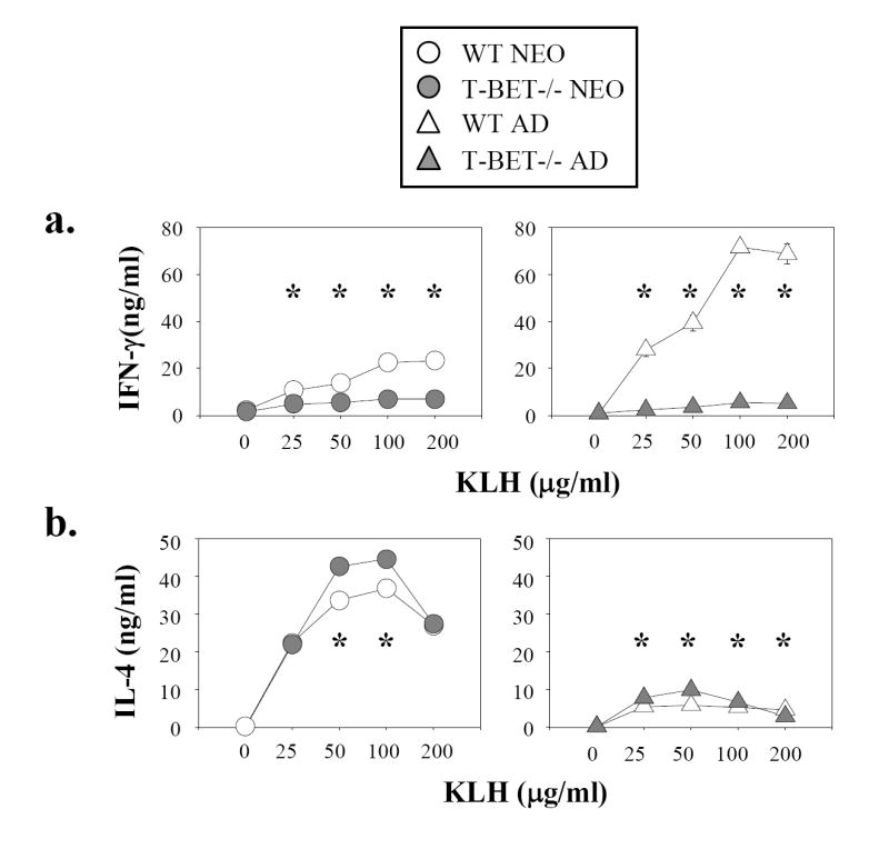
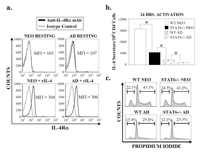
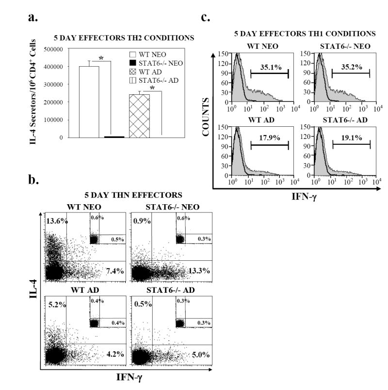
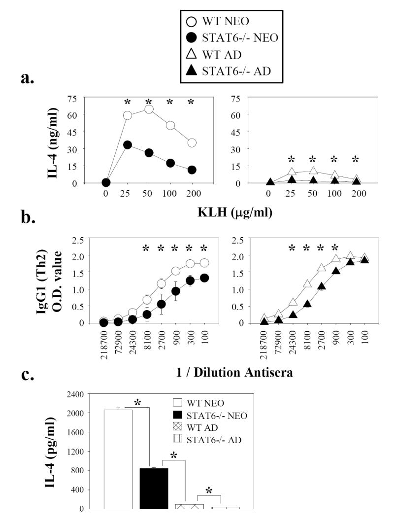
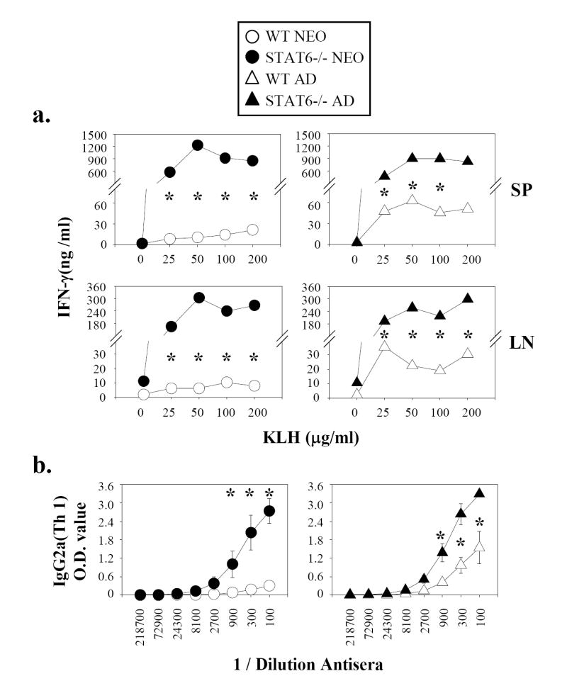
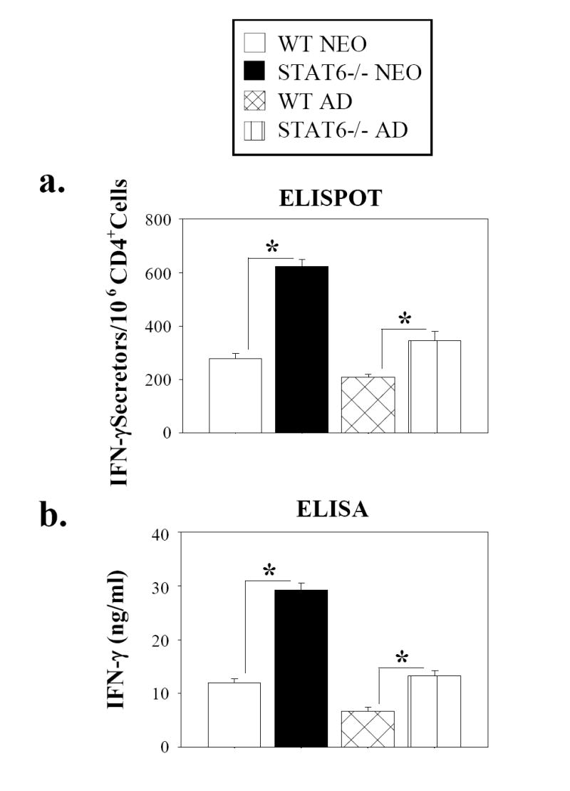
Murine neonatal immunity is typically Th2 biased. This is characterized by high-level IL-4 production at all phases of the immune response and poor IFN-gamma memory responses. The differential expression of Th1/Th2 cytokines by neonates and adults could arise if the critical regulators of Th differentiation and function, STAT6 and T-bet, operate differently during the neonatal period. To test this idea, the Th cell responses of wild-type, T-bet-deficient, or STAT6-deficient mice were compared in vitro and in vivo. The absence of these factors had similar qualitative effects on the development of effector function in neonates and adults, i.e., if a Th lineage was inhibited or enhanced in adult animals, a similar phenomenon was observed in neonates. However, there was a striking difference observed in the in vivo Th1 memory responses of STAT6-deficient mice initially immunized as neonates. Antigen-specific IFN-gamma production was increased 50-100-fold in STAT6-deficient neonates, achieving levels similar to those of STAT6-deficient adults. These findings demonstrate that STAT6 and T-bet signals are central in shaping Th responses in wild-type neonates, as in adult mice, and that the master regulators of Th cell development and function are already firmly established in early life.


Figures

Figure 1
Figure 1. T-bet positively regulates Th1 effector development and negatively regulates Th2 effector development of neonatal CD4+ T cells in vitro
CD4+ lymph node cells were isolated from pooled tissues from day 7 neonatal (n=12) and adult (n=2) wild-type or T-bet-deficient mice. The cells were activated with plate-bound anti-CD3 plus soluble anti-CD28 mAb under (a) ThN or (b) Th1-polarizing conditions. After 3 days, the cells were harvested, washed, and expanded in rIL-2 for an additional 2 days. Effector cells were then cultured in PMA, ionomycin, and monensin for 4 hr. The cells were then permeabilized and stained with mAb specific for IL-4 and IFN-γ or with isotype control antibodies (smaller inlaid graphs). The percentages of positively staining cells are indicated. The depicted experiment is representative of 3 independent experiments.
Figure 2
Figure 2. Neonatal Th1 immunity in vivo is impaired by T-bet-deficiency
Day 1 neonatal (n=12) and adult (n=4) wild-type or T-bet-deficient mice were immunized with DNP-KLH. After 4 weeks, the animals were re-immunized with DNP-KLH. 3 days later, CD4+ spleen cells were isolated from pooled tissues and were stimulated in vitro with syngeneic mitomycin-treated splenic APC plus DNP-KLH. After 72 hr, (a) IFN-γ or (b) IL-4 levels in the culture supernatants were determined by ELISA. The ELISA results are graphed as the mean ± the standard deviation of the triplicate ELISA values from a single representative experiment. This experiment was performed twice. * Statistically significant at p < 0.001. + Statistically significant at p < 0.01.
Figure 3
Figure 3. STAT6 is required for maximal early IL-4 production by neonatal CD4+ cells in vitro
CD4+ lymph node cells were isolated from pools of tissue from day 7 neonatal (n=15) and adult (n=2) wild-type or STAT6-deficient mice. (a) Freshly-isolated resting cells from neonatal or adult wild-type mice were stained with an anti-IL-4Rα mAb (black line) or with an isotype control mAb (gray line) either directly ex vivo or after 18 hr of culture with 0.5 ng/ml rIL-4. MFI = mean fluorescence intensity of the staining with αIL-4 mAb. (b) CD4+ cells were activated with plate-bound anti-CD3 plus soluble anti-CD28 mAb for 20 hr. The cells were then washed and restimulated with PMA and ionomycin for an additional 4 hr and the frequencies of IL-4-secreting cells were enumerated by ELISPOT. Each sample was plated in quadruplicate and the averages ± the standards are shown. * Statistically significant at p < 0.001. (c) CD4+ cells were activated with plate-bound anti-CD3 plus soluble anti-CD28 mAb for 24 hr. After ethanol fixation, cell cycle progression was analyzed via propidium iodide staining. The percentages of apoptotic cells (left marker) or cells in the S, G2, and M phases of the cell cycle are indicated (right marker). All results are representative of at least 2 independent experiments.
Figure 4
Figure 4. Neonatal Th2 effector cell development in vitro is STAT6-dependent
CD4+ cells from pools of tissue from day 7 neonatal (n=12) and adult (n=2) wild-type or STAT6-deficient mice were polyclonally activated under (a) Th2-polarizing, (b) neutral (ThN), or (c) Th1-polarizing conditions for 3 days. The cells were harvested, washed, and expanded in rIL-2 for 2 additional days. (a) Th2 effector cells were then processed to determine the frequencies of IL-4-secreting cells by ELISPOT, as described in Figure 3. * Statistically significant at p < 0.001. (b) ThN effectors were washed and cultured with PMA, ionomycin, and monensin for 4 hr. The cells were then permeabilized and stained for intracellular IL-4 and IFN-γ, as described in Figure 1. (c) Th1 effectors were washed and cultured with PMA, ionomycin, and monensin for 4 hr. The cells were then permeabilized and stained with antibodies specific for IFN-γ (filled histograms) or with control antibodies (thick black line). All experiments were performed at least twice.
Figure 5
Figure 5. Antigen-specific Th2 immune responses in vivo are reduced in STAT6-deficient neonates
Day 1 neonatal (n=8) and adult (n=8) wild-type or STAT6-deficient mice were immunized with DNP-KLH. 4 weeks later, the animals were re-immunized with DNP-KLH. (a) After 3 days, CD4+ spleen cells were isolated from pooled tissues from ½ of the immunized animals. The cells were restimulated with various concentrations of DNP-KLH in vitro and IL-4 levels in the culture supernatants were measured by ELISA after 72 hr, as described in Figure 2. (b) After 2 weeks, blood was collected from the second group of immunized mice and the serum levels of DNP-specific IgG1 were analyzed by ELISA. The ELISA results are graphed as the mean ± the standard deviation of the values obtained from individual animals. (c) To asssess primary antigen-specific Th2 responses generated in vivo, day 1 neonatal (n=12) and adult (n=2) wild-type or STAT6-deficient mice were immunized with DNP-KLH. One week later, CD4+ spleen cells were isolated, pooled, and were stimulated in vitro with mitomycin-treated syngeneic splenic APC plus 100 μg/ml DNP-KLH. After 48 hr, antigen-specific IL-4 production was measured by ELISA. All results are representative of at least 2 independent experiments. * Statistically significant at p < 0.001.
Figure 6
Figure 6. Neonatal Th1 memory responses in vivo are strongly enhanced by STAT6-deficiency
Day 1 neonatal and adult wild-type or STAT6-deficient mice were immunized and treated as described in Fig. 5. (a) Antigen-specific IFN-γ production and (b) hapten-specific serum IgG2a levels were measured by ELISA. All results are representative of at least 2 independent experiments. * Statistically significant at p < 0.001.
Figure 7
Figure 7. Antigen-specific Th1 primary responses are increased in STAT6-deficient neonatal mice, compared to their wild-type counterparts
Day 1 neonatal (n=12) and adult (n=2) wild-type or STAT6-deficient mice were immunized with DNP-KLH. One week later, CD4+ lymph node cells were isolated, pooled, and were stimulated in vitro with mitomycin-treated syngeneic splenic APC plus 100 μg/ml DNP-KLH. After 48 hr, (a) the cells were washed and transferred to ELISPOT wells to measure the frequencies of IFN-γ secreting cells and (b) antigen-specific IFN-γ production was measured by ELISA. Each experiment was performed at least twice. * Statistically significant at p < 0.001.
Figure 8
Figure 8. The characteristically poor Th1 memory responses of wild-type neonatal mice are not due to apoptosis induced by antigen re-exposure in vitro
(a) Day 1 neonatal (n=10) and adult (n=4) wild-type mice were immunized twice with DNP-KLH, and CD4+ spleen cells were restimulated in vitro with DNP-KLH, as described in Figure 2. Some cultures also contained either varying amounts of the caspase inhibitor Z-VAD-fmk. After 24 hr, IFN-γ levels in the culture supernatants were determined by ELISA. The depicted experiment is representative of two independent experiments.

Similar articles

Cited by

References

    1. Adkins B, Leclerc C, Marshall-Clarke S. Neonatal adaptive immunity comes of age. Nat Rev Immunol. 2004;4:553–564. - PubMed
    1. Singh RR, Hahn BH, Sercarz EE. Neonatal peptide exposure can prime T cells and, upon subsequent immunization, induce their immune deviation: implications for antibody vs. T cell-mediated autoimmunity. J Exp Med. 1996;183:1613–1621. - PMC - PubMed
    1. Bot A, Antohi S, Bot S, Garcia-Sastre A, Bona C. Induction of humoral and cellular immunity against influenza virus by immunization of newborn mice with a plasmid bearing a hemagglutinin gene. Int Immunol. 1997;9:1641–1650. - PubMed
    1. Adkins B, Du RQ. Newborn mice develop balanced Th1/Th2 primary effector responses in vivo but are biased to Th2 secondary responses. J Immunol. 1998;160:4217–4224. - PubMed
    1. Li L, Lee HH, Bell JJ, Gregg RK, Ellis JS, Gessner A, Zaghouani H. IL-4 utilizes an alternative receptor to drive apoptosis of Th1 cells and skews neonatal immunity toward Th2. Immunity. 2004;20:429–440. - PubMed

Publication types

LinkOut - more resources