Review
. 2006;8(2):207.
doi: 10.1186/ar1934. Epub 2006 Mar 23.

Magnetic resonance imaging in psoriatic arthritis: a review of the literature

Affiliations
Review

Magnetic resonance imaging in psoriatic arthritis: a review of the literature

Fiona McQueen et al. Arthritis Res Ther. 2006.

Abstract

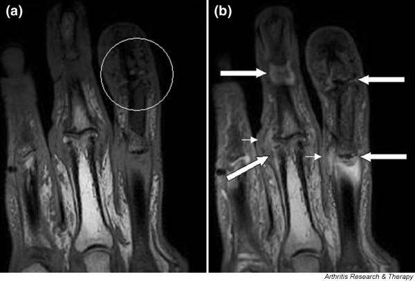
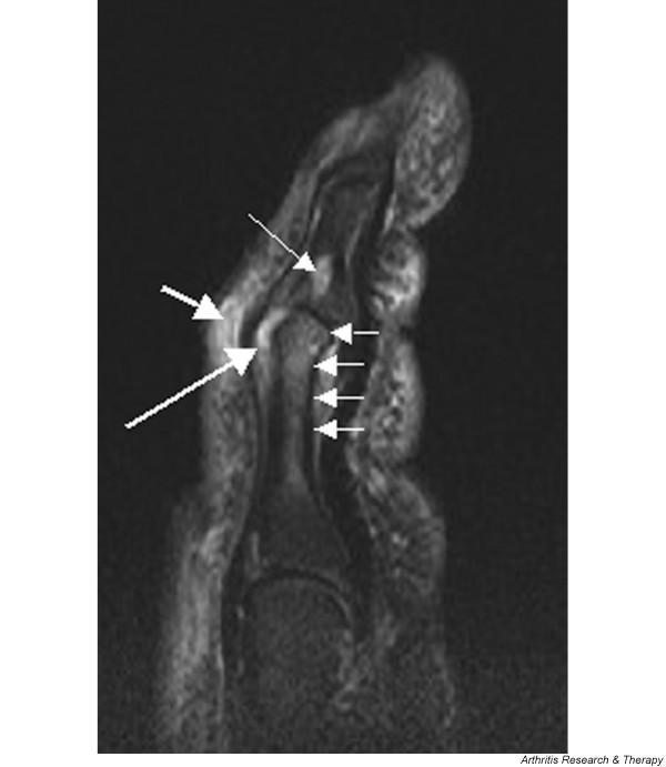
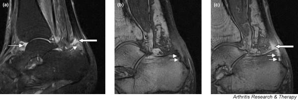
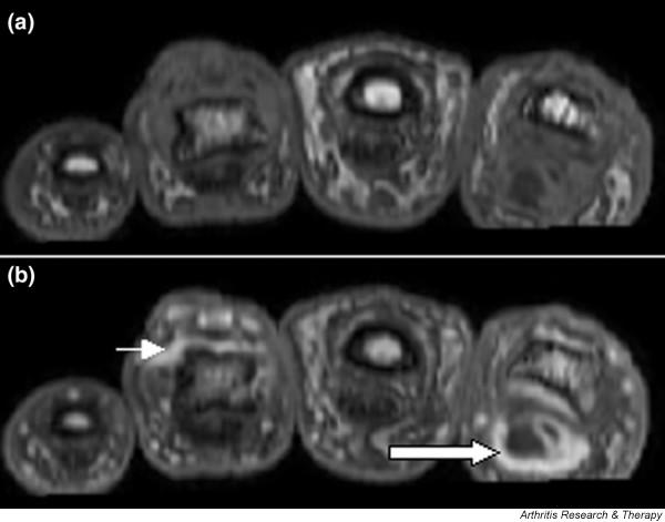
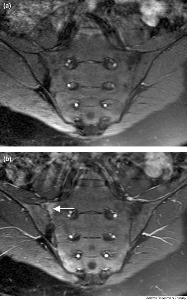
Psoriatic arthritis is a diverse condition that may be characterized by peripheral inflammatory arthritis, axial involvement, dactylitis and enthesitis. Magnetic resonance imaging (MRI) allows visualization of soft tissue, articular and entheseal lesions, and provides a unique picture of the disease process that cannot be gained using other imaging modalities. This review focuses on the literature on MRI in psoriatic arthritis published from 1996 to July 2005. The MRI features discussed include synovitis, tendonitis, dactylitis, bone oedema, bone erosions, soft tissue oedema, spondylitis/sacroiliitis and subclinical arthropathy. Comparisons have been drawn with the more extensive literature describing the MRI features of rheumatoid arthritis and ankylosing spondylitis.


Figures

Figure 1
Figure 1
Magnetic resonance images of fingers: psoriatic arthritis. Shown are T1-weighted (a) precontrast and (b) postcontrast coronal magnetic resonance images of the fingers in a patient with psoriatic arthritis. Enhancement of the synovial membrane at the third and fourth proximal interphalangeal (PIP) and distal interphalangeal (DIP) joints is seen, indicating active synovitis (large arrows). There is joint space narrowing with bone proliferation at the third PIP joint and erosions are present at the fourth DIP joint (white circle). Extracapsular enhancement (small arrows) is seen medial to the third and fourth PIP joints, indicating probable enthesitis. Note that this particular slice does not allow optimal visualization of all of the mentioned pathologies.
Figure 2
Figure 2
Magnetic resonance image of index finger: psoriatic arthritis (mutilans form). Shown is a T2 weighted fat suppressed sagittal image of the index finger in a patient with PsA (mutilans form). Focal increased signal (probable erosion) is seen at the base of the middle phalanx (long thin arrow). There is synovitis at the proximal interphalangeal joint (long thick arrow) plus increased signal in the overlying soft tissues indicating oedema (short thick arrow). There is also diffuse bone oedema (short thin arrows) involving the head of the proximal phalanx and extending distally down the shaft.
Figure 3
Figure 3
Sagittal magnetic resonance images of ankle region: psoriatic arthritis. (a) Short tau inversion recovery (STIR) image, showing high signal intensity at the Achilles tendon insertion (enthesitis, thick arrow) and in the synovium of the ankle joint (synovitis, long thin arrow). Bone marrow oedema is seen at the tendon insertion (short thin arrow). (b,c) T1 weighted images of a different section of the same patient, before (panel b) and after (panel c) intravenous contrast injection, confirm inflammation (large arrow) at the enthesis and reveal bone erosion at tendon insertion (short thin arrows).
Figure 4
Figure 4
Magnetic resonance images of fingers: psoriatic arthritis with dactylitis due to flexor tenosynovitis. Shown are T1 weighted axial (a) precontrast and (b) postcontrast magnetic resonance images of the fingers from a patient with psoriatic arthritis exhibiting flexor tenosynovitis at the second finger with enhancement and thickening of the tendon sheath (large arrow). Synovitis is seen in the fourth proximal interphalangeal joint (small arrow).
Figure 5
Figure 5
Magnetic resonance images of sacroiliac joints: psoriatic arthritis. Shown are T1-weighted semi-coronal magnetic resonance images through the sacroiliac joints (a) before and (b) after intravenous contrast injection. Enhancement is seen at the right sacroiliac joint (arrow), indicating active sacroiliitis.
Figure 6
Figure 6
Magnetic resonance images of lumbar and lower thoracic spine: psoriatic arthritis. (a) T1-weighted and (b) short tau inversion recovery (STIR) magnetic resonance images of the lumbar and lower thoracic spine. Signs of active inflammation are seen at several levels (arrows). In particular, anterior spondylitis is seen at level L1/L2 and an inflammatory Andersson lesion at the upper vertebral endplate of L3.

Similar articles

Cited by

References

    1. Helliwell PS, Taylor WJ. Classification and diagnostic criteria for psoriatic arthritis. Ann Rheum Dis. 2005;(Suppl II):ii3–ii7. doi: 10.1136/ard.2004.032318. - DOI - PMC - PubMed
    1. Ory PA, Gladman DD, Mease PJ. Psoriatic arthritis and imaging. Ann Rheum Dis. 2005;(Suppl II):ii55–ii57. doi: 10.1136/ard.2004.033928. - DOI - PMC - PubMed
    1. Antoni C, Dechant C, Lorenz PDH, Wendler J, Ogilvie A, Lueftl M, Kalden-Nemeth D, Kalden JR, Manger B. Open-label study of infliximab treatment for psoriatic arthritis: clinical and magnetic resonance imaging measurements of reduction of inflammation. Arthritis Care Res. 2002;47:506–512. doi: 10.1002/art.10671. - DOI - PubMed
    1. Backhaus M, Kamradt T, Sandrock D, Loreck D, Fritz J, Wolf KJ, Raber H, Hamm B, Burmester GR, Bollow M. Arthritis of the finger joints. A comprehensive approach comparing conventional radiography, scintigraphy, ultrasound and contrast-enhanced MRI. Arthritis Rheum. 1999;42:1232–1234. doi: 10.1002/1529-0131(199906)42:6<1232::AID-ANR21>3.0.CO;2-3. - DOI - PubMed
    1. Bollow M, Braun J, Hamm B, Eggens U, Schilling A, Konig H, Wolf KJ. Early sacroiliitis in patients with spondyloarthropathy: evaluation with dynamic gadolinium-enhanced MR imaging. Radiology. 1995;194:529–536. - PubMed