Comparative Study
. 2006 May;18(5):741-53.
doi: 10.1093/intimm/dxl011. Epub 2006 Mar 28.

Functional comparison of mouse CIRE/mouse DC-SIGN and human DC-SIGN

Affiliations
Comparative Study

Functional comparison of mouse CIRE/mouse DC-SIGN and human DC-SIGN

Irina Caminschi et al. Int Immunol. 2006 May.

Abstract

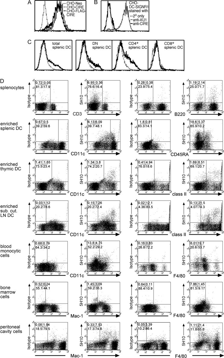
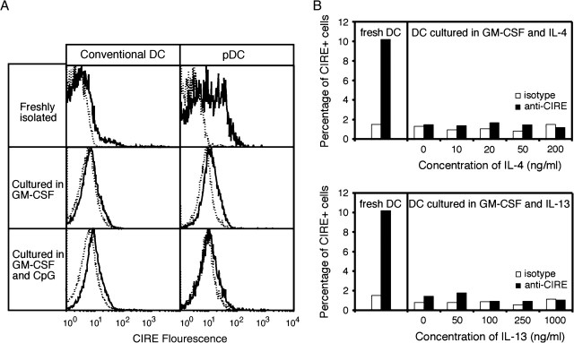
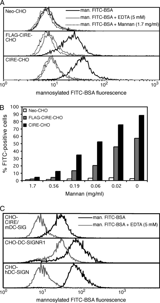
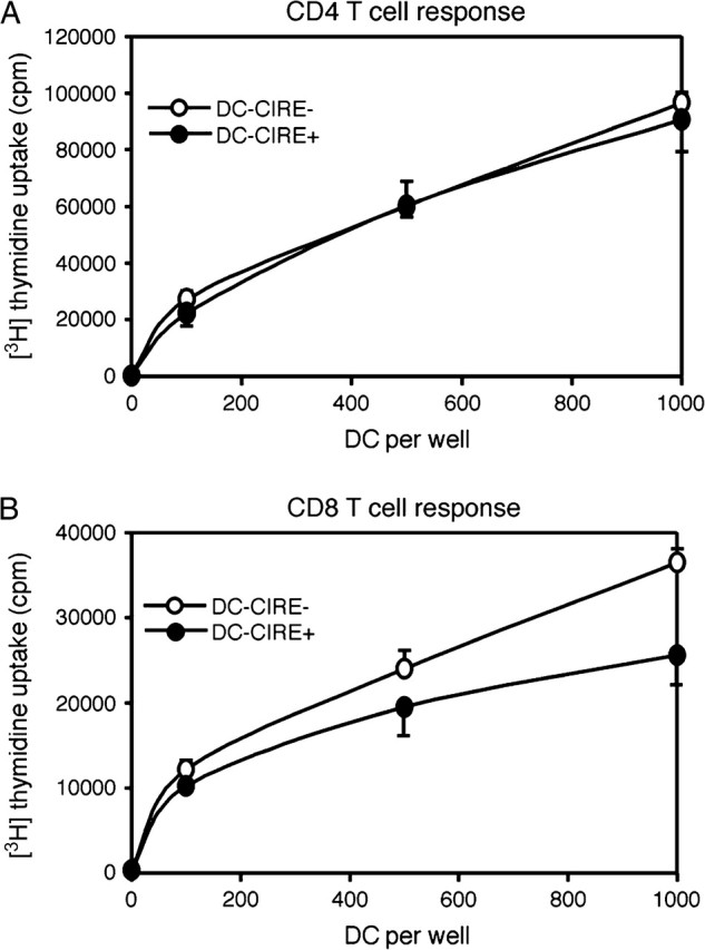
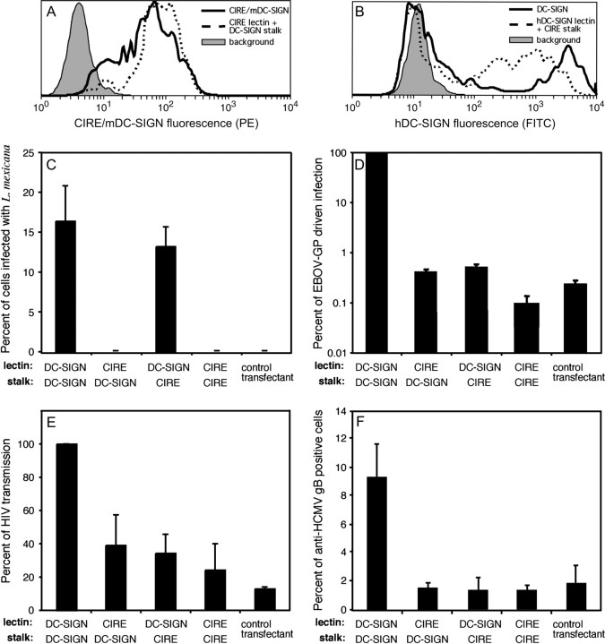
CIRE/mDC-SIGN is a C-type lectin we originally identified as a molecule differentially expressed by mouse dendritic cell (DC) populations. Immunostaining with a CIRE/mDC-SIGN-specific mAb revealed that CIRE/mDC-SIGN is indeed on the surface of some CD4+, CD4- 8- DCs and plasmacytoid pre-DCs, but not on CD8+ DCs. It has been proposed that CIRE/mDC-SIGN is the functional orthologue of human DC-SIGN (hDC-SIGN), a molecule that both enhances T cell responses and facilitates antigen uptake. We assessed if CIRE/mDC-SIGN and hDC-SIGN exhibit functional similarities. CIRE/mDC-SIGN is down-regulated upon activation, but unlike hDC-SIGN, incubation with IL-4 and IL-13 did not enhance CIRE/mDC-SIGN expression, indicating differences in gene regulation. Like hDC-SIGN, CIRE/mDC-SIGN bound mannosylated residues. However, we could detect no role for CIRE/mDC-SIGN in T cell-DC interactions and the protein did not bind to pathogens known to interact with hDC-SIGN, including Leishmania mexicana, cytomegalovirus, HIV and lentiviral particles bearing the Ebolavirus glycoprotein. The binding of CIRE/mDC-SIGN to hDC-SIGN ligands was not rescued when CIRE/mDC-SIGN was engineered to express the stalk region of hDC-SIGN. We conclude that there are significant differences in the fine specificity of the C-type lectin domains of hDC-SIGN and CIRE/mDC-SIGN and that these two molecules may not be functional orthologues.


Figures

Fig. 1.
Fig. 1.
Cytofluorometric analysis of CIRE/mDC-SIGN expression. (A) CHO cells expressing FLAG-CIRE/mDC-SIGN (CHO-FLAG-CIRE), CIRE/mDC-SIGN (CHO-CIRE) or the neomycin resistance gene alone (Neo-CHO) were stained with anti-CIRE/mDC-SIGN mAb and counterstained with goat anti-rat FITC antibody. (B) CHO expressing mouse SIGNR1 (where mouse SIGNR1 was genetically fused to express the AU1 sequence) was stained with 5H10. (C) Splenic DCs from SPF mice were isolated and identified by high forward light scatter and staining with anti-CD11c-Cy5. DCs were further stained with anti-CD4–Alexa and anti-CD8–FITC. Only in the samples depicted in panel C was the staining intensity for CIRE/mDC-SIGN amplified using Flow-Amp. The continuous line represents the anti-CIRE/mDC-SIGN staining on gated cells and the dotted line represents staining of isotype control antibody. Histographs are on a four-decade logarithmic scale. (D) Bone marrow cells, blood monocytes, peritoneal cavity cells, splenocytes, splenic cDCs, splenic pDCs, LNs–DCs and thymic DCs were isolated from germ-free mice. DCs were stained using anti-CD11c–FITC, anti-CD45RA–FITC or anti-class II (M5/114-FITC). Other cell types were stained with anti-F4/80–FITC, anti-CD3 (KT3-1.1-FITC), anti-B220 (RA3-6B2-FITC) and anti-Mac-1 (M1/70-FITC). Cells were stained using anti-CIRE/mDC-SIGN–biotin mAb, then detected in flow cytometry using streptavidin–PE. The CIRE/mDC-SIGN staining seen on transfectants (panel A) and primary cells (panel D) was not amplified using Flow-Amp. Percentages of cells in respective quadrants are displayed in the upper left corner.
Fig. 2.
Fig. 2.
CIRE/mDC-SIGN is down-regulated upon activation and is not up-regulated by IL-4 and IL-13. (A) Isolated splenic cDCs (98% < CD11hiCD45RA) and pDCs (98% < CD11cint CD45RA+) from SPF mice were purified by cell sorting and cultured in the presence of GM-CSF or GM-CSF and CpG-1668. The continuous line represents the anti-CIRE/mDC-SIGN staining and the dotted line is the background staining of an isotype control antibody. Histographs are on a logarithmic scale. The data are representative of minimum five independent experiments. (B) Freshly isolated DCs (>90% CD11c+) from SPF mice were cultured in the presence of GM-CSF plus various concentrations of IL-4 or IL-13 for 18 h. The level of CIRE expression was determined by flow cytometry. Open bars represent the control staining with an isotype-matched mAb. Data presented are representative of a minimum of four independent experiments.
Fig. 3.
Fig. 3.
CHO cells expressing CIRE/mDC-SIGN bind mannosylated protein in a calcium-dependent, mannan-inhibitable manner. CHO cells (105) expressing FLAG-CIRE/mDC-SIGN (FLAG-CIRE-CHO), CIRE/mDC-SIGN (CIRE-CHO) or neomycin resistance gene only (Neo-CHO) were re-suspended in various dilutions of mannan in ice-cold medium containing 2% FCS and Ca2+ and Mg2+ or in medium where the Ca2+ and Mg2+ were replaced by 5 mM EDTA. (A) Mannan and EDTA inhibit the binding of mannosylated FITC-conjugated BSA to CIRE/mDC-SIGN. (B) Mannan inhibits the binding of mannosylated FITC-conjugated BSA by CIRE/mDC-SIGN in a dose-dependent manner. Data are representative of a minimum of five independent experiments. (C) Human DC-SIGN and mouse SIGNR1 also bind mannosylated FITC-conjugated BSA in a Ca2+-dependent manner. Cells were then incubated with mannosylated FITC-conjugated BSA and fluorescence levels quantitated by flow cytometry. Data are representative of two independent experiments.
Fig. 4.
Fig. 4.
CIRE/mDC-SIGN+ DC does not preferentially activate T cells into proliferation. DCs from SPF mice were isolated and then sorted based on their expression of CD11c+CIRE/mDC-SIGN+ (purity: 75%) and CD11c+CIRE/mDC-SIGN (purity: 98%). DCs were incubated with 20 000 CD4 or CD8 T cells for 4 and 3 days, respectively, then pulsed with 1 μCi per well of [3H]thymidine for 6 h, harvested onto glass-fibre filters and thymidine incorporation was counted by liquid scintillation. All cultures were performed in quintets and are representative of two independent experiments.
Fig. 5.
Fig. 5.
CIRE/mDC-SIGN does not bind pathogens known to interact with hDC-SIGN. CHO cells were co-transfected with the neomycin resistance gene plus the cDNA coding for CIRE/mDC-SIGN, hDC-SIGN or the chimeric molecules (CIRE/mDC-SIGN lectin fused to hDC-SIGN stalk and hDC-SIGN lectin fused to CIRE/mDC-SIGN stalk). Transfectants were stained with 5H10 (anti-CIRE) or AZN-1 (anti-DC-SIGN) and counterstained with anti-rat–PE and anti-mouse–FITC, respectively. The level of fluorescence was determined by flow cytometry. Filled histograph denotes the background staining of control transfectants, lacking the binding site of the primary mAb. Continuous line represents the levels of CIRE/mDC-SIGN (A) and hDC-SIGN (B). Dotted line represents the levels of CIRE/mDC-SIGN-lectin fused to hDC-SIGN stalk (A) and hDC-SIGN lectin fused to CIRE/mDC-SIGN stalk (B). The above expression profiles of the CHO transfectants are representative of the expression pattern seen in the independently executed experiments seen in Fig. 5(C–F). CHO cells expressing CIRE/mDC-SIGN, hDC-SIGN or the chimeric molecules (CIRE/mDC-SIGN lectin fused to hDC-SIGN stalk and hDC-SIGN lectin fused to CIRE/mDC-SIGN stalk) or the neomycin resistance gene were tested for interaction with various pathogens. (C) Leishmania mexicana promastigotes were added at a multiplicity of infection of 5:1. Infection proceeded for 24 h at 33°C, before free parasites were removed, cells fixed in methanol and stained with Giemsa. The percent infected cells or cells with attached parasites were counted. The mean ± SEM of two independent experiments is shown. (D) Lentiviral pseudotypes bearing the EBOV-GP. The indicated CHO cell lines were inoculated with a luciferase reporter virus bearing EBOV-GP and luciferase activities in cell lysates were assessed 3 days after infection. The average of two independent experiments performed in quadruplicates is shown. Error bars indicate SEM. (E) HIV-1. Lectin-mediated HIV transmission was assessed by incubating transfectants with a HIV-1 NL4-3 variant harbouring the luciferase gene in place of nef, washing with culture medium and co-cultivation with receptor-positive target cells. Luciferase activities in cell lysates were determined 3 days after the start of the co-cultivation. The average ± SEM from two independent experiments carried out in quadruplicates is presented. (F) HCMV. Strain AD169 was added and incubated (1 h at 4°C in PBS/0.1% BSA/1 mM CaCl2/2 mM MgCl2) and percentage of transfectants to which AD169 attached was determined by FACS analysis using an anti-HCMV gB mAb. The data are presented as the mean ± SEM pooled from two independent experiments.
<b>Fig. 6</b>.
Fig. 6.
The chimeric molecule containing the CIRE/mDC-SIGN lectin domain fused to human DC-SIGN stalk appears to form multimers similarly to hDC-SIGN. Cell lysates from parental CHO cells (lane 1) or CHO cells expressing hDC-SIGN (lane 2), or the chimeric molecules [CIRE/mDC-SIGN lectin fused to hDC-SIGN stalk (lane 3) and hDC-SIGN lectin fused to CIRE/mDC-SIGN stalk (lane 4)] and CIRE/mDC-SIGN (lane 5) were separated on SDS-PAGE (10%) under reducing and non-reducing conditions, probed with mouse anti-human DC-SIGN mAb and anti-CIRE/mDC-SIGN and then revealed using anti-rat–HRP and anti-mouse–HRP antibody.

References

    1. Steinman R. M. 1991. The dendritic cell system and its role in immunogenicity. Annu. Rev. Immunol. 9:271. - PubMed
    1. Shortman K. and Liu Y. J. 2002. Mouse and human dendritic cell subtypes. Nat. Rev. Immunol. 2:151. - PubMed
    1. Vremec D., Pooley J., Hochrein H., Wu L., and Shortman K. 2000. CD4 and CD8 expression by dendritic cell subtypes in mouse thymus and spleen. J. Immunol. 164:2978. - PubMed
    1. Shortman K. and Wu L. 2001. Parentage and heritage of dendritic cells. Blood 97:3325. - PubMed
    1. Traver D., Akashi K., Manz M. et al.2000. Development of CD8alpha-positive dendritic cells from a common myeloid progenitor. Science 290:2152. - PubMed

Publication types

MeSH terms