. 2006 Jul 15;108(2):737-44.
doi: 10.1182/blood-2005-10-4135. Epub 2006 Mar 28.

Enhanced purification of fetal liver hematopoietic stem cells using SLAM family receptors

Affiliations

Enhanced purification of fetal liver hematopoietic stem cells using SLAM family receptors

Injune Kim et al. Blood. .

Abstract

Although adult mouse hematopoietic stem cells (HSCs) have been purified to near homogeneity, it remains impossible to achieve this with fetal HSCs. Adult HSC purity recently has been enhanced using the SLAM family receptors CD150, CD244, and CD48. These markers are expressed at different stages of the hematopoiesis hierarchy, making it possible to highly purify adult HSCs as CD150(+)CD48(-)CD244(-) cells. We found that SLAM family receptors exhibited a similar expression pattern in fetal liver. Fetal liver HSCs were CD150(+)CD48(-)CD244(-), and the vast majority of colony-forming progenitors were CD48(+)CD244(-)CD150(-) or CD48(+)CD244(+)CD150(-), just as in adult bone marrow. SLAM family markers enhanced the purification of fetal liver HSCs. Whereas 1 (11%) of every 8.9 Thy(low)Sca-1(+)lineage(-)Mac-1(+) fetal liver cells gave long-term multilineage reconstitution in irradiated mice, 1 (18%) of every 5.7 CD150(+)CD48(-)CD41(-) cells and 1 (37%) of every 2.7 CD150(+)CD48(-)Sca-1(+)lineage(-)Mac-1(+) fetal liver cells gave long-term multilineage reconstitution. These data emphasize the robustness with which SLAM family markers distinguish progenitors at different stages of the hematopoiesis hierarchy and enhance the purification of definitive HSCs from diverse contexts. Nonetheless, CD150, CD244, and CD48 are not pan-stem cell markers, as they were not detectably expressed by stem cells in the fetal or adult nervous system.


Figures
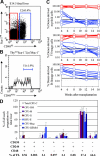
Figure 1.
Figure 1.
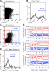
HSCs are contained within the CD48 fraction while colony-forming progenitors are almost entirely in the CD48+ fraction of fetal liver cells. (A) Twelve percent of fetal liver cells expressed CD48. (B) CD48 expression was heterogeneous in the ThylowSca-1+lineageMac-1+ HSC population, with 51% of cells being CD48+. For this analysis CD48, Thy-1, Sca-1, lineage marker, and Mac-1 staining were detected in the FITC, PE-Cy5, APC-Cy7 (PR), PE, and APC channels, respectively. (C) Recipient mice received transplants of 5750 CD48+ fetal liver cells (blue lines) or 44 250 CD48 fetal liver cells (red lines). Cell doses were based on the proportion of CD48+ versus CD48 cells in 50 000 fetal liver cells (which gives long-term multilineage reconstitution in nearly all recipient mice), as done in previous studies of marker expression by HSCs.6,14,24,26 Each line represents the frequency of donor-derived myeloid, B, or T cells in a single mouse. The black line at 0.3% represents the background threshold, meaning that reconstitution could not be detected in mice falling below this line. All long-term multilineage reconstituting activity was contained within the CD48 cell fraction (7 of 7 mice). CD48+ cells gave transient multilineage reconstitution (4 of 9 mice) or B-lineage-only reconstitution (5 of 9 mice). Data are from 1 of 2 independent experiments that gave similar results. (D) Fetal liver cells were divided based on their expression of all possible combinations of the CD48, CD244, and CD150 SLAM family receptors and sorted into methylcellulose to measure colony-forming activity. Each bar represents the percentage of all colony-forming progenitors of each type that fell within the indicated cell fraction. For example, around 80% of all CFU-GM, CFU-G, and CFU-M in the fetal liver fell within the CD48+CD244CD150 cell population. Error bars indicate SD.
Figure 2.
Figure 2.
Ter119+ erythroid lineage cells rarely express CD150, CD244, or CD48, while most other myeloid, B-, and T-lineage cells in the fetal liver express CD48. In the E14.5 fetal liver most myeloid lineage (Gr-1+ or Mac-1+), T-lineage (CD3+), and B-lineage (B220+) cells express CD48, but only around 20% of such cells express CD150 and/or CD244 (A, B). In contrast, no more than rare Ter119+ erythroid cells express any of these receptors (A, B). Error bars indicate SD.
Figure 3.
Figure 3.
Fetal liver HSCs are CD150+. (A) Two-and-a-half percent of fetal liver cells expressed CD150. (B) CD150 expression was heterogeneous within the Thylow Sca-1+lineageMac-1+ HSC population. For this analysis, CD150, Thy-1, Sca-1, lineage marker, and Mac-1 staining were detected in the FITC, PE-Cy5, APC-Cy7 (PR), PE, and APC channels, respectively. (C) Recipient mice received transplants of 1250 CD150+ fetal liver cells (blue lines) or 48 750 CD150 fetal liver cells (red lines). All multilineage reconstituting activity was contained within the CD150+ cell fraction (10 of 10 mice) not within the CD150 cell fraction (0 of 10 mice). The CD150 cell fraction showed transient B- and T-cell reconstitution (4 of 10 mice) or transient B-lineage reconstitution (3 of 10 mice), but no detectable myeloid lineage reconstitution.
Figure 4.
Figure 4.
Fetal liver HSCs are CD41. (A) CD150+CD48 cells accounted for 0.068% ± 0.01% of E14.5 fetal liver cells (note that these gates used for the analysis of HSC frequency were somewhat tighter than those used in Figure 1D for the analysis of colony-forming progenitors), and (B) 51% ± 7% of the cells in this population were CD41+. In the analysis shown in panel B, CD41, CD48, and CD150 staining were detected in the FITC, PE, and APC channels, respectively. (C) Three-and-one-fifth percent of fetal liver cells expressed CD41. (D) CD41 expression was detected on only 2.7% of cells within the ThylowSca-1+lineageMac-1+ HSC population. In this analysis CD41, Thy-1, Sca-1, lineage marker, and Mac-1 staining were detected in the FITC, PE-Cy5, APC-Cy7 (PR), PE, and APC channels, respectively. (E) Irradiated recipient mice received transplants of 1620 CD41+ fetal liver cells (blue lines) or 48 380 CD41 fetal liver cells (red lines). Most mice that received transplants of CD41 cells became long-term multilineage reconstituted (4 of 5; the other mouse was transiently multilineage reconstituted), but none of the mice that received transplants of CD41+ cells showed convincing reconstitution (0 of 8 mice).
Figure 5.
Figure 5.
Fetal liver HSCs are CD244. (A) Three-and-nine-tenths percent of fetal liver cells expressed CD244. (B) CD244 expression was detected in only 2.7% of ThylowSca-1+lineageMac-1+ cells. CD244, Thy-1, Sca-1, lineage marker, and Mac-1 staining were detected in the FITC, PE-Cy5, APC-Cy7 (PR), PE, and APC channels, respectively. (C) Irradiated recipient mice received transplants of 1950 CD244+ fetal liver cells (blue lines) or 48 050 CD244 fetal liver cells (red lines). All long-term multilineage reconstituting activity was contained within the CD244 cell fraction (5 of 7; the other 2 mice were reconstituted only in the B-cell lineage). None of the mice that received transplants of CD244+ cells (0 of 8) showed any donor cell reconstitution.

Similar articles

Cited by

References

    1. Muller AM, Medvinsky A, Strouboulis J, Grosveld F, Dzierzak E. Development of hematopoietic stem cell activity in the mouse embryo. Immunity. 1994;1: 291-301. - PubMed
    1. Cumano A, Dieterlen-Lievre F, Godin I. Lymphoid potential, probed before circulation in mouse, is restricted to caudal intraembryonic splanchnopleura. Cell. 1996;86: 907-916. - PubMed
    1. Medvinsky A, Dzierzak E. Definitive hematopoiesis is autonomously initiated by the AGM region. Cell. 1996;86: 897-906. - PubMed
    1. Gekas C, Dieterlen-Lievre F, Orkin SH, Mikkola HK. The placenta is a niche for hematopoietic stem cells. Dev Cell. 2005;8: 365-375. - PubMed
    1. Ottersbach K, Dzierzak E. The murine placenta contains hematopoietic stem cells within the vascular labyrinth region. Dev Cell. 2005;8: 377-387. - PubMed

Publication types

MeSH terms