Comparative Study
. 2006 Apr 24;94(8):1154-63.
doi: 10.1038/sj.bjc.6603067.

COX-2-mediated stimulation of the lymphangiogenic factor VEGF-C in human breast cancer

Affiliations
Comparative Study

COX-2-mediated stimulation of the lymphangiogenic factor VEGF-C in human breast cancer

A V Timoshenko et al. Br J Cancer. .

Abstract

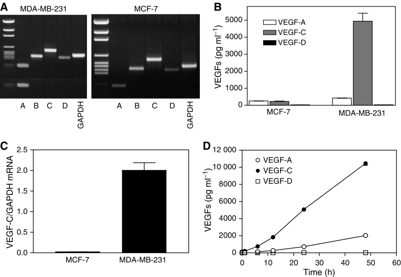
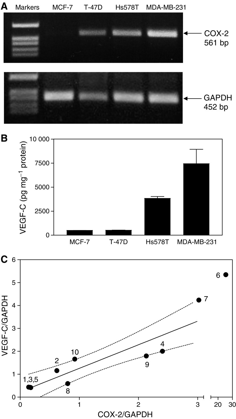
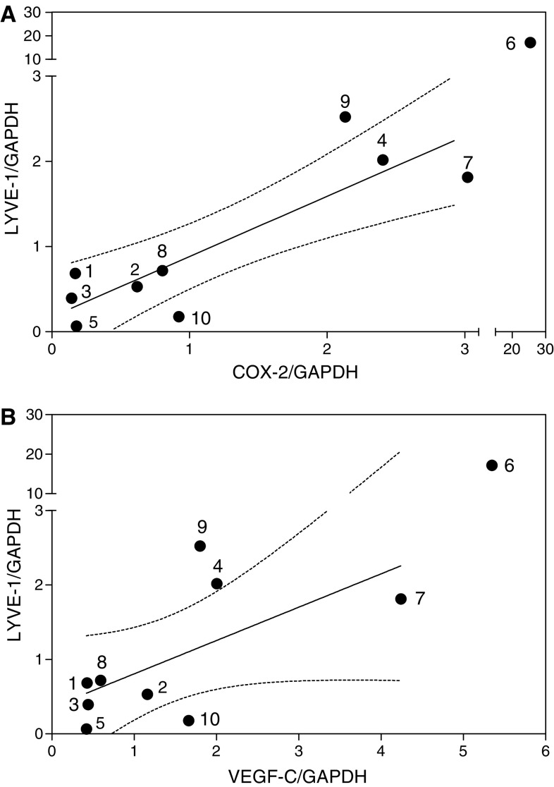
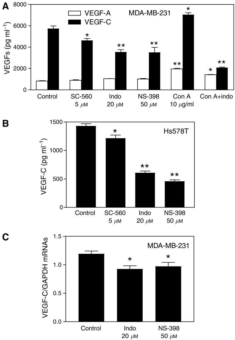
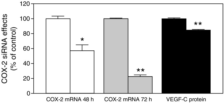
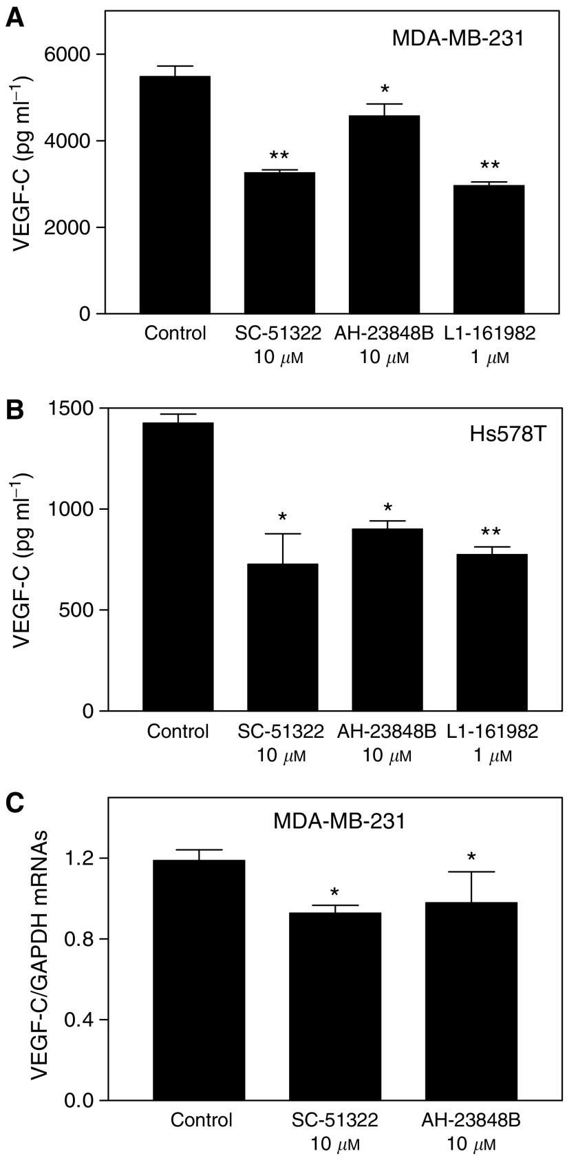
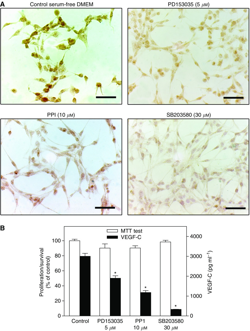
Increased expression of COX-2 or VEGF-C has been correlated with progressive disease in certain cancers. Present study utilized several human breast cancer cell lines (MCF-7, T-47D, Hs578T and MDA-MB-231, varying in COX-2 expression) as well as 10 human breast cancer specimens to examine the roles of COX-2 and prostaglandin E (EP) receptors in VEGF-C expression or secretion, and the relationship of COX-2 or VEGF-C expression to lymphangiogenesis. We found a strong correlation between COX-2 mRNA expression and VEGF-C expression or secretion levels in breast cancer cell lines and VEGF-C expression in breast cancer tissues. Expression of LYVE-1, a selective marker for lymphatic endothelium, was also positively correlated with COX-2 or VEGF-C expression in breast cancer tissues. Inhibition of VEGF-C expression and secretion in the presence of COX-1/2 or COX-2 inhibitors or following downregulation of COX-2 with COX-2 siRNA established a stimulatory role COX-2 in VEGF-C synthesis by breast cancer cells. EP1 as well as EP4 receptor antagonists inhibited VEGF-C production indicating the roles of EP1 and EP4 in VEGF-C upregulation by endogenous PGE2. Finally, VEGF-C secretion by MDA-MB-231 cells was inhibited in the presence of kinase inhibitors for Her-2/neu, Src and p38 MAPK, indicating a requirement of these kinases for VEGF-C synthesis. These results, for the first time, demonstrate a regulatory role of COX-2 in VEGF-C synthesis (and thereby lymphangiogenesis) in human breast cancer, which is mediated at least in part by EP1/EP4 receptors.


Figures

Figure 1
Figure 1
Expression of VEGF-A, -B, -C and -D mRNAs and VEGF-A, -C, and –D protein secretion by human breast cancer cell lines MDA-MB-231 and MCF-7. (A) RT–PCR-based amplification of VEGF mRNAs, showing that message for each VEGF class is detectable in both cell lines. (B) Accumulation of secreted VEGF-A, -C and -D proteins in serum-free culture media of both cell lines at 24 h showing high VEGF-C production by MDA-MB-231 cells. (C) Differences in expression of VEGF-C mRNA by MDA-MB-231 and MCF-7 cells as revealed by real time qPCR. (D) Kinetics of accumulation of VEGF-A, -C and -D protein in culture media of MDA-MB-231 cells. Data in (B, C, and D) represent mean±s.d. (n=4).
Figure 2
Figure 2
Relationship between COX-2 expression and VEGF-C synthesis/expression in human breast cancer cell lines and tissues. (A) Expression of COX-2 and GAPDH genes in MCF-7, T-47D, Hs578T and MDA-MB-231 human breast cancer cells as detected by RT–PCR. COX-2 expression is not detectable in MCF-7, weak in T-47D, moderate in Hs578T, and high in MDA-MB-231 cells. (B) Accumulation of VEGF-C in culture media of the above breast cancer cell lines at a 24 h correlates with their levels of COX-2 expression. Data represent mean±s.d. (n=4). (C) Correlation between relative levels of COX-2 mRNA and VEGF-C mRNA (standardized against GAPDH mRNA) expression in human breast cancer tissues (n=10) measured with real-time qPCR reveals a strong positive association between the two parameters (r=0.94, P=0.0002). Numbers on the graph present patient identifiers, and dotted lines present 95% confidence intervals of the regression line.
Figure 3
Figure 3
Relationship between LYVE-1 and COX-2 (A) or VEGF-C (B) mRNA expression in the same breast cancer tissues (n=10) used for data in Figure 2C. A positive association (r=0.75, P=0.017 and r=0.78, P=0.013, respectively) is revealed in both cases; dotted lines present 95% confidence interval of the regression line.
Figure 4
Figure 4
Effects of COX inhibitors on VEGF-C and VEGF-A secretion and VEGF-C expression in breast cancer cells. (A) Effects of COX-1 inhibitor SC-560, COX-1/-2 inhibitor indomethacin, and COX-2 inhibitor NS-398 on VEGF-C or VEGF-A production by COX-2 expressing MDA-MB-231 cells. (B) Effects of same inhibitors on VEGF-C production by COX-2 expressing Hs578T cells. COX-1 inhibitor caused a minor (but significant; P<0.05) reduction, whereas COX-2 or COX-1/-2 inhibitors caused a substantial (P<0.01) and similar reduction in VEGF-C production by both cell lines. However, these inhibitors have no inhibitory effect on VEGF-A production. Con A, known to stimulate PGE2 production by MDA-MB-231 cells, also stimulated VEGF-C as well as VEGF-A production, which were substantially (P<0.01) blocked with COX-1/2 inhibitor. (C) Effect of 24 h treatment with COX inhibitors (20 μM indomethacin or 50 μM NS-398 in serum-free culture) on VEGF-C mRNA expression in MDA-MB-231 cells. Data represent mean±s.d. (n=4). *P<0.05 and **P<0.001 in comparison to control untreated cells.
Figure 5
Figure 5
Effects of COX-2 siRNA treatment on COX-2 expression and VEGF-C production by MDA-MB-231 cells. The first bar in each series is control, that is, the treatment with scrambled siRNA, and the second bar is experimental, that is, the treatment with COX-2 siRNA. siRNA treatment was conducted in serum containing medium for 48 h. Cells were washed and cultured in serum free media for another 24 h to measure VEGF-C protein accumulation in the cell free media, and COX-2 mRNA levels in cells at both 48 and 72 h following the siRNA treatment. A significant suppression of COX-2 mRNA expression as well as VEGF-C secretion was noted in COX-2 siRNA treated cells. *P<0.01, **P<0.001.
Figure 6
Figure 6
Roles of EP receptors in the regulation of VEGF-C production and expression by COX-2 expressing human breast cancer cells. Cells were treated for 24 h with antagonists of EP1 receptors (SC-51322) and EP4 receptors (AH-23848B and L-161982) in serum-free DMEM. (A and B) Effects of EP receptor antagonists on VEGF-C accumulation in culture media of MDA-MB-231 cells (A) and Hs538T cells (B). A consistent and significant inhibition of VEGF-C production was observed with highly specific EP1 receptor antagonist SC-51322 and EP4 receptor antagonist L-161982. (C) Quantification of VEGF-C mRNA levels in MDA-MD-231 cells after the 24 h exposure to EP receptor antagonists. Data represent mean±s.d. (n=4). *P<0.05, **P<0.01.
Figure 7
Figure 7
Reduction of VEGF-C expression in MDA-MB-231 cells induced by kinase inhibitors for Her2/Neu (PD153035), Src (PP1) and p38 MAPK (SB203580). MDA-MB-231 cells were cultured for 24 h in serum-free DMEM supplemented with 0.2 % BSA. (A) imunohistochemistry; all the kinase inhibitors at the concentrations employed inhibited cytoplasmic immunostaining for VEGF-C. Scale bar represents 60 μm. (B) cell proliferation/survival (MTT test) and VEGF-C secretion; VEGF-C secretion, but not proliferation/survival, was inhibited by all the inhibitors. Data represent mean±s.d. (n=4), *P<0.001.

References

    1. Appleby SB, Ristimäki A, Neilson K, Narko K, Hla T (1994) Structure of the human cyclo-oxygenase-2 gene. Biochem J 302: 723–727 - PMC - PubMed
    1. Basu GD, Pathangey LB, Tinder TL, Gendler SJ, Mukherjee P (2005) Mechanisms underlying the growth inhibitory effects of the cyclo-oxygenase-2 inhibitor celecoxib in human breast cancer cells. Breast Cancer Res 7: R422–R435 - PMC - PubMed
    1. Beasley NJ, Prevo R, Banerji S, Leek RD, Moore J, van Trappen P, Cox G, Harris AL, Jackson DG (2002) Intratumoral lymphangiogenesis and lymph node metastasis in head and neck cancer. Cancer Res 62: 1315–1320 - PubMed
    1. Breyer RM, Bagdassarian CK, Myers SA, Breyer MD (2001) Prostanoid receptors: subtypes and signaling. Annu Rev Pharmacol Toxicol 41: 661–690 - PubMed
    1. Byeon JS, Jung HY, Lee YJ, Lee D, Lee GH, Myung SJ, Yang SK, Hong WS, Kim JH, Min YI, Kim JS (2004) Clinicopathological significance of vascular endothelial growth factor-C and cyclooxygenase-2 in esophageal squamous cell carcinoma. J Gastroenterol Hepatol 19: 648–654 - PubMed

Publication types

MeSH terms

Substances