. 2006 Jul 15;397(2):289-95.
doi: 10.1042/BJ20051938.

Dihydroceramide:sphinganine C-4-hydroxylation requires Des2 hydroxylase and the membrane form of cytochrome b5

Affiliations

Dihydroceramide:sphinganine C-4-hydroxylation requires Des2 hydroxylase and the membrane form of cytochrome b5

Ayako Enomoto et al. Biochem J. .

Abstract

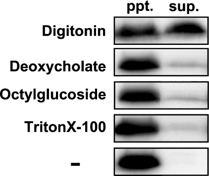
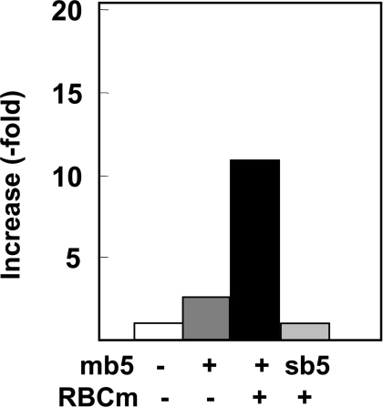
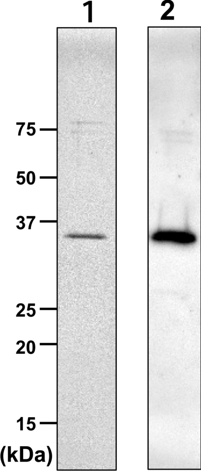
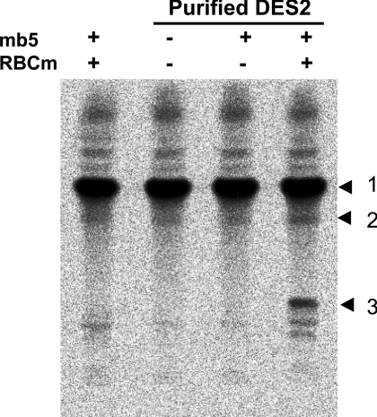
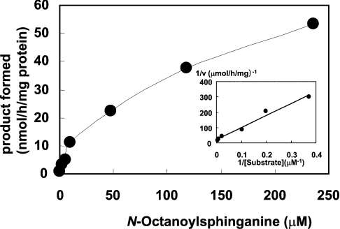
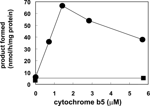
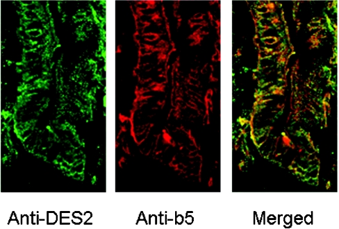
Des2 (degenerative spermatocyte 2) is a bifunctional enzyme that produces phytoceramide and ceramide from dihydroceramide. The molecular mechanism involved in C-4-hydroxylation has not been studied in detail. In the present paper, we report that C-4-hydroxylation requires an electron-transfer system that includes cytochrome b5 and that the hydroxylase activity is reconstituted in an in vitro assay with purified recombinant Des2. FLAG-tagged mouse Des2 was expressed in insect Sf9 cells and was purified by solubilization with digitonin and anti-FLAG antibody affinity column chromatography. The activity of dihydroceramide:sphinganine C-4-hydroxylase was reconstituted with the purified FLAG-Des2, mb5 (the membrane form of cytochrome b5) and bovine erythrocyte membrane. The apparent K(m) and V(max) of Des2 for the substrate N-octanoylsphinganine were 35 microM and 40 nmol x h(-1) x mg of protein(-1) respectively. The K(m) of the hydroxylase for mb5 was 0.8 microM. Interestingly, mb5 was not replaced with the soluble form of cytochrome b5, which lacks the C-terminal membrane-spanning domain. The erythrocyte membrane was separated into Triton X-100-soluble and -insoluble fractions, and the detergent-soluble fraction was replaced by the soluble or membrane form of b5R (NADH-cytochrome b5 reductase). The Triton-X-100-insoluble fraction contained trypsin-resistant factors. The Des2 protein is found in the endoplasmic reticulum and is assumed to have three membrane-spanning domains. The findings of the present study indicate that the hydroxylation requires complex formation between Des2 and mb5 via their membrane-spanning domains and electron transfer from NADH to the substrate via the reduction of mb5 by b5R.


Figures

Figure 1
Figure 1. Solubilization of FLAG–Des2 from the 600 g supernatant of transfected Sf9 cells
The fractions solubilized with various detergents at a final concentration of 1% (v/v) (see the Experimental section for details) were analysed by Western blotting with anti-FLAG antibody. The precipitates (ppt.) and supernatants (sup.) were separated by ultracentrifugation. − indicates no detergent used.
Figure 2
Figure 2. Effects of mb5 and bovine erythrocyte membrane on the C-4-hydroxylase activity in the fraction solubilized with 1% (v/v) digitonin
The solubilized protein (25 μg) was incubated in the presence of 5.6 μM mb5 alone, or with 85 μg of bovine erythrocyte membrane (RBCm). The addition of 5.5 μM sb5 did not increase the activity. Results are the means for duplicate assays.
Figure 3
Figure 3. SDS/PAGE and Western blotting of the purified FLAG–Des2
Lane 1, silver staining; lane 2, Western blotting with anti-FLAG antibody. Molecular masses are indicated in kDa.
Figure 4
Figure 4. Requirement of the bovine erythrocyte membrane for the C-4 hydroxylase activity
TLC autoradiography indicates that the C-4-hydroxylase activity requires Des2 (82 ng of protein), mb5 (5.7 μM) and the bovine erythrocyte membrane (85 μg of protein; RBCm). TLC was developed with chloroform/methanol/water (60:20:2, by vol.) and the radioactivity was visualized using a bio-imaging analyser. The numbers to the right of the Figure indicate the positions of the substrate and reaction products: 1, substrate N-octanoyldihydroceramide; 2, N-octanoylceramide, the product of δ4-desaturase; 3, N-octanoylphytoceramide, the product of C-4-hydroxylase.
Figure 5
Figure 5. Substrate isotherm and Lineweaver–Burk plot
The reaction mixture contained purified FLAG–Des2 (0.22 μg of protein), mb5 (1.4 μM) and bovine erythrocyte membrane (85 μg of protein). Other conditions were as described in the Experimental section. Km and Vmax of the hydroxylase for N-octanoylsphinganine were 34.8 μM and 40 nmol·h−1·mg of Des2 protein−1 respectively. Points are means for duplicate assays.
Figure 6
Figure 6. Effects of mb5 and sb5 on the C-4-hydroxylase activity
The reaction mixture contained purified FLAG–Des2 (0.3 μg of protein), bovine erythrocyte membrane (85 μg protein) and various amounts of mb5 or sb5. Results are means for duplicate assays for mb5 (●) and sb5 (■). The same effects of cytochrome b5 were observed when the digitonin-solubilized fraction or the Sf9 homogenates were used in place of the purified FLAG–Des2.
Figure 7
Figure 7. Restoration of C-4-hydroxylase activity with purified FLAG–Des2, mb5, b5R and the Triton X-100-insoluble fraction of the bovine erythrocyte membrane
Each reaction mixture contained purified FLAG–DES2 (0.37 μg), mb5 (1.4 μM), the Triton X-100-soluble fraction (14 μg; Triton sol. fr.) and the Triton X-100-insoluble fraction (12.8 μg; Triton insol. fr.). The amounts of these two fractions added to the assay contained proteins equivalent to the amount obtained from the same amount of bovine erythrocyte membrane (32 μg of protein). b5R activity added refers to the amount of b5R activity in the assay mixtures. The reductase activity was measured by monitoring the absorbance at 424 nm owing to the reduction of mb5 with NADH, and calculated using a molar absorption coefficient of 120 mM−1·cm−1 for the difference between the reduced and oxidized forms of mb5. trp, trypsin treatment; DE, DEAE column-bound fraction containing the membrane form of b5R; AMP, the membrane form of b5R purified using DEAE- and AMP–Sepharose column chromatography. a indicates the activity of the membrane form of b5R in the Triton X-100-soluble fraction before the trypsin treatment; b indicates the activity of the purified soluble form of b5R. Results are presented as means for duplicate assays. Note that the Triton X-100-soluble fraction contains trypsin-sensitive factors that can be replaced with the purified membrane or soluble forms of b5R and the Triton X-100-insoluble fraction contains a trypsin-resistant factor or factors. The activity reconstituted with all the purified proteins and the Triton X-100-insoluble fraction of the bovine erythrocyte membrane was still half of that obtained with the purified FLAG–DES2, mb5 and crude Triton X-100-soluble and -insoluble fractions, suggesting that the Triton X-100-soluble fraction contains trypsin-sensitive activation factors in addition to b5R.
Figure 8
Figure 8. Immunohistochemistry of the mouse small intestine using anti-(cytochrome b5) and anti-Des2 antibodies
Mouse small intestine was fixed and sectioned to thicknesses of 3 mm, as described in the Experimental section, and the frozen sections were stained with anti-(cytochrome b5) and anti-Des2 antibodies. Cytochrome b5 is evenly distributed in the epithelial cells and Des2 is localized in the epithelial cells near the crypt, indicating that the epithelial cells containing Des2 and cytochrome b5 possess the ability to biosynthesize phytoceramide (N-acyl-4-hydroxysphinganine).

Similar articles

Cited by

References

    1. Smith W. L., Merrill A. H., Jr Sphingolipid metabolism and signaling minireview series. J. Biol. Chem. 2002;277:25841–25842. - PubMed
    1. Merrill A. H., Jr De novo sphingolipid biosynthesis: a necessary, but dangerous, pathway. J. Biol. Chem. 2002;277:25843–25846. - PubMed
    1. Hakomori S. I. The glycosynapse. Proc. Natl. Acad. Sci. U.S.A. 2002;99:225–232. - PMC - PubMed
    1. Simons K., Vaz W. L. Model systems, lipid rafts, and cell membranes. Annu. Rev. Biophys. Biomol. Struct. 2004;33:269–295. - PubMed
    1. Holthuis J. C., Pomorski T., Raggers R. J., Sprong H., Van Meer G. The organizing potential of sphingolipids in intracellular membrane transport. Physiol. Rev. 2001;81:1689–1723. - PubMed

Publication types