. 2006 Mar 29:7:16.
doi: 10.1186/1471-2121-7-16.

Smad7 and protein phosphatase 1alpha are critical determinants in the duration of TGF-beta/ALK1 signaling in endothelial cells

Affiliations

Smad7 and protein phosphatase 1alpha are critical determinants in the duration of TGF-beta/ALK1 signaling in endothelial cells

Gudrun Valdimarsdottir et al. BMC Cell Biol. .

Abstract

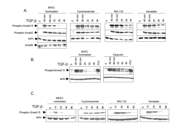
Background: In endothelial cells (EC), transforming growth factor-beta (TGF-beta) can bind to and transduce signals through ALK1 and ALK5. The TGF-beta/ALK5 and TGF-beta/ALK1 pathways have opposite effects on EC behaviour. Besides differential receptor binding, the duration of TGF-beta signaling is an important specificity determinant for signaling responses. TGF-beta/ALK1-induced Smad1/5 phosphorylation in ECs occurs transiently.

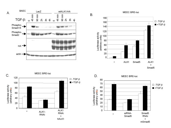
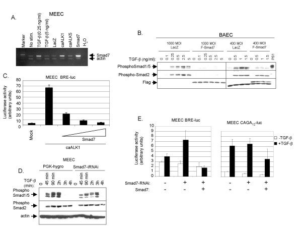
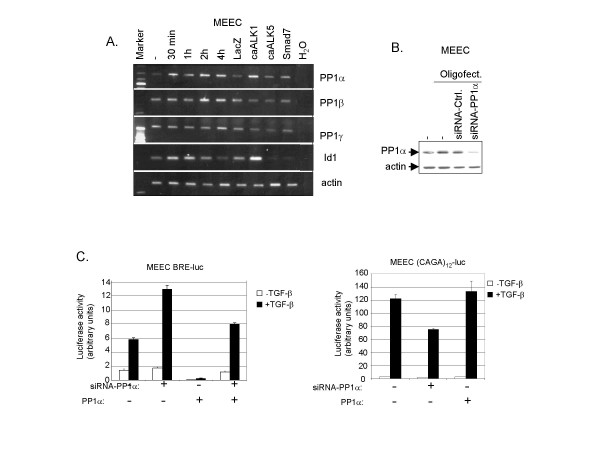
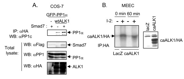
Results: The temporal activation of TGF-beta-induced Smad1/5 phosphorylation in ECs was found to be affected by de novo protein synthesis, and ALK1 and Smad5 expression levels determined signal strength of TGF-beta/ALK1 signaling pathway. Smad7 and protein phosphatase 1alpha (PP1alpha) mRNA expression levels were found to be specifically upregulated by TGF-beta/ALK1. Ectopic expression of Smad7 or PP1alpha potently inhibited TGF-beta/ALK1-induced Smad1/5 phosphorylation in ECs. Conversely, siRNA-mediated knockdown of Smad7 or PP1alpha enhanced TGF-beta/ALK1-induced signaling responses. PP1alpha interacted with ALK1 and this association was further potentiated by Smad7. Dephosphorylation of the ALK1, immunoprecipitated from cell lysates, was attenuated by a specific PP1 inhibitor.

Conclusion: Our results suggest that upon its induction by the TGF-beta/ALK1 pathway, Smad7 may recruit PP1alpha to ALK1, and thereby control TGF-beta/ALK1-induced Smad1/5 phosphorylation.


Figures

Figure 1
Figure 1
Negative regulation of TGF-β-induced Smad1/5 phosphorylation.(A) First panel. BAECs were stimulated with TGF-β (1 ng/ml) for different time periods at 37°C before lysis. Whole cell lysate was sonicated and fractionated by SDS-PAGE and blotted. The filters were incubated with PS1 antibody that specifically recognizes phosphorylated Smad1/5, PS2 antibody, which specifically recognizes phosphorylated Smad2, and antisera against Smad5. The blots were incubated with an actin antibody as a control for protein loading. Second panel. BAECs were treated with the protein synthesis inhibitor cyclohexamide (10 ng/ml) 30 minutes before they were stimulated with TGF-β. The filters were incubated with PS1, PS2 or actin antibodies. Third panel. BAECs were treated with the proteasome inhibitor, MG-132 (10 nM), for 30 min before they were stimulated with TGF-β. The filters were incubated with PS1, PS2 or actin antibodies. Last panel. BAECs were pre-treated with a phosphatase inhibitor, sodium orthovanadate (1 mM) 30 min prior to stimulation with TGF-β following the same procedure as in Fig. 1A. The filters were incubated with PS1, PS2 or actin antibody. (B) BAECs were pre-treated (right panel) or not (left panel) with the Ser/Thr phosphatase inhibitor calyculin (1 nM) 30 min prior to stimulation with TGF-β for different time periods before lysis. The filters were incubated with PS1 or actin antibody. (C) MEECs were pre-treated with cyclohexamide, MG-132, vanadate or not, and then stimulated with TGF-β for different time periods as in Fig. 1A.
Figure 2
Figure 2
ALK1 and Smad are critically important in Smad1/5 phosphorylation in ECs. (A) BAECs were adenovirally infected with wild-type ALK1/HA or LacZ construct at MOI of 1000. Fresh medium containing 10% FBS was added 16 h after infection. Eight hours later, the cells were starved overnight and then stimulated with TGF-β (1 ng/ml) for the indicated time periods and TGF-β-induced Smad1/5 phosphorylation was measured. Cells were lysed, sonicated and fractionated by SDS-PAGE. The gels were then subjected to immunoblotting. The filters were incubated with PS1, PS2, HA or actin antibody. (B) MEECs were transfected with BRE-luc in the absence or presence of ALK1 or Smad5, or both. After 48 h, cells were extensively washed. Then, cells were stimulated for 8 h with TGF-β, or not, and luciferase activity was measured. Values are corrected for transfection efficiency as measured by β-galactosidase activity. A representative experiment using triplicate samples is shown. (C) MEECs were transfected with (BRE)-luc in the absence or presence of ALK1-RNAi. After 48 h, cells were extensively washed. Then, cells were stimulated for 8 h with TGF-β, or not, and luciferase activity was measured. The luciferase activity upon RNAi-mediated knockdown of endogenous mouse ALK1 was rescued by co-transfecting a human ALK1 expression plasmid. Values are corrected for transfection efficiency as measured by β-galactosidase activity. A representative experiment using triplicate samples is shown. (D) MEECs were transfected with BRE-luc in the absence or presence of Smad5-RNAi. After 48 h, cells were extensively washed. Then, cells were stimulated for 16 h with TGF-β, or not, and luciferase activity was measured. The luciferase activity upon RNAi-mediated knockdown of endogenous mouse Smad5 was rescued by co-transfecting a mouse Smad5 plasmid. Values are corrected for transfection efficiency as measured by β-galactosidase activity. A representative experiment using triplicate samples is shown.
Figure 3
Figure 3
Smad7 is a potent inhibitor of ALK1-induced Smad1/5 phosphorylation in Ecs.(A) MEECs were stimulated with TGF-β or adenovirally infected at MOI of 500 with LacZ, caALK1, caALK5 and Smad7 (included as a positive control). Forty hours later, the noninfected cells were starved overnight and then stimulated with TGF-β, or not, for 90 min. Cells were lysed, and RNA was isolated and cDNA prepared. Expression of Smad7 was analysed by semi-quantitative RT-PCR. β-actin expression was measured to control for equal loading. The PCR products were loaded on 1% agarose gel and stained with ethidium bromide. (B) Left panel. BAECs were infected with adenoviral constructs of Flag-Smad7 or lacZ as a control with MOI of 1000. Forty hours later, BAECs were stimulated with a different dose of TGF-β at 37°C before lysis. Whole cell lysate was sonicated and fractionated by SDS-PAGE and blotted. The filters were incubated with PS1, PS2 or α-Flag antibody. Right panel. Differential Smad7 inhibition on TGF-β induced Smad1/5 phosphoylation and Smad2 phosphorylation in BAECs. BAECs were infected with adenoviral constructs of Flag-Smad7, or LacZ as a control, with MOI of 400. Forty hours after infection, BAECs were stimulated with 1 ng/ml of TGF-β for different time periods at 37°C before lysis. Whole cell lysate was sonicated and fractionated by SDS-PAGE and blotted. The filters were incubated with PS1, PS2 and Flag antibodies. (C) MEECs were co-transfected with caALK1 and BRE-luc with or without Smad7 at different concentrations. Luciferase activity was measured 48 h after transfection. Values are corrected for transfection efficiency as measured by β-galactosidase activity. A representative experiment using triplicate samples is shown. (D) Smad7-RNAi plasmid with a hygromycin cassette was stably transfected in MEECs and selected on hygromycin medium for 7 days. PGK-hygromycin selected cells were used as mock cells. The cells were serum-starved overnight and stimulated with TGF-β (1 ng/ml) at different time periods prior to lysis, sonication and fractionation by SDS-PAGE. Gels were then subjected to immunoblotting. The filters were incubated with PS1, PS2, or actin antibody. (E) Left panel. MEECs were transfected with BRE-luc in the absence or presence of Smad7-RNAi. Forty-eight hours after transfection the cells were washed extensively. Luciferase activity was measured after 8 hours of stimulation of TGF-β or not. Values are corrected for transfection efficiency as measured by β-galactosidase activity. A representative experiment using triplicate samples is shown. Right panel. MEECs were transfected with (CAGA)12-luc in the absence or presence of Smad7-RNAi. Forty-eight hours after transfection the cells were stimulated for 16 hours with TGF-β and luciferase activity measured. A representative experiment using triplicate samples, corrected for transfection efficiency, is shown.
Figure 4
Figure 4
ALK1-induced PP1α negatively regulates transcriptional activity downstream of TGF-β/ALK1.(A) TGF-β kinetics and upregulation of PP1 isoforms on mRNA level. MEECs were stimulated with TGF-β or adenovirally infected with LacZ, caALK1, caALK5 or Smad7 at MOI of 500. Forty hours later, including starvation overnight, the cells were lysed, RNA was isolated and cDNA was prepared. PCR-amplified products for PP1α, β and γ and Id1 are indicated on the right of the figure. β-actin was included as a loading control. (B) MEECs were transfected with siRNA-PP1α using oligofectamine in medium without serum. Twelve hours later the transfection medium was changed to medium with 10% FCS. Forty-eight hours later, the cells were lysed. Whole cell lysate was sonicated, fractionated on SDS-PAGE and subjected to immunoblotting. The filter was incubated with antibodies against PP1α and actin to measure loading of proteins in each sample. (C) Left panel. MEECs were transfected with BRE-luc in the absence or presence of PP1α. Sixteen hours later the cells (where indicated) were transfected with siRNA-PP1α using oligofectamine. Forty-eight hours later, luciferase activity was measured 6 h after stimulation with TGF-β or not. Values are corrected for transfection efficiency as measured by β-galactosidase activity. A representative experiment using triplicate samples is shown. Right panel. MEECs were transfected with (CAGA)12-luc in the absence or presence of PP1α. After 16 h, the cells was transfected with siRNA-PP1α (where indicated) using oligofectamine. Thirty-two hours later, cells were incubated with TGF-β for an additional 16 h, whereafter luciferase activity was measured. Values are corrected for transfection efficiency as measured by β-galactosidase activity. A representative experiment using triplicate samples is shown.
Figure 5
Figure 5
Smad7 recruits PP1α to ALK1. (A) COS-7 cells were transfected with eGFP-PP1α, wtALK1/HA and/or Flag-Smad7. After 48 h at 37°C, cells were lysed. Whole cell lysates were immunoprecipitatedwith HA antibody, fractionated by SDS-PAGE, and subjected to immunoblotting with PP1c antiserum. Total lysates were fractionated by SDS-PAGE and subjected to immunoblotting with antisera against Flag, PP1 and HA to check expression levels. (B) Left panel. Phosphatase inhibition assay. MEECs were adenovirally infected with the indicated constructs and immunopreciptated with HA antibody. Kinase assay was performed with 2.5 μCi of [32P]γ ATP per sample at 30°C for 30 min. Samples were washed in lysis buffer before they were incubated in a phosphatase buffer with or without the PP1 inhibitor, I-2 at 37°C for 60 min. The samples were separated by SDS-PAGE and phosphorylation quantified using phosphoimager. Right panel. Fraction of the total lysate was immunoblotted with HA antiserum to check expression levels of the receptor.

Similar articles

Cited by

References

    1. Shi Y, Massagué J. Mechanisms of TGF-β signaling from cell membrane to the nucleus. Cell. 2003;113:685–700. doi: 10.1016/S0092-8674(03)00432-X. - DOI - PubMed
    1. ten Dijke P, Hill CS. New insights into TGF-β-Smad signalling. Trends Biochem Sc. 2004;29:265–73. doi: 10.1016/j.tibs.2004.03.008. - DOI - PubMed
    1. Goumans M-J, Valdimarsdottir G, Itoh S, Rosendahl A, Sideras P, ten Dijke P. Balancing the activation state of the endothelium via two distinct TGF-β type I receptors. EMBO J. 2002;21:1743–53. doi: 10.1093/emboj/21.7.1743. - DOI - PMC - PubMed
    1. Oh SP, Seki T, Goss KA, Imamura T, Yi Y, Donahoe PK, Li L, Miyazono K, ten Dijke P, Kim S, Li E. Activin receptor-like kinase 1 modulates transforming growth factor-β 1 signaling in the regulation of angiogenesis. Proc Natl Acad Sci U S A. 2000;97:2626–31. doi: 10.1073/pnas.97.6.2626. - DOI - PMC - PubMed
    1. Chen YG, Massagué J. Smad1 recognition and activation by the ALK1 group of transforming growth factor-β family receptors. J Biol Chem. 1999;5:3672–7. doi: 10.1074/jbc.274.6.3672. - DOI - PubMed

Publication types

MeSH terms

LinkOut - more resources