. 2006 Apr 11;103(15):5846-51.
doi: 10.1073/pnas.0601536103. Epub 2006 Mar 29.

Experimental taphonomy shows the feasibility of fossil embryos

Affiliations

Experimental taphonomy shows the feasibility of fossil embryos

Elizabeth C Raff et al. Proc Natl Acad Sci U S A. .

Abstract

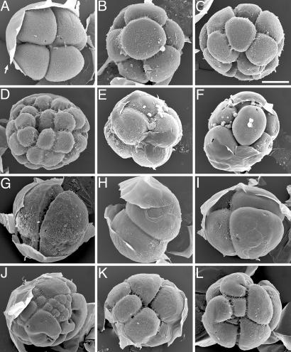
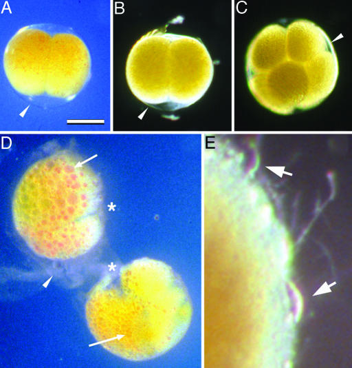
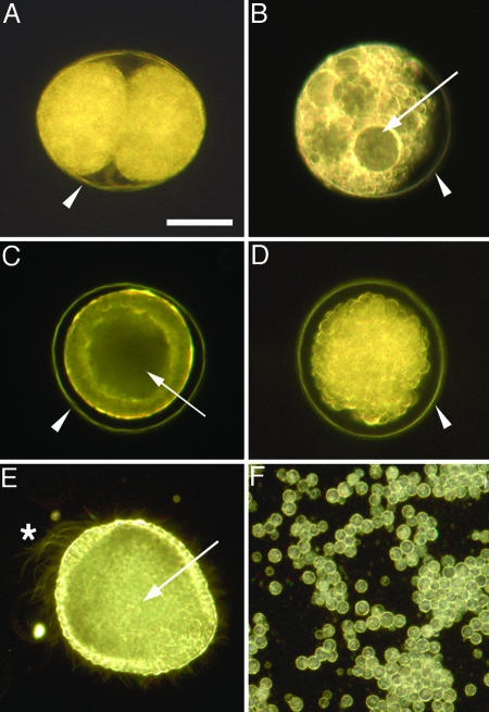
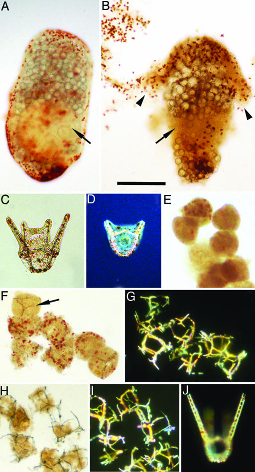
The recent discovery of apparent fossils of embryos contemporaneous with the earliest animal remains may provide vital insights into the metazoan radiation. However, although the putative fossil remains are similar to modern marine animal embryos or larvae, their simple geometric forms also resemble other organic and inorganic structures. The potential for fossilization of animals at such developmental stages and the taphonomic processes that might affect preservation before mineralization have not been examined. Here, we report experimental taphonomy of marine embryos and larvae similar in size and inferred cleavage mode to presumptive fossil embryos. Under conditions that prevent autolysis, embryos within the fertilization envelope can be preserved with good morphology for sufficiently long periods for mineralization to occur. The reported fossil record exhibits size bias, but we show that embryo size is unlikely to be a major factor in preservation. Under some conditions of death, fossilized remains will not accurately reflect the cell structure of the living organism. Although embryos within the fertilization envelope have high preservation potential, primary larvae have negligible preservation potential. Thus the paleo-embryological record may have strong biases on developmental stages preserved. Our data provide a predictive basis for interpreting the fossil record to unravel the evolution of ontogeny in the origin of metazoans.


Conflict of interest statement

Conflict of interest statement: No conflicts declared.

Figures

Fig. 1.
Fig. 1.
Cleavage-stage H. erythrogramma embryos under various experimental conditions. Fertilization envelopes (arrow in A) were manually peeled back after processing for scanning electron microscopy. (AD) Normal embryos at the 4-cell stage (A), 8-cell stage (B), 16-cell stage (C), and 32-cell stage (D). (E and F) Embryos from a culture placed in seawater containing 100 mM β-ME at the eight-cell stage. Embryos arrested at time of treatment and fixed after 12 days. (GJ) Embryos killed at mid-2- to 4-cell stage by various treatments and fixed after 8.5 h. Untreated controls had progressed to late blastula stage. (G) Embryo from a culture placed in 50% seawater. Embryos arrested at time of treatment; blastomeres expanded to fill the fertilization envelope. (HJ) Embryos placed in seawater containing 0.1% ammonium. Some embryos arrested immediately (H and I), and some underwent aberrant cleavage before arresting (J). (K) Embryo placed at 2-cell stage in N2-saturated seawater and fixed after 2 h. Cleavage continued during partial anoxia, but some blastomeres arrested or slowed cleavage rate. Untreated controls had progressed to the 16- to 32-cell stage. (L) Embryo fertilized under polyspermic conditions and fixed at 2.25 h (normal embryos would be 4- to 8-cell stage). (Scale bar: 100 μm.)
Fig. 2.
Fig. 2.
Cleavage-stage H. erythrogramma embryos under preserving and nonpreserving conditions. (A and B) Embryos killed at the two-cell stage by placing them in seawater containing 1% ammonium for 10 min, then transferring them to seawater containing 100 mM β-ME. They were photographed after 2 (A) or 10 (B) days. (C) Embryo killed at the eight-cell stage by transfer into seawater containing 100 mM β-ME, photographed after 12 days. (D) Embryos from the same two-cell stage culture as in A and B but returned to normal seawater after killing, photographed after 2 days. Embryos have undergone autolysis: cytoplasmic lipid and pigment have coalesced (arrows); cleavage furrows have degraded (asterisks); and fertilization envelopes are disintegrating (arrowhead). Autolysis is further advanced in the top embryo than in the bottom embryo. In the bottom embryo, the process is further advanced in the left-hand blastomere (arrow) than in the right-hand blastomere. (E) Decaying surface of an embryo from the set shown in A and B, returned to normal seawater after 4 days in reducing conditions, photographed 7 days later (total 11 days postdeath). Onset and progress of decay is slower than autolysis in embryos never exposed to reducing conditions. The fertilization envelope degrades and the cytoplasm of the embryo is then exposed to external decay processes, including attack by protists (arrows). (Scale bar: 200 μm for AD; 32 μm for E.)
Fig. 3.
Fig. 3.
Unhatched embryos of the indirect developing sea urchin L. pictus are preserved under reducing conditions but not after hatching. Where present, the fertilization envelope is indicated by an arrowhead (AD). Embryos in A, B, D, and F were killed by 12 min in seawater containing 1% ammonium, then treated as described. (A) Embryo killed at two-cell stage, then placed in seawater containing 100 mM β-ME, photographed after 26 days. Blastomere arrangement and structure are well preserved. (B) Embryo killed at two-cell stage, then placed in normal seawater, photographed after 5 h. The fertilization envelope is intact, but the embryo has swollen to fill the space within the fertilization envelope and is undergoing autolysis; cell structure has already disappeared (compare Fig. 2D). (C) Live unhatched blastula (17 h postfertilization) focused to show columnar cell shape of the blastula wall and the internal hollow blastocoel (arrow). (D) Unhatched blastula killed at the same stage as C, then placed in seawater containing 100 mM β-ME, photographed after 1 day. Embryo is intact within the fertilization envelope, but the cells have rounded up. Focusing through the embryo revealed the blastocoel has collapsed. (E) Live hatched blastula (24 h postfertilization) focused at the internal hollow blastocoel (arrow). The embryo was slightly flattened under a coverslip to immobilize it; the beating cilia are visible, including the large apical tuft cilia at the animal pole (asterisk). Differentiated epithelial cell shape at the animal and vegetal poles can be seen, and some internalized micromeres near the vegetal end of the blastocoel can be seen. (F) Cells remaining from a culture of hatched blastulae killed at the same stage as E, then placed in seawater containing 100 mM β-ME, photographed after 5 h. Cells rounded up and disaggregated. (Scale bar: 50 μm.)
Fig. 4.
Fig. 4.
Reducing conditions do not preserve Heliocidaris larvae. (A and B) H. erythrogramma. (A) Normal 2-day larva focused to show the internal pentameral adult rudiment (arrow); large lipid droplets that serve as “yolk” are also visible (19). (Metamorphosis would occur at day 4.) (B) Larva from a culture killed at 2 days by treatment with 1% ammonium for 20 min, then placed in seawater containing 100 mM β-ME, photographed after 2 days. Larvae become fragile, lose structural integrity, and disintegrate. In the specimen shown, adult rudiment structure is lost (arrow) and pigmented epithelium (arrowheads) is detaching. (CJ) H. tuberculata. (C) Normal 48-h pluteus; skeletal rods in arms and body are visible within the transparent pigmented epithelium. (D) Normal 36-h pluteus, starting stage for experiments in EJ. Photographed with Nomarski optics to display internal skeletal structure; hindgut is visible in body of larva. (EI) Embryos from the same culture as D, killed by treatment with 1% ammonium for 20 min, then placed in seawater containing 100 mM (E) or 10 mM β-ME (F and G) or returned to normal seawater (H and I), photographed after 1 day. (E) In strong reducing conditions, structural integrity and morphology are rapidly lost; skeletal elements rapidly disappear. (In sample shown, no skeletal elements were detected under polarizing optics.) (F) In less stringent reducing conditions, structural integrity and pigmentation disappear more slowly. Pluteus morphology and skeletal organization are lost, but skeletal elements (arrow) are stable. (G) Same field as F under polarizing optics to display skeletal elements. (H) In normal seawater, cytoplasm is subject to both internal and external decay processes, including predation by protists, and rapidly disappears. Skeletal organization is lost, but skeletal elements are preserved for prolonged periods. (I) Same field as H under polarizing optics. (J) Normal 3-day pluteus from untreated controls under polarizing optics. (Scale bar: 200 μm.).

References

    1. Xiao S., Zhang Y., Knoll A. H. Nature. 1998;391:553–558.
    1. Chen J., Oliveri P., Li C.-W., Zhou G.-Q., Gao F., Hagadorn J. W., Peterson K. J., Davidson E. H. Proc. Natl. Acad. Sci. USA. 2000;97:4457–4462. - PMC - PubMed
    1. Chen J., Oliveri P., Gao F., Dornbos S. Q., Li C. W., Bottjer D. J., Davidson E. H. Dev. Biol. 2002;248:182–196. - PubMed
    1. Condon D., Zhu M., Bowring S. A., Wang W., Yang A., Jin Y. Science. 2005;308:95–98. - PubMed
    1. Xiao S., Yuan X., Knoll A. H. Proc. Natl. Acad. Sci. USA. 2000;97:13684–13689. - PMC - PubMed

Publication types

LinkOut - more resources