. 2006 Jun;17(6):2572-80.
doi: 10.1091/mbc.e06-01-0052. Epub 2006 Mar 29.

Engrailed-1 negatively regulates beta-catenin transcriptional activity by destabilizing beta-catenin via a glycogen synthase kinase-3beta-independent pathway

Affiliations

Engrailed-1 negatively regulates beta-catenin transcriptional activity by destabilizing beta-catenin via a glycogen synthase kinase-3beta-independent pathway

Liora Bachar-Dahan et al. Mol Biol Cell. 2006 Jun.

Abstract

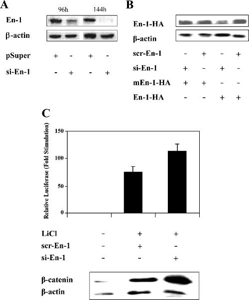
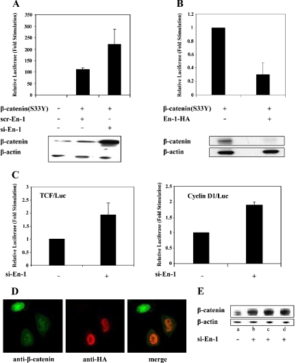
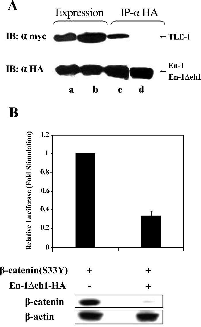
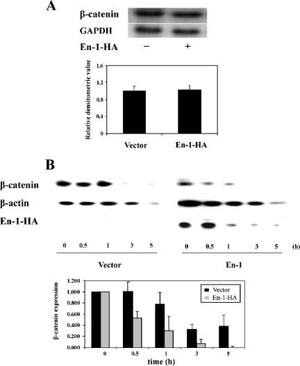
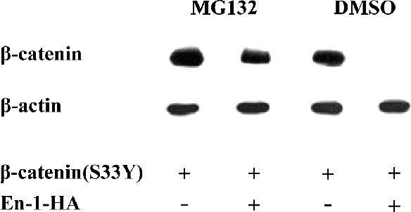
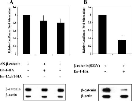
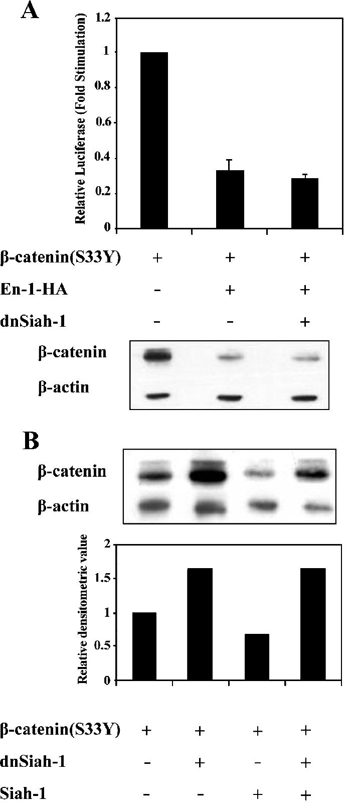
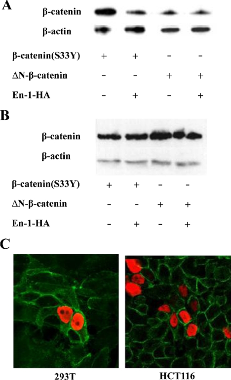
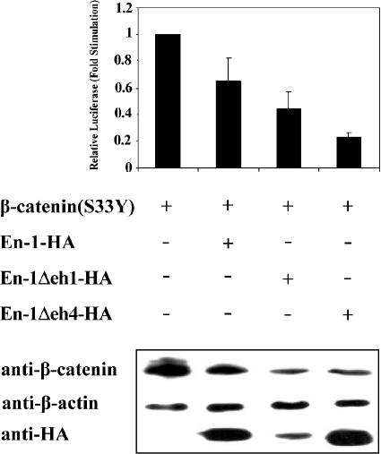
The Wnt signaling pathway plays a major role in development, and upon deregulation it is implicated in neoplasia. The hallmark of the canonical Wnt signal is the protection of beta-catenin from ubiquitination and proteasomal degradation induced by glycogen synthase kinase (GSK)-3beta inhibition. The stabilized beta-catenin translocates to the nucleus where it binds to T-cell factor/lymphoid enhancer factor (TCF/LEF) transcription factors, activating the expression of Wnt target genes. In the absence of Wnt signal, TCF/LEF bind to Groucho (Gro)/TLE corepressors and repress Wnt target genes. Gro/TLE bind also to Engrailed (En) transcription factors mediating En-repressive activity on En target genes. Here, we present data suggesting that En-1 serves also as a negative regulator of beta-catenin transcriptional activity; however, its repressive effect is independent of Gro/TLE. Our data suggest that En-1 acts by destabilizing beta-catenin via a proteasomal degradation pathway that is GSK-3beta-independent. Moreover, because En-1-mediated beta-catenin degradation is also Siah independent, our data imply that En-1 exerts its repressive effect by a novel mechanism negatively controlling the level of beta-catenin.


Figures

Figure 1.
Figure 1.
Silencing of En-1 expression stimulates β-catenin transcriptional activity. (A) Si-En-1 decreases the expression of the native En-1. 293T cells were cotransfected with si-En-1 or empty pSUPER (4 μg). At the indicated times after transfection, 100 μg of cell lysates were analyzed by Western blotting using anti-En-1 or anti-β-actin antibody. (B) si-En-1 decreases the level of expression of ectopically expressed En-1. 293T cells were cotransfected with si-En-1 or scr-En-1 (2.9 μg) and En-1-HA or mEn-1-HA (0.01 μg). Seventy-two hours after transfection, 100 μg of cell lysates was analyzed by Western blotting using anti-HA or anti-β-actin antibody. (C) LiCl-activated β-catenin transcriptional activity is stimulated in the presence of si-En-1. 293T cells were cotransfected with si-En-1, scr-En-1, or empty vector (2.5 μg), TCF/Luc reporter (1 μg) and β-Gal (0.1 μg). Twenty-four hours after transfection, cells were treated with 30 mM LiCl and after 48 h, cell lysates were measured for the levels of luciferase and β-Gal activities. Data are presented as mean values and standard deviations for at least three independent experiments done in duplicate, compared with the level of luciferase activity obtained in the absence of LiCl treatment presented as 1 (top). Cell lysates (100 μg) were subjected to Western analysis using anti-β-catenin or anti-β-actin antibody (bottom).
Figure 2.
Figure 2.
En-1 represses β-catenin(S33Y) transcriptional activity, concomitantly with decreasing β-catenin level. (A) Silencing of the native En-1 augments β-catenin(S33Y)-transcriptional activity. 293T cells were cotransfected with β-catenin(S33Y) or empty vector (0.1 μg), si-En-1 or scr-En-1 (2.5 μg each), TCF/Luc reporter (1 μg), and β-Gal (0.1 μg). Luciferase and β-Gal levels were measured as described in the legend to Figure 1. Cell lysates (100 μg) were subjected to Western analysis using anti-β-catenin or β-actin antibody. (B) Ectopically expressed En-1 represses β-catenin(S33Y)-transcriptional activity. 293T cells were cotransfected with β-catenin(S33Y) or empty vector (0.1 μg), En-1-HA or empty vector (1 μg), Cyclin D1/Luc (1 μg), and β-Gal (0.1 μg). Luciferase and β-Gal levels were measured as described in Figure 1. Cell lysates (30 μg) were subjected to Western analysis using anti-β-catenin or anti-β-actin antibody. (C) En-1 represses the constitutively active β-catenin in SW480 cell line. SW480 cells were transiently transfected with si-En-1 or empty vector (2.5 μg), TCF/Luc or CyclinD1/Luc reporter (1 μg), and Renilla luciferase vector (0.5 μg). Forty-eight hours after transfection, the luciferase activity was measured, and the luciferase levels were normalized according to Renilla luciferase activity. (D) Immunohistochemistry assays showing that En-1 decreases the level of native β-catenin. SW480 cells were transiently transfected with En-1-HA, and 48 h after transfection cells were subjected to immunofluorescence analysis with anti-β-catenin and anti-HA antibody, followed by FITC-conjugated anti-mouse antibody and rhodamine anti-rabbit antibody, respectively. Green, β-catenin expression; red, En-1-HA expression. (E) Silencing of the endogenous En-1 augments the level of native β-catenin in SW480 cells. SW480 cells were stably transfected with si-En-1 (clones b–d) or empty pSUPER (a), and the level of native β-catenin was quantitated by Western analysis using anti-β-catenin or anti-β-actin antibody.
Figure 3.
Figure 3.
En-1 repressive activity is TLE-independent. (A) En-1Δeh1-HA lacks the ability to bind to TLE-1. 293T cells were cotransfected with TLE-1-Myc (2 μg) and either En-1-HA (a and c) or En-1Δeh1-HA (b and d) (2 μg). Lysates were immunoprecipitated using anti-HA antibody, and Western blots were treated with anti-Myc or anti-HA antibody before (a and b) or after (c and d) immunoprecipitation. (B) En-1Δeh1-HA represses β-catenin-transcriptional activity. 293T cells cotransfected with β-catenin(S33Y) (0.1 μg), En-1Δeh1-HA or empty vector (1 μg), Cyclin D1/Luc (1 μg), and β-Gal (0.1 μg), and the luciferase and β-Gal levels were measured as described in the legend to Figure 1 (top). Cell lysates (30 μg) were subjected to Western analysis using anti-β-catenin or anti-β-actin antibody (bottom).
Figure 4.
Figure 4.
En-1 affects the level of β-catenin by a posttranslational mode. (A) En-1 does not affect the level of β-catenin transcription. 293T cells were cotransfected with β-catenin(S33Y) and either En-1 or empty vector. Forty-eight hours after transfection, Northern analysis was performed by hybridizing the blots to either β-catenin or GAPDH probe. A representative gel is shown (top). Densitometric analysis of six independent experiments is shown (bottom). Each bar denotes the mean ± SD, compared with the level of GAPDH expression. (B) En-1 accelerates the rate of β-catenin(S33Y) degradation after cycloheximide addition. 293T cells were cotransfected with β-catenin(S33Y) and either En-1-HA or empty vector. Five hours after transfection, 80 μg/ml cycloheximide was added, and at the indicated hours postcycloheximide addition, lysates were subjected to Western analysis using anti-β-catenin, anti-HA, or anti-β-actin antibody. Representative gel is shown (top). The mean of the densitometric analysis of three independent experiments is shown (bottom). Each bar denotes the level of β-catenin divided by the level of β-actin at each time point, compared with the level of β-catenin at time zero, taken as 1.
Figure 5.
Figure 5.
En-1 mediated β-catenin destabilization effect is blocked in the presence of the proteasome inhibitor MG132. 293T cells transfected with β-catenin(S33Y) (0.1 μg) and either En-1-HA or empty vector (1 μg) were treated with 25 μM MG132 or dimethyl sulfoxide. Four hours later, 50 μg of cell lysates were analyzed by Western blot analysis using anti-β-catenin or anti-β-actin antibody.
Figure 6.
Figure 6.
The N terminus of β-catenin is indispensable for En-1–destabilizing effect. (A) 293T cells were cotransfected with ΔN-β-catenin (0.1 μg), En-1-HA, En-1Δeh1-HA, or empty vector (1 μg), Cyclin D1/Luc (1 μg), and β-Gal (0.1 μg). (B) 293T cells were cotransfected with β-catenin(S33Y) (0.1 μg), En-1-HA or empty vector (1 μg), Cyclin D1/Luc (1 μg), and β-Gal (0.1 μg). Luciferase and β-Gal levels were measured as described in the legend to Figure 1 (top). Cell lysates (30 μg) were subjected to Western analysis using anti-β-catenin or anti-β-actin antibody (bottom).
Figure 7.
Figure 7.
En-1–mediated β-catenin destabilization is Siah-1 independent. (A) The presence of dnSiah-1 does not affect En-1–repressive activity. 293T cells were transfected with β-catenin(S33Y) (0.1 μg), En-1-HA or empty vector (1 μg), dnSiah-1(1 μg) or empty vector, Cyclin D1/Luc (1 μg), and β-Gal (0.1 μg), and the luciferase and β-Gal levels were measured as described in the legend to Figure 1 (top). Cell lysates (30 μg) were subjected to Western analysis using anti-β-catenin or anti-β-actin antibody (bottom). (B) The presence of dnSiah-1 blocks Siah-1–mediated β-catenin degradation. 293T cells were transfected with β-catenin(S33Y) (0.1 μg), Siah-1 (0.1 μg) or empty vector, and dnSiah-1(1 μg) or empty vector. Cell lysates (30 μg) were subjected to Western analysis using anti-β-catenin or β-actin antibody (top). Densitometric analysis of the levels of β-catenin compared with β-actin levels is shown (bottom).
Figure 8.
Figure 8.
En-1 does not affect the stability of membrane-bound β-catenin. 293T cells were cotransfected with β-catenin(S33Y) or ΔNβ-catenin, and either En-1-HA or empty vector. The level of β-catenin was measured in cytosolic (A) or membrane (B) fraction by Western analysis, using anti-β-catenin or β-actin antibody. (C) 293T or HCT116 cells were transiently transfected with En-1-HA, and 48 h after transfection cells were subjected to immunofluorescence analysis with anti-β-catenin and anti-HA antibodies, followed by FITC-conjugated anti-mouse antibody and rhodamine-conjugated anti-rabbit antibody, respectively. Green, β-catenin expression; red, En-1-HA expression.
Figure 9.
Figure 9.
En-1–repressive effect on Wnt signal is not exerted via En-1 target genes. 293T cells were cotransfected with β-catenin(S33Y) (0.1 μg), and En-1-HA, En-1Δeh1-HA, En-1Δeh4-HA, or empty vector (1 μg), Cyclin D1/Luc (1 μg), and β-Gal (0.1 μg). Luciferase and β-Gal levels were measured as described in Figure 1. Cell lysates (30 μg) were subjected to Western analysis using anti-β-catenin, anti-HA, or anti-β-actin antibody.

References

    1. Aberle H., Bauer A., Stappert J., Kispert A., Kemler R. beta-catenin is a target for the ubiquitin-proteasome pathway. EMBO J. 1997;16:3797–3804. - PMC - PubMed
    1. Akiyama T. Wnt/beta-catenin signaling. Cytokine Growth Factor Rev. 2000;11:273–282. - PubMed
    1. Araki I., Nakamura H. Engrailed defines the position of dorsal dimesencephalic boundary by repressing diencephalic fate. Development. 1999;126:5127–5135. - PubMed
    1. Bejsovec A., Martinez Arias A. Roles of wingless in patterning the larval epidermis of Drosophila. Development. 1991;113:471–485. - PubMed
    1. Brabletz T., Jung A., Reu S., Porzner M., Hlubek F., Kunz-Schughart L. A., Knuechel R., Kirchner T. Variable beta-catenin expression in colorectal cancers indicates tumor progression driven by the tumor environment. Proc. Natl. Acad. Sci. USA. 2001;98:10356–10361. - PMC - PubMed

Publication types