. 2006 Jun;17(6):2780-8.
doi: 10.1091/mbc.e05-10-0973. Epub 2006 Mar 29.

RINT-1 regulates the localization and entry of ZW10 to the syntaxin 18 complex

Affiliations

RINT-1 regulates the localization and entry of ZW10 to the syntaxin 18 complex

Kohei Arasaki et al. Mol Biol Cell. 2006 Jun.

Abstract

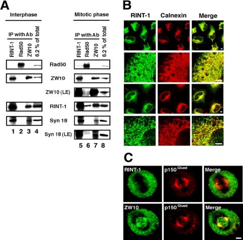
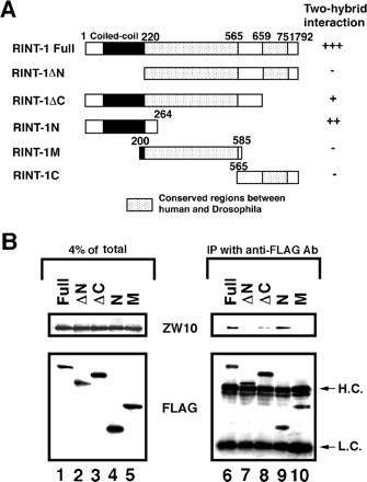
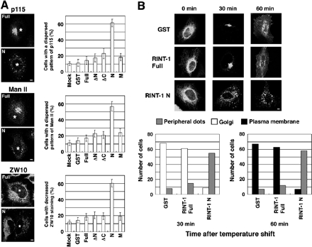
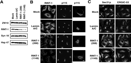
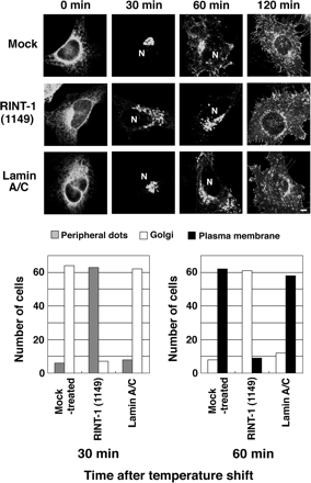
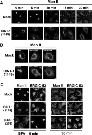
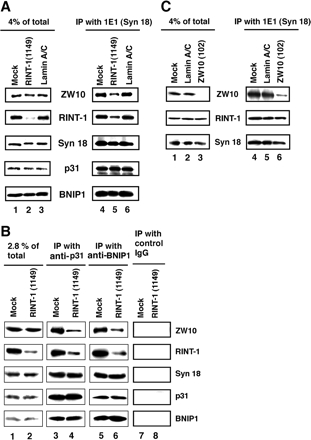
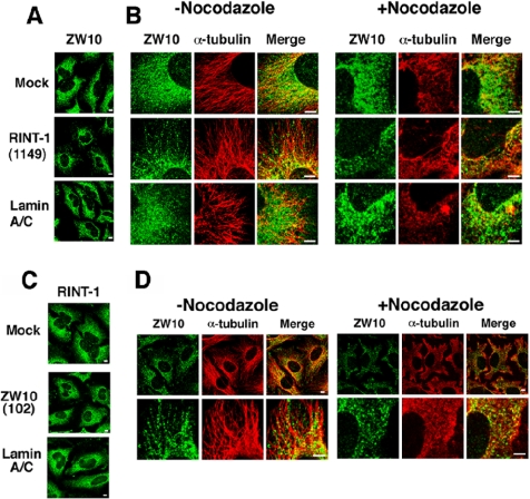
RINT-1 was first identified as a Rad50-interacting protein that participates in radiation-induced G2/M checkpoint control. We have recently reported that RINT-1, together with the dynamitin-interacting protein ZW10 and others, is associated with syntaxin 18, an endoplasmic reticulum (ER)-localized SNARE involved in membrane trafficking between the ER and Golgi. To address the role of RINT-1 in membrane trafficking, we examined the effects of overexpression and knockdown of RINT-1 on Golgi morphology and protein transport from the ER. Overexpression of the N-terminal region of RINT-1, which is responsible for the interaction with ZW10, caused redistribution of ZW10. Concomitantly, ER-to-Golgi transport was blocked and the Golgi was dispersed. Knockdown of RINT-1 also disrupted membrane trafficking between the ER and Golgi. Notably, silencing of RINT-1 resulted in a reduction in the amount of ZW10 associated with syntaxin 18, concomitant with ZW10 redistribution. In contrast, no redistribution or release of RINT-1 from the syntaxin 18 complex was observed when ZW10 expression was reduced. These results taken together suggest that RINT-1 coordinates the localization and function of ZW10 by serving as a link between ZW10 and the SNARE complex comprising syntaxin 18.


Figures

Figure 1.
Figure 1.
RINT-1 is associated with ZW10 and syntaxin 18 throughout the cell cycle. (A) Lysates were prepared from interphasic or mitotic HeLa cells and immunoprecipitated with an antibody against RINT-1 (lanes 1 and 5), Rad50 (lanes 2 and 6), or ZW10 (lanes 3 and 7). The immunoprecipitated proteins were separated by SDS-PAGE and analyzed by immunoblotting with the indicated antibodies (lanes 1–3 and 5–7). Input (6.2% of total) was also analyzed (lanes 4 and 8). LE, long exposure. (B) HeLa cells were treated without (top two rows) or with 30 μg/ml digitonin (bottom two rows) for permeabilization and double-stained with antibodies against RINT-1 (first column) and calnexin (second column). Merged images are shown on the right. Bar, 5 μm. (C) Mitotic HeLa cells were fixed and double-stained with RINT-1 (top row) or ZW10 (bottom row) and p150Glued. Merged images are shown on the right. Bar, 5 μm.
Figure 2.
Figure 2.
The N-terminal region of RINT-1 interacts with ZW10. (A) Schematic representation of RINT-1 and its deletion constructs. The interaction of RINT-1 or its deletion constructs with ZW10 in yeast cells was evaluated by filter assays for β-galactosidase. Blue color was detected within 1 h (+++), 2 h (++) and 16 h (+). Expression of RINT-1C in yeast cells was confirmed by immunoblotting, whereas this fragment was not expressed in 293T cells (unpublished data). (B) 293T cells were transfected with the plasmids encoding the indicated FLAG-tagged constructs. At 24 h after transfection, cell lysates were prepared, and FLAG-tagged proteins were immunoprecipitated with an anti-FLAG antibody. The precipitated proteins were analyzed by immunoblotting with antibodies against ZW10 and FLAG (lanes 6–10). Lanes 1–5 show the amounts of ZW10 and FLAG-tagged proteins in 4% of the lysate. H.C. and L.C. denote immunoglobulin heavy and light chains, respectively.
Figure 3.
Figure 3.
Overexpression of the N-terminal region of RINT-1 perturbs membrane trafficking between the ER and Golgi. (A) HeLa cells were transfected with the plasmids encoding GST or the indicated FLAG-tagged constructs. After 24 h, the cells were fixed and double-stained. Staining for p115 (top two panels), Man II (middle two panels), and ZW10 (bottom two panels). Asterisks indicate cells overexpressing FLAG-RINT-1 full-length (Full) or FLAG-RINT-1N (N). Bar, 5 μm. The quantitative results are shown on the right. Error bars, SE of the mean for three experiments. (B) The plasmid for VSVG-GFP was cotransfected with the plasmid for GST, as a control, FLAG-RINT-1 full-length, or FLAG-RINT-1N into HeLa cells, and the protein transport assay was performed. To identity cells expressing GST or FLAG-tagged constructs, the fixed cells were stained with an anti-GST or anti-FLAG antibody followed by a Texas Red–conjugated secondary antibody. Only GFP fluorescence is shown. Bar, 5 μm. The quantitative results are shown at the bottom. “Peripheral dots” denotes the dispersed dotlike distribution pattern of VSVG-GFP. When VSVG-GFP was found to be concentrated in the perinuclear area, this localization pattern was categorized into “Golgi.” When VSVG-GFP was detected on the plasma membrane, this pattern was counted as “Plasma membrane.” In many of the cells categorized into “Plasma membrane,” VSVG-GFP was detected not only on the plasma membrane but also in the perinuclear area.
Figure 4.
Figure 4.
Silencing of RINT-1 causes dispersion of proteins located in compartments of the early secretory pathway. (A) HeLa cells were transfected without (mock) or with lamin A/C siRNA, RINT-1 (268), or RINT-1 (1149). At 72 h after transfection, the cells were solubilized in phosphate-buffered saline with 0.5% SDS. Equal amounts of total proteins were separated by SDS-PAGE and analyzed by immunoblotting with the indicated antibodies. (B) Immunofluorescence microscopic analysis of mock-treated cells, and cells transfected with lamin A/C siRNA, RINT-1 (268), or RINT-1 (1149). At 72 h after transfection, the cells were fixed and double-stained with antibodies against RINT-1 and p115. Expanded images for p115 are shown on the right. Bar, 5 μm. (C) Cells were treated as described in B and double-stained with antibodies against Sec31p and ERGIC-53. Bar, 5 μm.
Figure 5.
Figure 5.
Delay in VSVG-GFP transport from the ER in cells with reduced expression of RINT-1. HeLa cells were successively transfected with siRNA and the plasmid for VSVG-GFP. Transport of VSVG-GFP from the ER was monitored as described in Materials and Methods. Typical images at 0, 30, 60, and 120 min after the temperature shift to 32°C are shown. N indicates the position of the nucleus. Bar, 5 μm. The quantitative results are shown at the bottom.
Figure 6.
Figure 6.
Retrograde transport is not significantly impaired in cells with reduced expression of RINT-1. (A) HeLa cells were transfected without (mock) or with RINT-1 (1149). At 72 h after transfection, the cells were incubated with 10 μM BFA for the indicated times, fixed, and stained with an antibody against Man II. Bar, 5 μm. (B) At 54 h after transfection, the cells were further transfected with the plasmids for Sar1p mutants and incubated for 18 h. Redistribution of Man II to the ER occurred in almost all RINT-1–depleted and control cells irrespective of the expression levels of Sar1p mutants (unpublished data). Bar, 5 μm. (C) RINT-1 or β-COP expression was knocked down by RNAi. The expression level of β-COP was decreased by 86% (unpublished data). The cells were incubated with 10 μM BFA for 30 min, fixed, and double-stained with antibodies against Man II and ERGIC-53. Bar, 5 μm.
Figure 7.
Figure 7.
Silencing of RINT-1 causes a decrease in the amount of ZW10 associated with syntaxin 18. (A) HeLa cells were transfected without (mock) or with RINT-1 (1149) or lamin A/C siRNA. At 72 h after transfection, lysates were prepared and immunoprecipitated with an anti-syntaxin 18 antibody. The precipitated proteins were detected by immunoblotting with the indicated antibodies (lanes 4–6). Input (4% of total) was also analyzed (lanes 1–3). (B) Cell lysates were prepared as described in A, and immunoprecipitated with an anti-p31 (lanes 3 and 4), anti-BNIP1 (lanes 5 and 6), or control IgG (lanes 7 and 8). Input (2.8% of total) was also analyzed (lanes 1 and 2). (C) HeLa cells were transfected without (mock) or with lamin A/C siRNA or ZW10 (102). At 72 h after transfection, lysates were prepared and immunoprecipitated with an anti-syntaxin 18 antibody. The precipitated proteins were analyzed by immunoblotting with the indicated antibodies (lanes 4–6). Input (4% of total) was also analyzed (lanes 1–3).
Figure 8.
Figure 8.
Silencing of RINT-1 causes redistribution of ZW10. (A and B) HeLa cells were transfected without (mock) or with RINT-1 (1149) or lamin A/C siRNA. At 72 h after transfection, the cells were treated with or without 10 μg/ml nocodazole for 90 min. The cells were fixed and double-stained with antibodies against ZW10 and α-tubulin. Bar, 5 μm. (C and D) HeLa cells transfected without (mock) or with ZW10 (102) or lamin A/C siRNA were incubated for 72 h and stained for RINT-1. As observed previously (Hirose et al., 2004), p115 was modestly dispersed in ZW10 (102)-transfected cells (unpublished data), confirming the depletion of ZW10. Alternatively, the cells were incubated with or without 10 μg/ml nocodazole for 90 min, fixed, and double-stained with antibodies against ZW10 and α-tubulin. Expanded images are shown (bottom row). Bar, 5 μm.

References

    1. Andag U., Neumann T., Schmitt H. D. The coatomer-interacting protein Dsl1p is required for Golgi-to-endoplasmic reticulum retrieval in yeast. J. Biol. Chem. 2001;276:39150–39160. - PubMed
    1. Andag U., Schmitt H. D. Dsl1p, an essential component of the Golgi-endoplasmic reticulum retrieval system in yeast, uses the same sequence motif to interact with different subunits of the COPI vesicle coat. J. Biol. Chem. 2003;278:51722–51734. - PubMed
    1. Bannykh S.. I., Rowe T., Balch W. E. The organization of endoplasmic reticulum export complexes. J. Cell Biol. 1996;135:19–35. - PMC - PubMed
    1. Belgareh-Touze N., Corral-Debrinski M., Launhardt H., Galan J. M., Munder T., Le Panse S., Haguenauer-Tsapis R. Yeast functional analysis: identification of two essential genes involved in ER to Golgi trafficking. Traffic. 2003;4:607–617. - PubMed
    1. Bonifacino J. S., Glick B. S. The mechanism of vesicle budding and fusion. Cell. 2004;116:153–166. - PubMed

Publication types

MeSH terms