. 2006 Jun;17(6):2592-603.
doi: 10.1091/mbc.e05-10-0909. Epub 2006 Mar 29.

SMAP2, a novel ARF GTPase-activating protein, interacts with clathrin and clathrin assembly protein and functions on the AP-1-positive early endosome/trans-Golgi network

Affiliations

SMAP2, a novel ARF GTPase-activating protein, interacts with clathrin and clathrin assembly protein and functions on the AP-1-positive early endosome/trans-Golgi network

Waka Natsume et al. Mol Biol Cell. 2006 Jun.

Abstract

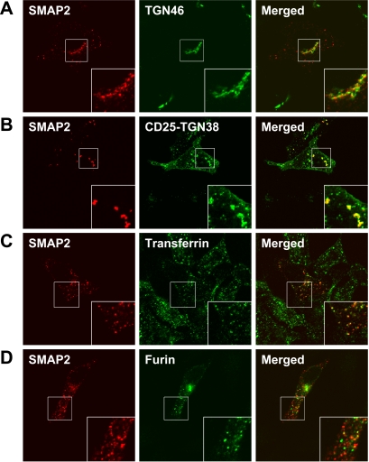
We recently reported that SMAP1, a GTPase-activating protein (GAP) for Arf6, directly interacts with clathrin and regulates the clathrin-dependent endocytosis of transferrin receptors from the plasma membrane. Here, we identified a SMAP1 homologue that we named SMAP2. Like SMAP1, SMAP2 exhibits GAP activity and interacts with clathrin heavy chain (CHC). Furthermore, we show that SMAP2 interacts with the clathrin assembly protein CALM. Unlike SMAP1, however, SMAP2 appears to be a regulator of Arf1 in vivo, because cells transfected with a GAP-negative SMAP2 mutant were resistant to brefeldin A. SMAP2 colocalized with the adaptor proteins for clathrin AP-1 and EpsinR on the early endosomes/trans-Golgi-network (TGN). Moreover, overexpression of SMAP2 delayed the accumulation of TGN38/46 molecule on the TGN. This suggests that SMAP2 functions in the retrograde, early endosome-to-TGN pathway in a clathrin- and AP-1-dependent manner. Thus, the SMAP gene family constitutes an important ArfGAP subfamily, with each SMAP member exerting both common and distinct functions in vesicle trafficking.


Figures

Figure 1.
Figure 1.
The aa sequence of the SMAP2 protein and depiction of its functional domains. (A) Alignment of the aa sequences of the SMAP2 and SMAP1 proteins. The aa sequence of SMAP2 has been predicted as BC052413, which is registered in GenBank/NCBI. We authenticated the sequence of BC052413 by independently isolating and sequencing cDNA clones. The aa sequence of SMAP1 has been described in our previous study (Tanabe et al., 2005). SMAP2 and SMAP1 encode 428 and 440 aa proteins, respectively. Their identical aa residues are indicated by white letters on a black background. The classical and atypical CHC-binding motifs correspond to LLGLD (aa 187-191) and DLL (aa 212-214), respectively. (B) Functional domains of the SMAP2 protein. These domains include a GAP activity domain (aa 1-163), a clathrin-interacting domain (aa 163-231), and a CALM-interacting domain (aa 339-395). The regions that are highly conserved between SMAP2 and SMAP1, namely, the aa 16-125, aa 173-212, aa 244-277, and aa 369-428 regions, are also indicated (by bars). The aa numbers refer to those in SMAP2.
Figure 2.
Figure 2.
GAP activity of SMAP2 on Arf1 and Arf6. (A) Comparison of the GAP activity of SMAP2 with that of SMAP1 and GAP1. Six micrograms each of the SMAP2 (aa 1-163), SMAP1 (aa 1-255), or GAP1 (aa 1-246) proteins were incubated in vitro with 1 μg of GTP-loaded Arf1 or Arf6 for 60 min. In “mock,” no GAP was added to the reaction. After separating the GDP from GTP on thin-layer chromatograms, the degree of GTP hydrolysis was calculated. This is shown as a percentage of GDP over GDP+GTP. Each reaction was independently performed three times and the averages as well as SD values are shown. (B and C) Concentration- and incubation time–dependent GAP activity of the SMAP2 protein. Six (○) or 3 μg (•) of SMAP2 protein, or 6 μg of mutated SMAP2R56Q protein (▵) were incubated with 1 μg of GTP-loaded Arf1 (B) or Arf6 (C) for the indicated times. After separating the GDP from GTP on thin-layer chromatograms, the degree of GTP hydrolysis was calculated and is shown as a percentage as in A. Each data point shows the averages and SD values of three independent experiments.
Figure 3.
Figure 3.
Interaction of SMAP2 protein with the clathrin molecule. (A) Coimmunoprecipitation of SMAP2 and CHC. Cos-7 cells were transfected with Myc-SMAP2, HA-SMAP2, or HA-SMAP2CBm as indicated, and their cell lysates were immunoprecipitated (IP) by anti-Myc, anti-HA, or anti-CHC antibodies as indicated. The precipitates were then subjected to electrophoresis, transferred to a filter, and probed by the indicated antibody (IB). (B) Subcellular localization of the SMAP2 and CHC proteins. Cos-7 cells were transfected with cDNAs encoding the SMAP2 (aa 1-428), with deletion mutants of SMAP2 that corresponded to aa 163-428 or aa 231-428, or with the CBm mutant of SMAP2, respectively. In the CBm mutant, all the aa residues in the LLGLD (aa 187-191) and DLL (aa 212-214) motifs were replaced by alanine. The cells were then fixed and subjected to double immunofluorescence analysis. The HA-tagged SMAP2 and its mutant proteins were detected by anti-HA antibody (red fluorescence), whereas the endogenous clathrin molecules were detected by anti-CHC antibody (green fluorescence). Merged images are shown in the right-hand column. In the insets, portions of the cells are enlarged.
Figure 4.
Figure 4.
Interaction of SMAP2 with CALM. (A) SMAP2 was tested for its ability to interact with CALM. Yeast cells were cotransformed by the indicated combinations of bait (pGBKT7) and prey (pGADT7) vectors and grown on nonselective (−Trp, −Leu) and selective (−Trp, −Leu, −His, −Ade, 2.5 mM 3-amino-1,2,4-triazole) media, respectively. The cDNAs tested were the intact SMAP2 (aa 1-428), a portion of SMAP2 (aa 339-428), and the intact CALM, respectively. (B) Subcellular localization of SMAP2 and CALM proteins. Cos-7 cells were cotransfected with CALM and SMAP2 (aa 1-428) or deletion mutants of SMAP2 that corresponded to aa 163-395 or aa 339-428, respectively, fixed, and processed for double immunofluorescence analysis. The HA-tagged SMAP2 or deletion proteins were detected by anti-HA antibody (red fluorescence), whereas the Myc-tagged CALM protein was detected by anti-Myc antibody (green fluorescence). Merged images are shown in the right-hand column. In the insets, portions of the cells are enlarged.
Figure 5.
Figure 5.
Subcellular localization of SMAP2 and coat proteins. HeLa cells were transfected with HA-SMAP2 (aa 1-428), fixed, and processed for double immunofluorescence analysis. SMAP2 was detected by anti-HA antibody (red fluorescence), whereas endogenous CHC or COPI were detected by their respective specific antibodies (green fluorescence). Merged images are shown in the right-hand column. In the insets, portions of the cells are enlarged.
Figure 6.
Figure 6.
Subcellular localization of SMAP2 and clathrin adaptors. HeLa cells were transfected with HA-SMAP2 (aa 1-428), fixed, and processed for double immunofluorescence analysis. SMAP2 was detected by anti-HA antibody (red fluorescence), whereas AP-1, AP-2, AP-3, EpsinR, and GGA1 were detected by their respective specific antibodies (green fluorescence). AP-1, AP-2, and AP-3 are endogenous molecules while EpsinR and GGA1 are the transfected Myc-EpsinR and Myc-GGA1 molecules, respectively. Merged images are shown in the right-hand column. In the insets, portions of the cells are enlarged. It must be noted that the juxtanuclear structures seen in Figure 5 were out of focus in the SMAP2 photos here.
Figure 7.
Figure 7.
Effect of SMAP2R56Q expression and BFA treatment on the subcellular distribution of AP-1 and COPI. HeLa cells were transfected with HA-SMAP2R56Q (aa 1-428; A and B) or HA-GAP1C45A (C), treated with or without BFA, and then fixed and processed for double immunofluorescence analysis. SMAP2R56Q or GAP1C45A were detected by anti-HA antibody, whereas endogenous AP-1 or COPI were detected by their respective specific antibodies. The arrows and arrowheads indicate SMAP2R56Q-expressing and nonexpressing cells, respectively.
Figure 8.
Figure 8.
Subcellular localization of SMAP2 and its possible cargo protein. (A) HeLa cells were transfected with HA-SMAP2 (aa 1-428), fixed, and processed for double immunofluorescence analysis. SMAP2 was detected by anti-HA antibody (red fluorescence), whereas endogenous human TGN46 was detected by anti-TGN46 antibody (green fluorescence). (B) HeLa cells were cotransfected with HA-SMAP2 and the chimeric CD25-murine TGN38 molecule, fixed, and processed for double immunofluorescence analysis. SMAP2 (red fluorescence) and CD25-TGN38 (green fluorescence) were detected by their respective specific antibodies. (C) HeLa cells were transfected with HA-SMAP2, incubated with dye-conjugated transferrin, fixed, and processed for double immunofluorescence analysis. SMAP2 was detected by anti-HA antibody (red fluorescence), whereas transferrin was visible as green fluorescence. (D) HeLa cells were cotransfected with HA-SMAP2 and Myc-furin convertase, fixed, and processed for double immunofluorescence analysis. SMAP2 and furin convertase were detected by anti-HA antibody (red fluorencence) and anti-Myc antibody (green fluorescence), respectively. In A–D, merged images are shown in the right-hand column. In the insets, portions of the cells are enlarged.
Figure 9.
Figure 9.
Effect of SMAP2 overexpression on the transport of CD25-TGN38 molecule. Cos-7 cells were cotransfected by either one of HA-SMAP2 (A and B), HA-SMAP2R56Q (C and D), or HA-SMAP2CBm (E and F) together with the CD25-TGN38 chimeric cDNA. The cells were then incubated for 15 or 45 min with the anti-CD25 antibody that triggers the internalization of the chimeric molecule. The cells were fixed and processed for double immunofluorescence. In A, C, and E, the SMAP2 protein was detected by the anti-HA antibody, whereas the chimeric protein was detected by the anti-CD25 antibody in B, D, and F. The photos presented in A to F represent the cells after 15 min incubation. The arrows indicate the SMAP2-expressing cells. In G, among the 100 cells that were positive for the SMAP2 staining, the numbers of cells that showed the TGN accumulation or no accumulation of TGN38 were counted, respectively. The percent inhibition corresponds to the cells that were SMAP2-positive but did not accumulate TGN38 at the TGN. Independent transfections were repeated three times, and the averages and SD values of percent inhibition are shown. In a separate experiment, we confirmed by immunoblot analysis that the expression level of HA-SMAP2, HA-SMAP2R56Q, and HA-SMAP2CBm did not vary significantly among the transfected cells.

Similar articles

Cited by

References

    1. Aoe T., Huber I., Vasudevan C., Watkins S. C., Romero G., Cassel D., Hsu V. W. The KDEL receptor regulates a GTPase-activating protein for ADP-ribosylation factor 1 by interacting with its non-catalytic domain. J. Biol. Chem. 1999;274:20545–20549. - PubMed
    1. Bernards A. GAPs galore! A survey of putative Ras superfamily GTPase activating proteins in man and Drosophila. Biochim. Biophys. Acta. 2003;1603:47–82. - PubMed
    1. Chiba N., Watanabe T., Nomura S., Tanaka Y., Minowa M., Niki M., Kanamaru R., Satake M. Differentiation dependent expression and distinct subcellular localization of the protooncogene product, PEBP2beta/CBFbeta, in muscle development. Oncogene. 1997;14:2543–2552. - PubMed
    1. Claing A., Perry S. J., Achiriloaie M., Walker J. K., Albanesi J. P., Lefkowitz R. J., Premont R. T. Multiple endocytic pathways of G protein-coupled receptors delineated by GIT1 sensitivity. Proc. Natl. Acad. Sci. USA. 2000;97:1119–1124. - PMC - PubMed
    1. Cremona O. Live stripping of clathrin-coated vesicles. Dev. Cell. 2001;1:592–594. - PubMed

Publication types