. 2006 Jun 2;281(22):15258-67.
doi: 10.1074/jbc.M512953200. Epub 2006 Mar 29.

Tumor necrosis factor-induced toxic liver injury results from JNK2-dependent activation of caspase-8 and the mitochondrial death pathway

Affiliations

Tumor necrosis factor-induced toxic liver injury results from JNK2-dependent activation of caspase-8 and the mitochondrial death pathway

Yongjun Wang et al. J Biol Chem. .

Abstract

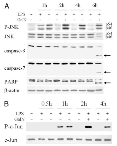
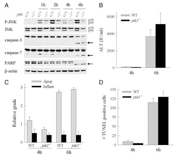
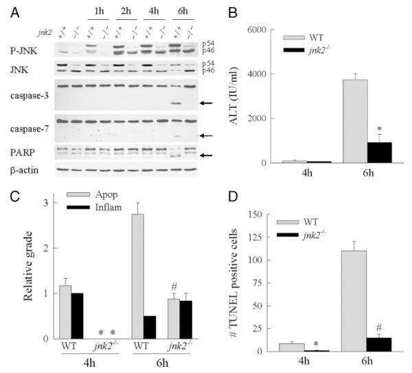
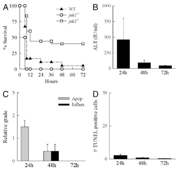
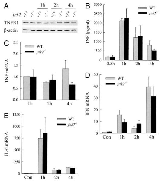
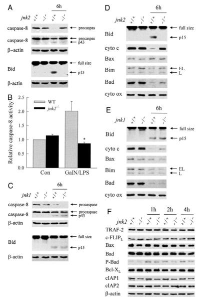
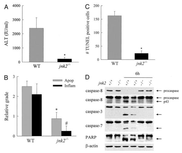
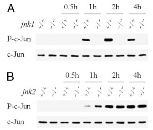
In vitro studies of hepatocytes have implicated over-activation of c-Jun N-terminal kinase (JNK) signaling as a mechanism of tumor necrosis factor-alpha (TNF)-induced apoptosis. However, the functional significance of JNK activation and the role of specific JNK isoforms in TNF-induced hepatic apoptosis in vivo remain unclear. JNK1 and JNK2 function was, therefore, investigated in the TNF-dependent, galactosamine/lipopolysaccharide (GalN/LPS) model of liver injury. The toxin GalN converted LPS-induced JNK signaling from a transient to prolonged activation. Liver injury and mortality from GalN/LPS was equivalent in wild-type and jnk1-/- mice but markedly decreased in jnk2-/- mice. This effect was not secondary to down-regulation of TNF receptor 1 expression or TNF production. In the absence of jnk2, the caspase-dependent, TNF death pathway was blocked, as reflected by the failure of caspase-3 and -7 and poly(ADP-ribose) polymerase cleavage to occur. JNK2 was critical for activation of the mitochondrial death pathway, as in jnk2-/- mice Bid cleavage and mitochondrial translocation and cytochrome c release were markedly decreased. This effect was secondary to the failure of jnk2-/- mice to activate caspase-8. Liver injury and caspase activation were similarly decreased in jnk2 null mice after GalN/TNF treatment. Ablation of jnk2 did not inhibit GalN/LPS-induced c-Jun kinase activity, although activity was completely blocked in jnk1-/- mice. Toxic liver injury is, therefore, associated with JNK over-activation and mediated by JNK2 promotion of caspase-8 activation and the TNF mitochondrial death pathway through a mechanism independent of c-Jun kinase activity.


Figures

FIGURE 1
FIGURE 1. GalN/LPS co-administration leads to prolonged JNK activation before the onset of caspase cleavage
A, protein was isolated from the livers of C57BL/6 mice that were untreated or treated with LPS or GalN/LPS for the number of hours indicated. Samples were immunoblotted with antibodies against phospho-JNK (P-JNK), total JNK, caspase-3 and -7, PARP, and β-actin. The p54 and p46 JNK bands are labeled, and arrows indicate the caspase-3 and -7 and PARP cleavage products. B, hepatic JNK activity was determined in untreated and LPS- or GalN/LPS-treated livers by an in vitro kinase activity assay employing c-Jun as substrate. JNK activity was assayed by immunoblots for phosphorylated c-Jun (P-c-Jun) as described under “Experimental Procedures.” Stripped membranes were immunoblotted with an anti-total c-Jun antibody to check the equivalency of protein loading. Results are representative of three independent experiments.
FIGURE 2
FIGURE 2. Liver injury from GalN/LPS is unaffected by loss of jnk1
A, total hepatic protein was isolated from wild-type (+/+) or jnk1−/− (−/−) mice at the indicated hours after treatment with GalN/LPS and immunoblotted with phospho-JNK (P-JNK), total JNK, caspase-3 and -7, PARP, and β-actin antibodies. The p54 and p46 JNK bands are labeled, and the caspase-3 and -7 and PARP cleavage products are indicated by arrows. Findings are representative of three independent experiments. B, serum ALT levels from wild-type (WT) and jnk1−/− mice at 4 and 6 h after GalN/LPS administration. C, graded histological scoring of the degree of apoptosis (Apop) and inflammation (Inflam) in the same livers. D, numbers of TUNEL positive cells per high power field in the identical liver sections. Data in B–D are from three independent experiments and total five–eight animals per data point.
FIGURE 3
FIGURE 3. Liver histology and TUNEL staining in wild-type and jnk1 and jnk2 knock-out mice after GalN/LPS treatment
Shown are hematoxylin- and eosin-stained sections of wild-type (A), jnk1−/− (B), and jnk2−/− (C) mice at 6 h after GalN/LPS treatment. TUNEL staining at the same time point in wild-type (D), jnk1−/− (E), and jnk2−/− (F) mice (magnification 400×) is also shown.
FIGURE 4
FIGURE 4. jnk2−/− mice are protected from GalN/LPS-induced liver injury
A, protein was isolated from the livers of wild-type (+/+) or jnk2−/− (−/−) mice at the indicated hours after treatment with GalN/LPS and immunoblotted with phospho-JNK (P-JNK), total JNK, caspase-3 and -7, PARP, and β-actin antibodies. The p54 and p46 JNK bands are labeled, and arrows indicate the caspase-3 and -7 and PARP cleavage products. Findings are representative of three independent experiments. B, serum ALT levels from wild-type (WT) and jnk2−/− mice at 4 and 6 h after GalN/LPS treatment. Results are from 4 independent experiments representing a total of 4 mice at 4 h and 8 mice at 6 h per data point (*, p < 0.0001 compared with wild type). C, graded histological scoring of the degree of apoptosis (Apop) and inflammation (Inflam) in the same livers (p < 0.01 (*) and p < 0.001 (#) compared with wild type). D, numbers of TUNEL-positive cells per high power field in the identical liver sections (p < 0.01 (*) and p < 0.00001 (#) compared with wild-type mice).
FIGURE 5
FIGURE 5. jnk2−/− mice have increased long-term survival after GalN/LPS treatment
A, percentages of survival of wild-type (WT), jnk1−/−, and jnk2−/− mice at the indicated hours after administration of GalN/LPS. Data are from 3 independent experiments that include a total of 19 wild-type, 10 jnk1−/−, and 25 jnk2−/− mice (p < 0.01 for jnk2−/− as compared with wild type or jnk1−/−). B, serum ALT levels in wild-type mice surviving for the indicated hours after GalN/LPS treatment. C, graded histological scoring of the degree of apoptosis (Apop) and inflammation (Inflam) in the livers of the same animals. D, numbers of TUNEL-positive cells per high power field in the identical liver sections. Data in B–D are from four independent experiments and total three-eight animals per data point.
FIGURE 6
FIGURE 6. TNFR1 expression and cytokine induction are equivalent in wild-type and jnk2−/− mice
A, total hepatic protein was isolated from the livers of wild-type (+/+) or jnk2−/− (−/−) mice at the indicated hours after treatment with GalN/LPS and immunoblotted with TNFR1 and β-actin antibodies. B, levels of serum TNF at the times shown after GalN/LPS treatment. Serum TNF was undetectable in untreated mice. C–E, total liver RNA was isolated from wild-type and jnk2 knock-out mouse mice untreated and treated with GalN/LPS for 1, 2, and 4 h. Real-time RT-PCR was performed with primers for TNF, interferon γ (IFN), interleukin-6 (IL-6), and β-actin as described under “Experimental Procedures.” Levels of cytokine mRNA were normalized to that of β-actin. TNF mRNA was undetectable in untreated livers, and the TNF values are expressed relative to levels in 1-h-treated wild-type animals (C). Levels of interferon γ (D) and interleukin-6 (E) mRNA were normalized to the level in untreated wild-type livers. The results are from three independent experiments.
FIGURE 7
FIGURE 7. Ablation of jnk2 but not jnk1 inhibits caspase-8 activation, Bid cleavage, and mitochondrial cytochrome c release
A, the cytosolic protein fraction was isolated from the livers of wild-type (+/+) and jnk2 knock-out (−/−) mice that were untreated or treated with GalN/LPS for 6 h. Samples were immunoblotted with antibodies against caspase-8, Bid, and β-actin. The top two panels represent different exposures of the same caspase-8 immunoblot. B, levels of capsase-8 activity in untreated control (Con) and 5-h GalN/LPS-treated wild-type (WT) and jnk2−/− animals (*, p < 0.02 compared with wild-type mice). C, immunoblots of cytosolic protein fractions from untreated or 6-h GalN/LPS-treated wild-type and jnk1−/− mice for caspase-8, Bid, and β-actin. D and E, mitochondrial protein fractions were isolated from the same livers as for the cytosolic fractions from wild-type, jnk2 (D), and jnk1 (E) knock-out mice and immunoblotted with antibodies against Bid, cytochrome c (cyto c), and cytochrome oxidase (cyto ox). F, total hepatic protein was isolated from wild-type (+/+) or jnk2 (−/−) knock-out mice that were untreated or treated with GalN/LPS for the indicated number of hours. Western blot analysis was performed with antibodies directed against TRAF-2, c-FLIP, Bax, Bad, phospho-Bad (P-Bad), Bcl-XL, cellular inhibitor of apoptosis 1 (cIAP1) and 2 (cIAP2), and β-actin. Arrows in A, C, D, and E indicate procaspase-8 and its p43 cleavage product, full size Bid and its cleaved form tBid, and the extra long (EL) and long (L) forms of Bim. Findings are representative of three independent experiments.
FIGURE 8
FIGURE 8. jnk2−/− mice are protected from GalN/TNF-induced liver injury
A, serum ALT levels from wild-type (WT) and jnk2−/− mice at 6 h after GalN/TNF treatment (*, p < 0.04 as compared with wild type). B, graded histological scoring of the degree of apoptosis (Apop) and inflammation (Inflam) in the same livers (p < 0.01 (*) and p < 0.02 (#) compared with wild type). C, numbers of TUNEL positive cells per high power field in the identical liver sections (*, p < 0.002 compared with wild-type mice). D, immunoblots of total protein isolated from the livers of wild-type (+/+) and jnk2 knock-out (−/−) mice that were untreated or treated with GalN/TNF for 6 h. Samples were immunoblotted with antibodies against caspase-8, -3, and -7, PARP, and β-actin. The top two panels represent different exposures of the same caspase-8 immunoblot. Results are from three independent experiments with three-five animals per data point.
FIGURE 9
FIGURE 9. GalN/LPS-induced JNK c-Jun kinase activity is jnk1- but not jnk2-dependent
Hepatic JNK activity was determined in wild-type (+/+) and jnk1 (A) or jnk2 (B) knock-out (−/−) mice that were untreated or treated with GalN/LPS for the number of hours shown. JNK activity is reflected in the levels of phosphorylated c-Jun (P-c-Jun) on immunoblots. Stripped membranes were immunoblotted for total c-Jun to indicate equal protein loading.

Similar articles

Cited by

References

    1. Czaja MJ, Xu J, Alt E. Gastroenterology. 1995;108:1849–1854. - PubMed
    1. Yin M, Wheeler MD, Kono H, Bradford BU, Gallucci RM, Luster MI, Thurman RG. Gastroenterology. 1999;117:942–952. - PubMed
    1. Teoh N, Field J, Sutton J, Farrell G. Hepatology. 2004;39:412–421. - PubMed
    1. Zhou W, Zhang Y, Hosch MS, Lang A, Zwacka RM, Engelhardt JF. Hepatology. 2001;33:902–914. - PubMed
    1. Su F, Schneider RJ. Proc. Natl. Acad. Sci. U. S. A. 1997;94:8744–8749. - PMC - PubMed

Publication types

MeSH terms