. 2006 Mar 29;26(13):3465-73.
doi: 10.1523/JNEUROSCI.4907-05.2006.

Persistent sodium current in layer 5 neocortical neurons is primarily generated in the proximal axon

Affiliations

Persistent sodium current in layer 5 neocortical neurons is primarily generated in the proximal axon

Nadav Astman et al. J Neurosci. .

Abstract

In addition to the well described fast-inactivating component of the Na+ current [transient Na+ current (INaT)], neocortical neurons also exhibit a low-voltage-activated, slowly inactivating "persistent" Na+ current (INaP), which plays a role in determining neuronal excitability and synaptic integration. We investigated the Na+ channels responsible for INaP in layer 5 pyramidal cells using cell-attached and whole-cell recordings in neocortical slices. In simultaneous cell-attached and whole-cell somatic recordings, no persistent Na+ channel activity was detected at potentials at which whole-cell INaP operates. Detailed kinetic analysis of late Na+ channel activity in cell-attached patches at 36 degrees C revealed that somatic Na+ channels do not demonstrate "modal gating" behavior and that the probability of single late openings is extremely low (<1.4 x 10(-4) or <0.02% of maximal open probability of INaT). Ensemble averages of these currents did not reveal a sustained component whose amplitude and voltage dependence could account for INaP as seen in whole-cell recordings. Local application of TTX to the axon blocked somatically recorded INaP, whereas somatic and dendritic application had little or no effect. Finally, simultaneous current-clamp recordings from soma and apical dendrite revealed that Na+ plateau potentials originate closer to the axon. Our data indicate that the primary source of INaP is in the spike initiation zone in the proximal axon. The focal axonal presence of regenerative subthreshold conductance with voltage and time dependence optimal to manipulate integration of synaptic input, spike threshold, and the pattern of repetitive firing provides the layer 5 pyramidal neuron with a mechanism for dynamic control of its gain.


Figures

Figure 1.
Figure 1.
Properties of INaP elicited in layer 5 pyramidal neurons by slow voltage ramps. A, IR-DIC image of a typical, visually identified layer 5 pyramidal cell in 300-μm-thick coronal slice of the neocortex. B, Black trace, Instantaneous I–V curve during a slow (35 mV/s) voltage ramp from −70 to 0 mV (access resistance, 5 MΩ; linear leak current subtracted). K+ currents were blocked by using Cs+ as the main intracellular monovalent cation, and Ca2+ currents were blocked by adding 200 μm Cd2+ to the bath. Blue trace, Instantaneous I–V curve in the presence of TTX (10−6 m). Note that within the voltage range of −70 to −35 mV, virtually all voltage-dependent conductances are blocked. Icat activates at potentials more positive than −35 mV. Digital subtraction of traces before and after TTX application yields the instantaneous I–V curve of INaP (red trace). C, Activation of INaP is not dependent on polarity of the voltage ramp. TTX-sensitive current has similar amplitude and voltage dependence during the depolarizing (top) and hyperpolarizing (bottom) voltage ramps. During the interval between the ramps, the membrane was held at −70 mV. Before hyperpolarizing ramp, the membrane potential was stepped and held at −35 mV for 200 ms. D, Conductance transform of INaP elicited by depolarizing (red) and hyperpolarizing (green) ramps reveals that half-activation voltage and activation steepness factor are similar and they do not depend on direction of voltage change.
Figure 2.
Figure 2.
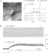
Somatic Na+ channels do not open at subthreshold voltages. A, Left, IR-DIC image illustrating simultaneous whole-cell, cell-attached recording from layer 5 pyramidal cell. Middle, Transient openings of Na+ channels elicited by 40 ms depolarizing voltage pulses from Vr to Vr + 50 mV applied to the patch pipette. Right, Superimposed, Upstroke-aligned action potentials elicited by applying suprathreshold current pulse through whole-cell pipette and capacitance- and leak-subtracted sweeps of transient Na+ channels openings elicited by these action potentials and recorded via the cell-attached pipette. The patch contained 28 channels. B, In the same recording, no channel openings were detected when the cell was depolarized by 250-ms-long current steps to the subthreshold potentials at which INaP begins to operates. Shown are 20 whole-cell current (bottom) and voltage (middle) sweeps along with the corresponding patch current traces (top).
Figure 3.
Figure 3.
Evaluation of the number of Na+ channels in multichannel cell-attached patch recordings. A, Analysis of peak current variability. Left, Fluctuations in Na+ channel current peak amplitudes during repetitive depolarizations. Seven consecutive capacitance- and leak current-subtracted sweeps from multi-Na+ channel patch digitized at 20 kHz through a low-pass filter of 3.5 kHz (−3 dB) are shown. The holding potential was Vr − 40 mV, and the 5-ms-long depolarizing steps to Vr + 60 mV were applied every 200 ms. Recording at 36 ± 1°C. Right, Fluctuations in Na+ current peak amplitudes during a series of 750 depolarizing steps as in the left panel. The right axis is calibrated in single-channel unitary amplitudes, and dashed lines mark multiples of these. Note that in a series of repetitive depolarizations, the number of Na+ channel openings that overlap during the peak of the current varies from a minimum of three to maximum of nine. B, Variance analysis of Na+ current decay. Left column, Seven inverted individual current traces as in A (thick line), superimposed on the ensemble average of 750 sweeps (mean current [I(t)] thin line). Middle column, Differences between each individual trace and the mean current. Right column, The squares of these differences. The ensemble average of 750 such traces represents the variance of the current, σ2(t) (C, inset). C, The ratio between the variance and the mean current [σ2(t)/I(t)] plotted as a function of the mean current [I(t)]. The straight line represents a linear fit of the portion of σ2(t)/I(t) that corresponds to the current decay with Equation 5 (see Results). Note that the x and y axes intercepts of the fitted line correspond to the number of channels in the patch and the unitary current amplitude, respectively. The open circle is the mean unitary current amplitude as measured directly (1.36 ± 0.01 pA; n = 185). Inset, σ2(t) and I(t) traces. Error bars represent SD.
Figure 4.
Figure 4.
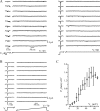
Late Na+ channel openings occur rarely in somatic patches at physiological temperature. A, In somatic cell-attached recording from a representative neuron, brief late openings are rare and there are no noninactivating gating mode episodes. The patch contained 12 Na+ channels (as determined by peak current fluctuation analysis; see above). Patch membrane was held at Vr − 40 mV and stepped to just suprathreshold voltage of Vr + 25 mV (left) or to Vr + 60 mV (right) for 500 ms. Channel currents were filtered at 3.5 kHz (−3 dB) and digitized at 20 kHz. Note that, in this patch, approximately one of the channels would have to be persistently open in one of every eight depolarizations for INaP/peak INaT amplitude ratio to reach 1% value. B, Late somatic Na+ channel openings are rare during slow voltage ramps. Ten consecutive current traces recorded from a patch that contained 14 channels under conditions similar to those described for A. Transient openings of Na+ channels were elicited by 40 ms depolarizing pulses from Vr to Vr + 70 mV, which were followed by 500 ms ramps from Vr to Vr + 70 mV to test the probability of late Na+ channel openings. Note that, as with step depolarizations, during voltage ramps, late openings of somatic Na+ channel do not accumulate into a significant persistent current component. C, Voltage dependence of open probability of Na+ channels during slow voltage ramps (n = 5 patches). To obtain the open probability value, the voltage range covered by ramp was divided into 14, 5-mV-wide bins, and the total open time during each bin was divided by the product of bin duration and the number of channels in the patch. Error bars represent SD.
Figure 5.
Figure 5.
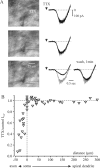
Local application of TTX to the proximal axon blocks whole-cell INaP, whereas somatic and dendritic applications have little or no effect. A, Whole-cell somatic voltage-clamp recording of INaP, and the effect of application of 0.1 μm TTX near the apical dendrite (75 μm), near the soma, and near the proximal axon (−10 μm). Superimposed are current traces before (gray) and after TTX application (black) at a time indicated by arrow. Currents were elicited by applying the 2-s-long voltage ramps from −70 to 0 mV. Note that blockade of INaP by TTX applied near the axon was partially reversible. B, The graph showing the relative effect of TTX on INaP amplitude as a function of application site. The dots are pooled data from nine neurons including that shown in A. Each dot represents a ratio of INaP amplitude at −35 mV before and after TTX application.
Figure 6.
Figure 6.
Na plateau potentials are underlaid by axonal INaP. A, Current-clamp manifestation of INaP in layer 5 neuron: a prolonged, regenerative Na plateau potential triggered by a brief, just suprathreshold somatic current pulse (10 ms, 200 pA). K+ currents were blocked by using Cs+ as the main intracellular monovalent cation, and Ca2+ currents were blocked by adding 200 μm Cd2+ to the bath. Membrane potential was kept at about −65 mV by injection of holding current of −50 pA. B, Plateau potentials recorded simultaneously from the soma (thin traces) and the dendrite (thick traces; distance, 80 μm). Note that the slow decline of the plateau is associated with a crescendo of voltage oscillations that grew to trigger action potentials of increasing upstroke velocity and amplitude. C, Voltage oscillations are generated close to soma and spread into dendrites. Two cycles of voltage oscillation recorded simultaneously from the soma (thin traces) and the dendrite (thick traces; distance, 80 μm) to show that the dendritic oscillation is phase-shifted (∼16°) and smaller in amplitude compared with the somatic one. In a different pyramidal cell (dendritic recording distance, 220 μm), this phase shift is larger (∼30°), consistent with hypothesis that the oscillations initiate around soma and spread passively into the dendrites. D, Hyperpolarizing current pulses (120 pA) of incrementing duration delivered through the somatic pipette during Na plateau, to test the threshold of all-or-none repolarization. Pulses of >40 ms were effective, and the voltage threshold, as determined from six trials, was approximately −58 mV. E, Simultaneous recordings from the soma (thin traces) and the dendrite (thick traces; distance, 200 μm) to show that the minimal duration of hyperpolarizing current pulses required to elicit regenerative repolarization is longer in dendrite than in the soma. The apparent voltage threshold of all-or-none repolarization is significantly lower in the dendrite (−69 ± 8 mV; n = 24) compared with the soma (−60 ± 8 mV; n = 43; p < 0.01).

Similar articles

Cited by

References

    1. Alzheimer C (1994). A novel voltage-dependent cation current in rat neocortical neurones. J Physiol (Lond) 479:199–205. - PMC - PubMed
    1. Alzheimer C, Schwindt PC, Crill WE (1993a). Postnatal development of a persistent Na+ current in pyramidal neurons from rat sensorimotor cortex. J Neurophysiol 69:290–292. - PubMed
    1. Alzheimer C, Schwindt PC, Crill WE (1993b). Modal gating of Na+ channels as a mechanism of persistent Na+ current in pyramidal neurons from rat and cat sensorimotor cortex. J Neurosci 13:660–673. - PMC - PubMed
    1. Andreasen M, Lambert JD (1999). Somatic amplification of distally generated subthreshold EPSPs in rat hippocampal pyramidal neurones. J Physiol (Lond) 519:85–100. - PMC - PubMed
    1. Angelides KJ, Elmer LW, Loftus D, Elson E (1988). Distribution and lateral mobility of voltage-dependent sodium channels in neurons. J Cell Biol 106:1911–1925. - PMC - PubMed

Publication types

LinkOut - more resources