. 2006 Mar 29;26(13):3474-81.
doi: 10.1523/JNEUROSCI.4338-05.2006.

B-ephrin reverse signaling is required for NMDA-independent long-term potentiation of mossy fibers in the hippocampus

Affiliations

B-ephrin reverse signaling is required for NMDA-independent long-term potentiation of mossy fibers in the hippocampus

John N Armstrong et al. J Neurosci. .

Abstract

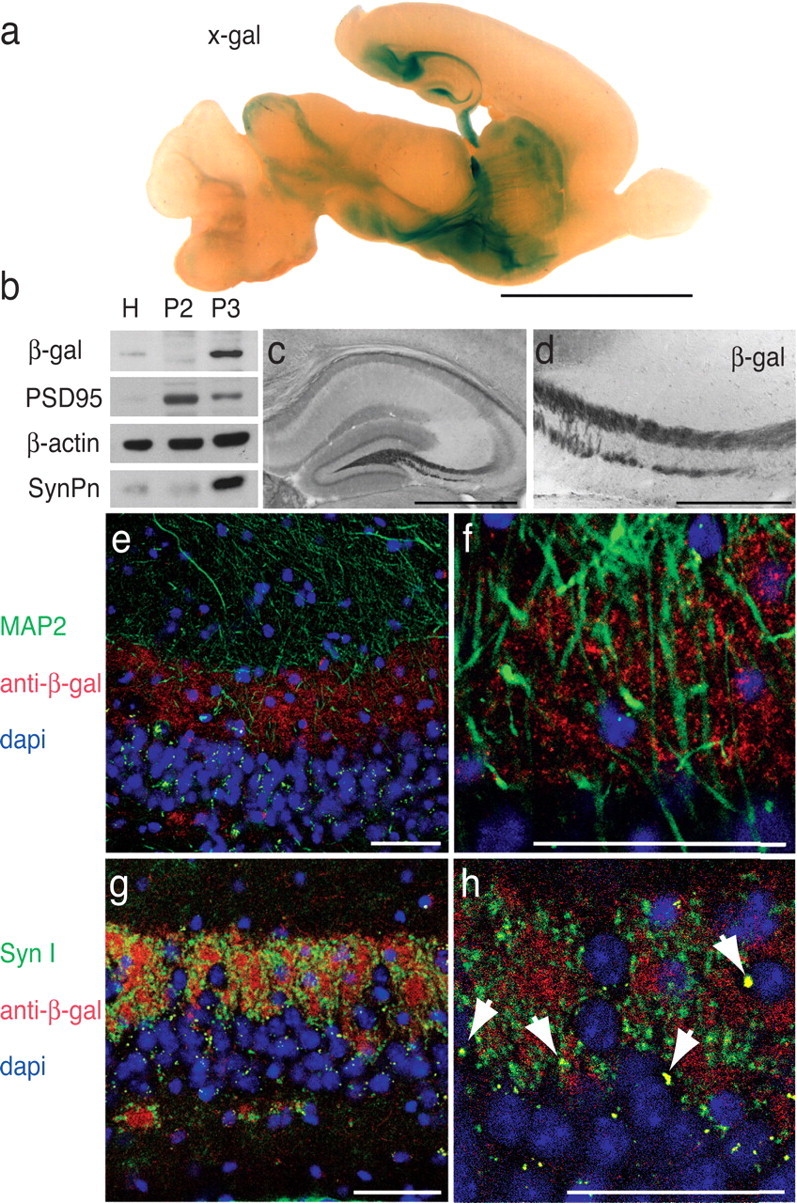
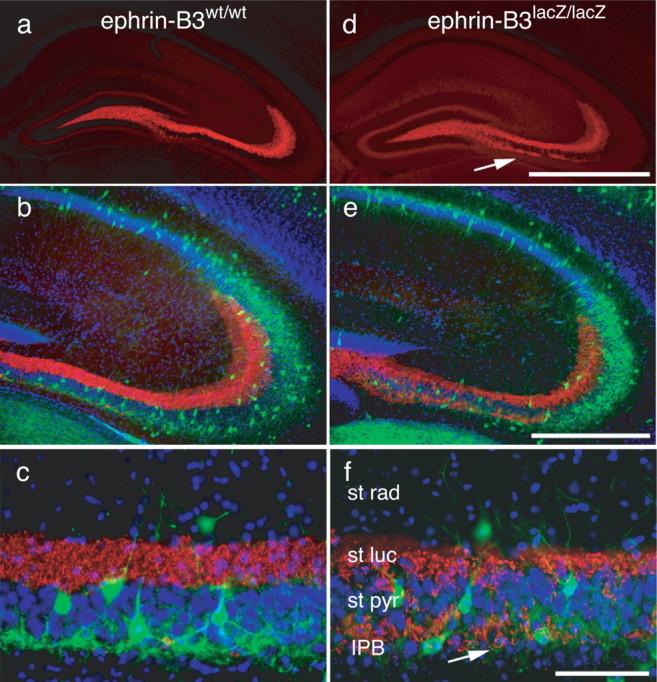
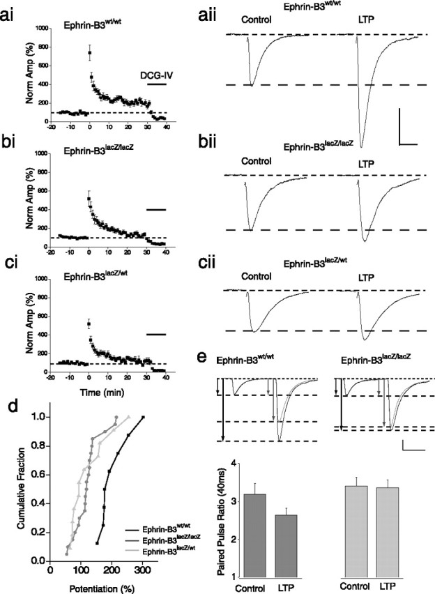
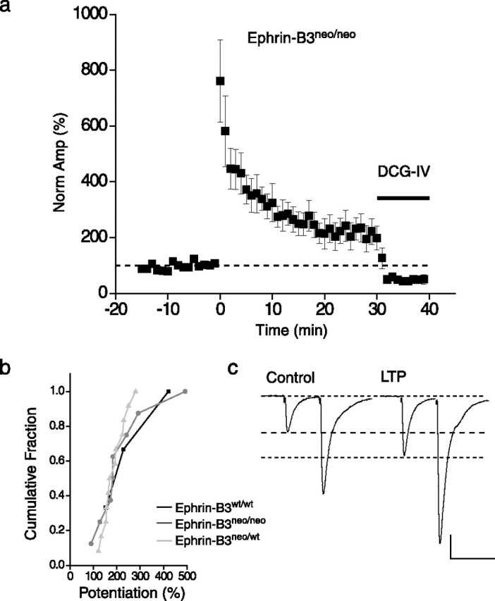
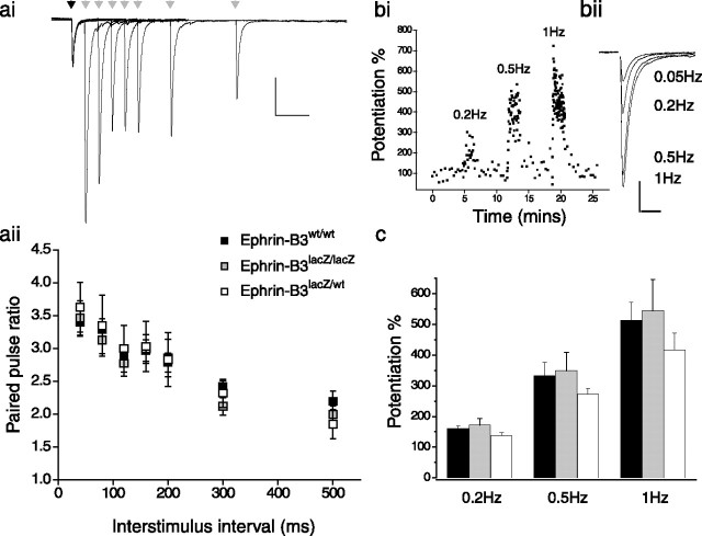
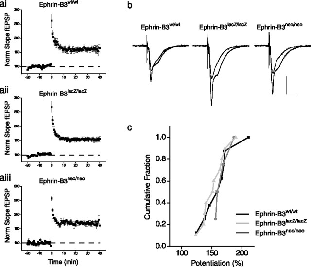
The mossy fiber to CA3 pyramidal neuron synapse in the hippocampus displays an atypical form of NMDA receptor-independent long-term potentiation (LTP). Plasticity at this synapse is expressed in the presynaptic terminal as an elevated probability of neurotransmitter release. However, evidence indicates that postsynaptic mechanisms and trans-synaptic signaling through an association between postsynaptic EphB receptors and presynaptic B-ephrins are necessary for the induction of LTP. Here we show that ephrin-B3 protein is highly expressed in mossy fiber axons and terminals. There are specific deficits in mossy fiber LTP in mice in which the cytoplasmic C-terminal signaling domain of the ephrin-B3 protein is replaced with beta-galactosidase. These deficits are not observed in ephrin-B3 null mutant mice because of functional redundancy by virtue of other B-ephrins. These results indicate that B-ephrin reverse signaling into the presynaptic mossy fiber bouton is required for the induction of NMDA receptor-independent LTP at this synapse.


Figures

Figure 1.
Figure 1.
Ephrin-B3 is localized to mossy fiber axons and synaptic terminals. a, Parasagittal section from ephrin-B3lacZ/lacZ mice stained with X-gal. In the hippocampus, X-gal reaction product was observed throughout the mossy fiber termination zones in the dentate hilus and stratum lucidum of CA3. Additional reaction product was clearly apparent in the striatum and in discrete fiber bundles in the ventral midbrain and inferior colliculus. Scale bar, 5 mm. b, Western blot analysis of hippocampal protein from ephrin-B3lacZ/lacZ mice. A single membrane containing protein isolated from total hippocampus (H), small synaptosomes (P2), and large mossy fiber synaptosomes (P3) was blotted with antibodies raised against β-galactosidase to confirm the presence of the ephrin-B3–β-galactosidase fusion protein in mossy fiber synapses. Additional blotting was performed with antibodies raised against PSD95, β-actin, or synaptoporin (SynPn) to confirm that the P3 fraction contained mossy fiber synapses. c, d, Anti-β-galactosidase immunoreactivity in hippocampal sections taken from ephrin-B3lacZ/lacZ mice further confirm that the ephrin-B3–β-galactosidase fusion protein was present in mossy fiber termination zones in the dentate hilus and stratum lucidum of CA3. Anti-β-galactosidase immunoreactivity was also apparent in the infrapyramidal cell region of the CA3. Scale bars: c, 1 mm; d, 300 μm. In stratum lucidum, triple-immunofluorescent labeling and confocal microscopy confirmed that immunoreactivity for ephrin-B3–β-galactosidase fusion protein (red) was not colocalized with the postsynaptic dendritic marker MAP2 (green) (e, f) but was colocalized with the presynaptic marker synapsin I (green) (g, h); colocalization appears yellow (see arrows in h). Scale bars: e–h, 50 μm.
Figure 2.
Figure 2.
Immunohistological analysis of hippocampi from ephrin-B3lacZ/lacZ mice. a, d, Hippocampal sections from wild-type and ephrin-B3lacZ/lacZ mice stained with presynaptic marker synaptoporin (red) to highlight the mossy fiber bundle. b, e, Triple staining with synaptoporin, DAPI (blue) to highlight cell body layers, and parvalbumin (green) to highlight a subpopulation of interneurons. c, f, Higher magnification of the CA3 region in wild-type and ephrin-B3lacZ/lacZ mice. No gross morphological disturbances in the main mossy fiber bundle and pyramidal neuron layers in CA3 region were observed in ephrin-B3lacZ/lacZ, but there was a significant increase in the extent of the infrapyramidal blade of the mossy fiber projection. st rad, Stratum radiatum; st luc, stratum lucidum; st pyr, stratum pyramidale. Scale bars: a, d, 1 mm; b, e, 500 μm; c, f, 100 μm.
Figure 3.
Figure 3.
Mossy fiber LTP is disrupted in ephrin-B3lacZ/lacZ mice. ai, Averaged time course of mossy fiber LTP for all recordings from ephrin-B3wt/wt mice. Magnitude of LTP was calculated as the percentage increase in the size of the mossy fiber EPSC 20–30 min after the tetanus compared with 15 min control period before induction. Black bar represents application of group II mGluR agonist at the end of each experiment. aii, Representative EPSC traces from one experiment in ephrin-B3wt/wt mice before and after the induction of LTP Calibration: 10 ms, 200 pA. bi, Averaged time course of mossy fiber LTP in ephrin-B3lacZ/lacZ mice. bii, Representative EPSC traces from one experiment during control and 20–30 min after tetanic stimulation. ci, cii, Averaged time course of mossy fiber LTP and representative EPSC traces from ephrin-B3lacZ/wt mice. d, Cumulative probability distribution of mossy fiber LTP measured as the percentage potentiation 25–30 min after tetanus compared with control period. LTP in ephrin-B3lacZ/lacZ and heterozygous littermates was significantly lower than in wild-type littermate controls. e, Paired-pulse ratio measured at an interstimulus interval of 40 ms is reduced after LTP in wild-type mice signifying an increase in release probability of the synapse. Top panel shows representative traces from ephrin-B3wt/wt and ephrin-B3lacZ/lacZ before and after LTP. The traces are scaled to the first EPSC in the pair to highlight the paired-pulse ratio. Calibration: 50 ms, 200 pA; 50 ms, 510 pA for ephrin-B3wt/wt after LTP. Bottom panel summarized paired-pulse data for all recordings. A significant change in paired-pulse ratio is observed after LTP in ephrin-B3wt/wt mice but no change in recordings from ephrin-B3lacZ/lacZ mice.
Figure 4.
Figure 4.
Magnitude of mossy fiber LTP is not altered in ephrin-B3neo/neo hypomorphic null mice. a, Time course of LTP in ephrin-B3neo/neo mice averaged for all recordings. b, Cumulative probability histogram for mossy fiber LTP recordings from ephrin-B3neo/neo mice and heterozygote and wild-type littermates. Magnitude of LTP measured 20–30 min after tetanus is not significantly different between the three genotypes. c, Representative EPSC traces from ephrin-B3neo/neo before and after induction of mossy fiber LTP. Calibration: 50 ms, 200 pA.
Figure 5.
Figure 5.
Short-term mossy fiber synaptic plasticity is not impaired in ephrin-B3lacZ/lacZ mice. ai, Representative mossy fiber EPSCs evoked using paired-pulse protocols at several interstimulus intervals in ephrin-B3lacZ/lacZ mice. Calibration: 100 ms, 200 pA. aii, Paired-pulse ratios calculated as EPSC2/EPSC1 for a range of interstimulus intervals for ephrin-B3lacZ/lacZ and wild-type and heterozygous littermates. There was no significant difference observed at any of the intervals tested (40–500 ms) in recordings from any of the genotypes. b, Time course of frequency facilitation from one ephrin-B3lacZ/lacZ recording. Frequency of afferent stimulation was increased from 0.05 to 0.2, 0.5, and 1 Hz without a change in stimulation intensity. bii, Representative EPSC traces at each stimulation frequency. Calibration: 20 ms, 200 pA. c, Summary of frequency facilitation at the three stimulation frequencies for ephrin-B3wt/wt (black), ephrin-B3lacZ/lacZ (gray), and ephrin-B3lacZ/wt (white) mice.
Figure 6.
Figure 6.
Magnitude of NMDA receptor-mediated LTP at the Schaffer collateral to CA1 pyramidal neuron synapse induced by theta burst stimulation is normal in ephrin-B3 mutant mice. ai–aiii, Time course of CA1 LTP in ephrin-B3 mutant and wild-type mice. Control wild-type littermate recordings from both groups showed no significant difference in the magnitude of LTP and were therefore grouped together. b, Representative CA1 fEPSPs before and after the induction of LTP for ephrin-B3wt/wt, ephrin-B3lacZ/lacZ, and ephrin-B3neo/neo mice. Calibration: 20 ms, 0.4 mV. c, Cumulative distribution histogram of CA1 LTP from ephrin-B3 mutants.
Figure 7.
Figure 7.
Mossy fiber synapses from ephrin-B3lacZ/lacZ mice can be potentiated normally by forskolin. a, Time course of facilitation of mossy fiber synapse by the extracellular application of forskolin to the slice averaged for all recordings from ephrin-B3lacZ/lacZ mice. Significant potentiation is observed at 20–30 min after washout of forskolin, which was not significantly different from that observed in ephrin-B3wt/wt littermates. b, Representative EPSC traces before and after forskolin enhancement from ephrin-B3lacZ/lacZ recordings. Calibration: 20 ms, 200 pA. c, Diagram representation of ephrin-B reverse signaling at mossy fiber terminals and dominant-negative role of the ephrin-B3–β-galactosidase fusion protein in ephrin-B3lacZ/lacZ mice.

Similar articles

Cited by

References

    1. Acsady L, Kamondi A, Sik A, Freund T, Buzsaki G (1998). GABAergic cells are the major postsynaptic targets of mossy fibers in the rat hippocampus. J Neurosci 18:3386–3403. - PMC - PubMed
    1. Alle H, Jonas P, Geiger JR (2001). PTP and LTP at a hippocampal mossy fiber-interneuron synapse. Proc Natl Acad Sci USA 98:14708–14713. - PMC - PubMed
    1. Bergemann AD, Zhang L, Chiang MK, Brambilla R, Klein R, Flanagan JG (1998). Ephrin-B3, a ligand for the receptor EphB3, expressed at the midline of the developing neural tube. Oncogene 16:471–480. - PubMed
    1. Castillo PE, Janz R, Sudhof TC, Tzounopoulos T, Malenka RC, Nicoll RA (1997). Rab3A is essential for mossy fibre long-term potentiation in the hippocampus. Nature 388:590–593. - PubMed
    1. Castillo PE, Schoch S, Schmitz F, Sudhof TC, Malenka RC (2002). RIM1alpha is required for presynaptic long-term potentiation. Nature 415:327–330. - PubMed

Publication types

MeSH terms