. 2006 Mar 29;26(13):3482-90.
doi: 10.1523/JNEUROSCI.4171-05.2006.

Early and simultaneous emergence of multiple hippocampal biomarkers of aging is mediated by Ca2+-induced Ca2+ release

Affiliations

Early and simultaneous emergence of multiple hippocampal biomarkers of aging is mediated by Ca2+-induced Ca2+ release

John C Gant et al. J Neurosci. .

Abstract

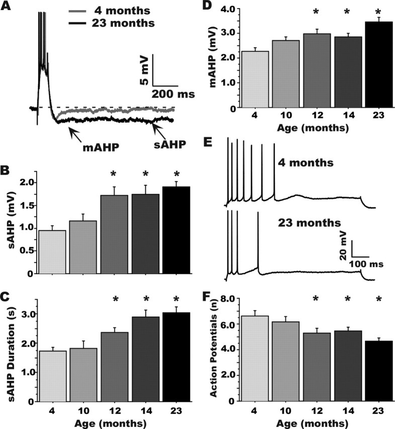
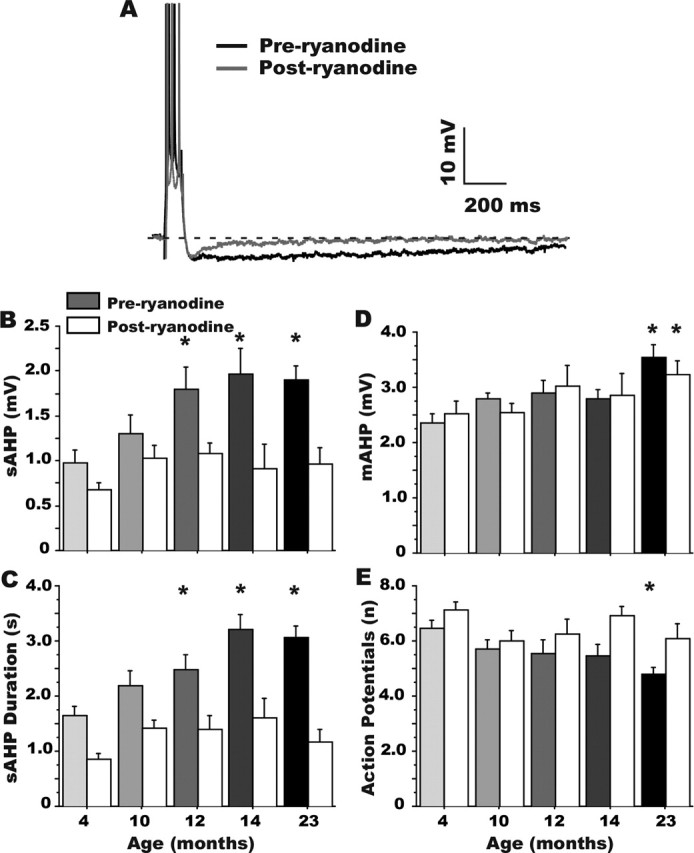
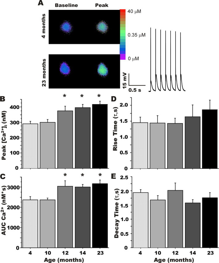
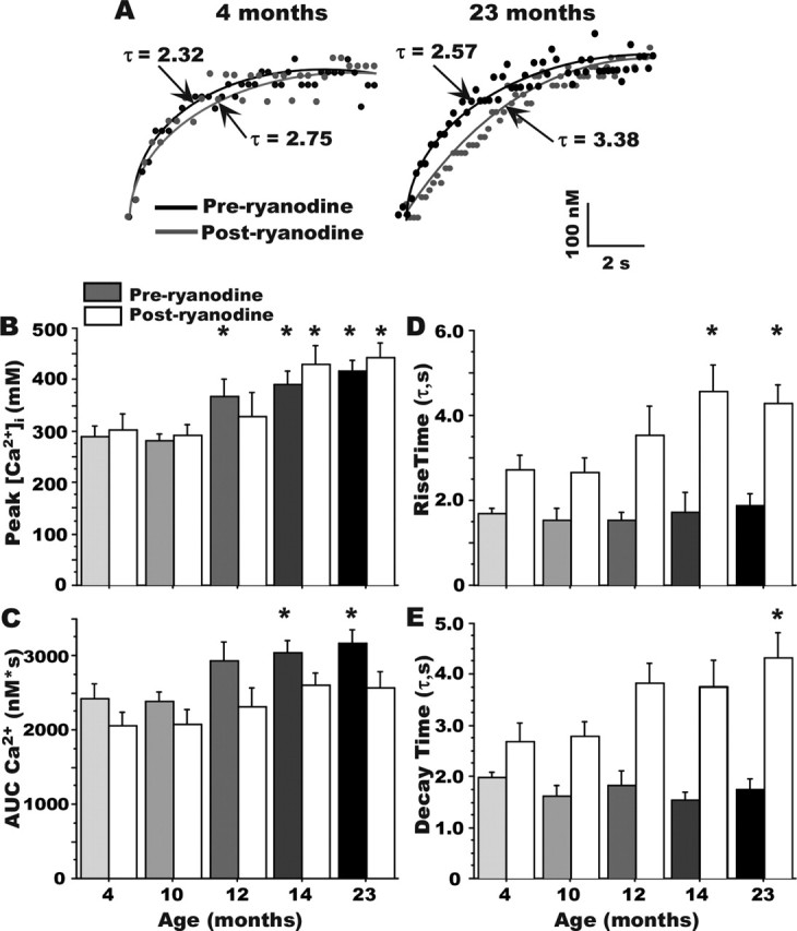
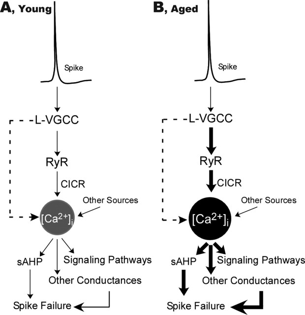
Age-dependent changes in multiple Ca2+-related electrophysiological processes in the hippocampus appear to be consistent biomarkers of aging, and several also correlate with cognitive decline. These findings have led to the hypothesis that a common mechanism of Ca2+ dyshomeostasis underlies aspects of aging-dependent brain impairment. However, some key predictions of this view remain untested, including that multiple Ca2+-related biomarkers should emerge concurrently during aging and their onset should also precede/coincide with initial signs of cognitive decline. Moreover, blocking a putative common source of dysregulated Ca2+ should eliminate aging differences. Here, we tested these predictions using combined electrophysiological, imaging, and pharmacological approaches in CA1 neurons to determine the ages of onset (across 4-, 10-, 12-, 14-, and 23-month-old F344 rats) of several established biomarkers, including the increases in the slow afterhyperpolarization, spike accommodation, and [Ca2+]i rise during repetitive synaptic stimulation. In addition, we tested the hypothesis that altered Ca2+-induced Ca2+ release (CICR) from ryanodine receptors, which can be triggered by L-type Ca2+ channels, provides a common source of dysregulated Ca2+ in aging. Results showed that multiple aging biomarkers were first detectable at about the same age (12 months of age; approximately midlife), sufficiently early to influence initial cognitive decline. Furthermore, selectively blocking CICR with ryanodine slowed the Ca2+ rise during synaptic stimulation more in aged rat neurons and, notably, reduced or eliminated aging differences in the biomarkers. Thus, this study provides the first evidence that altered CICR plays a role in driving the early and simultaneous emergence in hippocampus of multiple Ca2+-related biomarkers of aging.


Figures

Figure 1.
Figure 1.
Age course of alterations in Ca2+-dependent AHPs in CA1 neurons during aging. A, Representative examples of AHPs recorded in CA1 neurons from a young-adult (4 months) and an aged (23 months) animal illustrating where measurements were taken (arrows). B–D, Quantitative group changes in sAHP amplitude and duration and mAHP amplitude, respectively. E, Representative examples of spike frequency accommodation in a 4-month-old (top) and a 23-month-old (bottom) animal. F, Group results on measures of spike-frequency accommodation (number of action potentials generated during an 800 ms depolarization). *Significantly different from the 4-month-old group at p < 0.05. Means ± SEM are shown. Note that the earliest detectable changes occurred for all variables at 12 months of age.
Figure 2.
Figure 2.
Ryanodine eliminates the aging effect on the sAHP. A, Representative examples of the effect of 20 μm ryanodine on the AHP recorded from a 23-month-old rat CA1 neuron. Only a subgroup of the cells presented in Figure 1 were treated with ryanodine, and thus the aging effect was also analyzed separately for that subgroup, both before (grayscale bars) and after (white bars) ryanodine treatment (B–E). Note that ryanodine reduced measures of sAHP amplitude (A, B), sAHP duration (C), and accommodation (E) more in aged animals and eliminated the aging effect in each measure. However, the mAHP (D) was unaffected by ryanodine. *Different at p < 0.05 from the 4-month-old group appropriate for preryanodine and postryanodine comparisons. Means ± SEM are shown.
Figure 3.
Figure 3.
Rapid ratiometric Ca2+ imaging. A, Representative pseudocolor ratiometric images of bis-Fura-2-filled CA1 pyramidal neurons from a young-adult (4 months; top) and an aged (23 months; bottom) animal. Somatic regions are displayed at rest (left) and during repetitive synaptic stimulation (peak; right). The inset shows the first second of the postsynaptic response during 7 Hz RSS above action potential threshold in a young-adult neuron. Group comparisons revealed a significant effect of aging on peak [Ca2+]i and the area under the curve (AUC) of the somatic Ca2+ response (B, C), but no significant differences in the rising and decaying phases of the [Ca2+]i response (D, E, respectively). *Different from the 4-month-old group at p < 0.05. Means ± SEM are shown.
Figure 4.
Figure 4.
Effect of ryanodine on kinetics of the [Ca2+]i response. A, Examples of the effects of ryanodine in slowing the rising phase of somatic Ca2+ at the onset of RSS in a 4-month-old (left) and 23-month-old (right) animal. B, Ryanodine did not significantly affect peak Ca2+ levels measured during RSS. Grayscale bars, Preryanodine. White bars, Postryanodine. Aging effect persists after ryanodine (asterisks). C, Ryanodine eliminated the effect of aging on measures of the AUC. Although the rising (D) and decaying (E) time constants of Ca2+ did not differ with aging, ryanodine treatment significantly lengthened both time constants. All aging comparisons are made within a treatment condition (i.e., preryanodine only or postryanodine only). Note that peak Ca2+ levels are increased with aging in cells before the ryanodine treatment (asterisks above grayscale bars) and also after ryanodine treatment (asterisks above white bars). *Different from the 4-month-old group appropriate for preryanodine and postryanodine comparisons at p < 0.05. Means ± SEM are shown.
Figure 5.
Figure 5.
Schematic model of proposed interactions of L-VGCC and altered CICR in driving Ca2+-related processes in the hippocampus of young (A) and aged (B) animals. The increased L-VGCC activity and enhanced CICR are in series, amplifying the impact of Ca2+ influx on multiple functions. The thickness of arrows schematically represents the relative weight of contribution to [Ca2+]i levels and responses. Despite similar spike-mediated depolarization, with aging, the contribution is increased at several stages. The dashed arrows indicate a possible direct parallel contribution of L-VGCCs to [Ca2+]i.

Similar articles

Cited by

References

    1. Aitken DH, Meaney MJ (1989). Temporally graded, age-related impairments in spatial memory in the rat. Neurobiol Aging 10:273–276. - PubMed
    1. Alford S, Frenguelli BG, Schofield JG, Collingridge GL (1993). Characterization of Ca2+ signals induced in hippocampal CA1 neurones by the synaptic activation of NMDA receptors. J Physiol (Lond) 469:693–716. - PMC - PubMed
    1. Bach ME, Barad M, Son H, Zhuo M, Lu YF, Shih R, Mansuy I, Hawkins RD, Kandel ER (1999). Age-related defects in spatial memory are correlated with defects in the late phase of hippocampal long-term potentiation in vitro and are attenuated by drugs that enhance the cAMP signaling pathway. Proc Natl Acad Sci USA 96:5280–5285. - PMC - PubMed
    1. Barnes CA (1994). Normal aging: regionally specific changes in hippocampal synaptic transmission. Trends Neurosci 17:13–18. - PubMed
    1. Barnes CA, Rao G, Shen J (1997). Age-related decrease in the N-methyl-d-aspartateR-mediated excitatory postsynaptic potential in hippocampal region CA1. Neurobiol Aging 18:445–452. - PubMed

Publication types