. 2006 Mar 29;26(13):3541-50.
doi: 10.1523/JNEUROSCI.2488-05.2006.

Nuclear factor kappaB deficiency is associated with auditory nerve degeneration and increased noise-induced hearing loss

Affiliations

Nuclear factor kappaB deficiency is associated with auditory nerve degeneration and increased noise-induced hearing loss

Hainan Lang et al. J Neurosci. .

Abstract

Degeneration of the spiral ganglion neurons (SGNs) of the auditory nerve occurs with age and in response to acoustic injury. Histopathological observations suggest that the neural degeneration often begins with an excitotoxic process affecting the afferent dendrites under the inner hair cells (IHCs), however, little is known about the sequence of cellular or molecular events mediating this excitotoxicity. Nuclear factor kappaB (NFkappaB) is a transcription factor involved in regulating inflammatory responses and apoptosis in many cell types. NFkappaB is also associated with intracellular calcium regulation, an important factor in neuronal excitotoxicity. Here, we provide evidence that NFkappaB can play a central role in the degeneration of SGNs. Mice lacking the p50 subunit of NFkappaB (p50(-/-) mice) showed an accelerated hearing loss with age that was highly associated with an exacerbated excitotoxic-like damage in afferent dendrites under IHCs and an accelerated loss of SGNs. Also, as evidenced by immunostaining intensity, calcium-buffering proteins were significantly elevated in SGNs of the p50(-/-) mice. Finally, the knock-out mice exhibited an increased sensitivity to low-level noise exposure. The accelerated hearing loss and neural degeneration with age in the p50(-/-) mice occurred in the absence of concomitant hair cell loss and decline of the endocochlear potential. These results indicate that NFkappaB activity plays an important role in protecting the primary auditory neurons from excitotoxic damage and age-related degeneration. A possible mechanism underlying this protection is that the NFkappaB activity may help to maintain calcium homeostasis in SGNs.


Figures

Figure 1.
Figure 1.
Age-related accelerated hearing loss in p50−/− mice. Physiological measurements in p50 −/− and WT mice in the three age groups are shown. A, Mean CAP thresholds ± SEM obtained from groups at 1, 3, and 8 months of age (n = 9 for each group). Asterisks indicate statistically significant differences at the indicated frequency (ANOVA, *p < 0.05; **p < 0.01). At 1 month of age, CAP thresholds in the knock-outs were elevated by 6–10 dB relative to the wild-type group at all frequencies. At 3 months of age, thresholds in knock-outs were elevated by 30 dB at higher frequencies and by 20 dB at lower frequencies. By 8 months of age, most p50−/− mice no longer responded to auditory stimuli at the higher frequencies, whereas the WT mice retained CAP responses at most frequencies tested. B, Difference in mean threshold shifts compared with 1-month-old WT mean thresholds.
Figure 2.
Figure 2.
The deficiency in the p50 subunit of NFκB does not affect OHC and lateral wall function compared with WT mice. A, DPOAEs obtained with constant level (L1 = L2 = 70 dB SPL) primaries from eight WT and eight knock-out mice at 3 months of age. Error bars represent SEM. The differences of DPOAE amplitudes between WT and knock-out mice were not statistically significant (ANOVA, p > 0.05). B, Mean EP values obtained from basal turns from the same animals shown in Fig. 1. Data are expressed as mean ± SEM (n = 9 for each group). No EP reduction with age was found in either the wild-type or the p50−/− mice (ANOVA, p > 0.05). EP differences between WT and knock-out mice were not statistically significant at ages of 1, 3, and 8 months, respectively (ANOVA, p > 0.05).
Figure 3.
Figure 3.
Afferent dendrites beneath IHCs in WT and p50−/− mice. Cochleas from a total of 15 WT and p50−/− mice at ages of 1 or 3 months were observed by electron microscopy (Table. 1). A, Electron microscopic features of the basal half of an IHC and its subcellular synaptic region from the basal turn of a 3-month-old WT mouse. An inner pillar cell (IPC) and border cell (BC) surround the IHC and subadjacent nerves consisting of intermingled afferent inner radial fibers (IR; black arrow) and efferent spiral fibers (white arrow). B, Vacuole-like spaces replace the afferent terminals of the inner radial nerves in a 3-month-old p50−/− mouse. The efferent inner spiral fibers appear intact. The p50−/− mice show membranous structures (black arrow), presumably representing residue from degenerated cell organelles. The cytoplasm in the base of the IHC consists of numerous small vesicles infiltrated with mitochondria and short profiles of cisternae. Efferent terminals (white arrow) appear normal. C, An IHC from another 3-month-old p50−/− mouse shows edematous-appearing extracellular spaces between the IHC and supporting cells (black asterisk). D, An IHC from a 1-month-old knock-out shows similar edematous-appearing spaces underlying the IHC. E, An OHC and underlying Deiters’s cell (DC) from the basal turn of a 3-month-old p50−/− mouse shows no pathologic change. The same cochlea is shown in B. F, The stria vascularia (StV) from the basal turn of a 3-month-old p50−/− mouse (same cochlea as in B) shows no pathologic change. Scale bars: A–E, 2 μm; F, 3 μm.
Figure 4.
Figure 4.
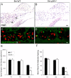
Age-related accelerated degeneration of SGN and nerve fibers in p50−/− mice. SGNs and nerve fibers were counted in sections through the basal turn from WT and p50−/− mice. A, B, Hematoxylin and eosin (H&E)-stained sections of the basal turns from 8-month-old wild-type and knock-out mice show the profiles of SGNs with granular cytoplasm and the large spherical nuclei (arrows). The diminished number of SGNs in the basal turn is clearly evident in 8-month-old p50−/− mice. C, D, Peripheral axons of auditory fibers in 8-month-old WT and knock-out mice. The nerve fibers were labeled with neurofilament 200 (green), and nuclei were counterstained with PI (red). Photographs of tangential sections were taken through the osseous spiral lamina in the basal turn showing the groups of fibers. A significant loss of nerve fibers was seen in the basal turn of an 8-month-old p50−/− mouse. E, SGN counts from 1-, 3-, and 8-month-old wild-type and knock-out mice (n = 5 for each group). Data are expressed as mean ± SEM. The density of SGNs in the 8-month-old group was one-half that of the wild-type and was significantly different (ANOVA, **p < 0.01). There were no significant differences in the SGN densities between WT and p50−/− mice in the 1- and 3-month-groups. F, Average numbers of neurofilament 200-positive nerve fibers in each habenular opening from 1-, 3-, and 8-month-old WT and p50−/− mice (n = 4 for each group). Data are expressed as mean ± SEM. The number of nerve fibers per habenular opening in the 8-month-old group is significantly decreased compared with that of the wild-type mice (ANOVA, *p < 0.05). Scale bars: (in B) A, B, 40 μm; (in D) C, D, 5 μm.
Figure 5.
Figure 5.
Deficiency of the p50 subunit of NFκB has little effect on hair-cell survival. Data are represented as mean ± SEM. OHC and IHC densities at defined cochlear locations in 1- and 8-month-old WT and p50−/− mice are shown. Hair-cell counts were obtained from four cochleas of 1-month-old WT mice, four cochleas of 1-month-old knock-outs, six cochleas of 8-month-old WT mice, and six cochleas of 8-month-old knock-outs. All three rows of OHCs were included in the OHC counts. A, Low-magnification view of the surface preparation of the basilar membrane showing filamentous actin-positive stereocilia bundles (green) on IHCs and OHCs as well as apical region of supporting cells. SGNs were stained red with anti-neurofilament 200. The image shows the apical half of the cochlea from a 1-month-old p50−/− mouse. B, C, Higher-magnification view of the surface preparation shows the nuclei of IHCs and OHCs stained with PI (red). Images were taken from the mid-apical turn (B) and mid-basal turn of an 8-month-old p50−/− mouse. Missing OHCs are shown with arrowheads. D, A significant loss of OHCs was present in the basal turns of the 8-month-old WT and knock-out mice as compared with 1-month-old WT mice (ANOVA, p < 0.01). However, OHC losses were similar in knock-outs and WT mice of the same age (ANOVA, p > 0.05). E, No significant loss of IHCs with age was found in either WT or p50−/− mice (ANOVA, p > 0.05). IHC density differences between wild-type and knock-out mice were not statistically significant at ages of 1 and 8 months, respectively (ANOVA, p > 0.05). Scale bars: A, 250 μm; (in C) B, C, 10 μm.
Figure 6.
Figure 6.
The immunoreactivity for a set of calcium-buffering proteins is markedly increased in p50−/− mice. Immunohistochemical staining for NCS1, PMCA3, calbindin D28K, and synaptophysin (green) was performed on paraffin sections of 8-month-old WT and p50−/− mice. Nuclei were counterstained with PI (red). Ganglion cells were identified by their large spherical nucleus (white arrows). All images were taken from cochlear apical turns. A, E, SGNs show a uniform cytoplasm staining for NCS1. B, F, Anti-PMCA3 shows punctuate staining in cytoplasm of SGNs. C, G, Anti-calbindin D28K shows uniform cytoplasm and weak nuclei staining in SGNs. D, H, SGNs reveal a punctuate staining pattern for synaptophysin. Scale bar: (in H,) A–H, 20 μm. J–L, The relative intensities of immunostaining for NCS1, PMCA3, calbindin D28K, and synaptophysin in both the apical and basal cochlea are significantly increased in p50−/− mice compared with WT mice. (ANOVA, *p < 0.05; **p < 0.01). Data are represented as mean ± SEM.
Figure 7.
Figure 7.
Increased susceptibilities to noise-induced hearing loss in p50−/− mice. A, An intensity series of CAP responses from a WT mouse (left) showed no threshold shifts immediately after a 2 h wideband exposure to a low-level noise (70 dB SPL). However, the CAP responses from a p50−/− mouse were shifted by ∼10 dB at 16 kHz after a similar exposure. B, CAP I/O functions plotting the CAP peak amplitude as a function of the intensity of acoustic probe tones at 16 kHz. Data are from the animal shown in A. The CAP I/O function obtained from a p50−/− ear shows a reduced slope after the 2 h noise exposure. C, CAP threshold shifts in p50−/− and WT mice after noise exposure. Data are mean ± SEM (n = 4 for each group). ANOVA, *p < 0.05, **p < 0.01.

Similar articles

Cited by

References

    1. Adamson CL, Reid MA, Davis RL (2002). Opposite actions of brain-derived neurotrophic factor and neurotrophin-3 on firing features and ion channel composition of murine spiral ganglion neurons. J Neurosci 22:1385–1396. - PMC - PubMed
    1. Aleyasin H, Cregan SP, Iyirhiaro G, O’Hare MJ, Callaghan SM, Slack RS, Park DS (2004). Nuclear factor-κB modulates the p53 response in neurons exposed to DNA damage. J Neurosci 24:2963–2973. - PMC - PubMed
    1. Anniko M, Arnold W, Stigbrand T, Strom A (1995). The human spiral ganglion. ORL J Otorhinolaryngol Relat Spec 57:68–77. - PubMed
    1. Arundine M, Tymianski M (2003). Molecular mechanisms of calcium-dependent neurodegeneration in excitotoxicity. Cell Calcium 34:325–337. - PubMed
    1. Bao J, Lei D, Du Y, Ohlemiller KK, Beaudet AL, Role LW (2005). Requirement of nicotinic acetylcholine receptor subunit β2 in the maintenance of spiral ganglion neurons during aging. J Neurosci 25:3041–3045. - PMC - PubMed

Publication types