

Hypothalamic deep brain stimulation in positron emission tomography
- PMID: 16571767
- PMCID: PMC6673857
- DOI: 10.1523/JNEUROSCI.4609-05.2006
Hypothalamic deep brain stimulation in positron emission tomography
Abstract
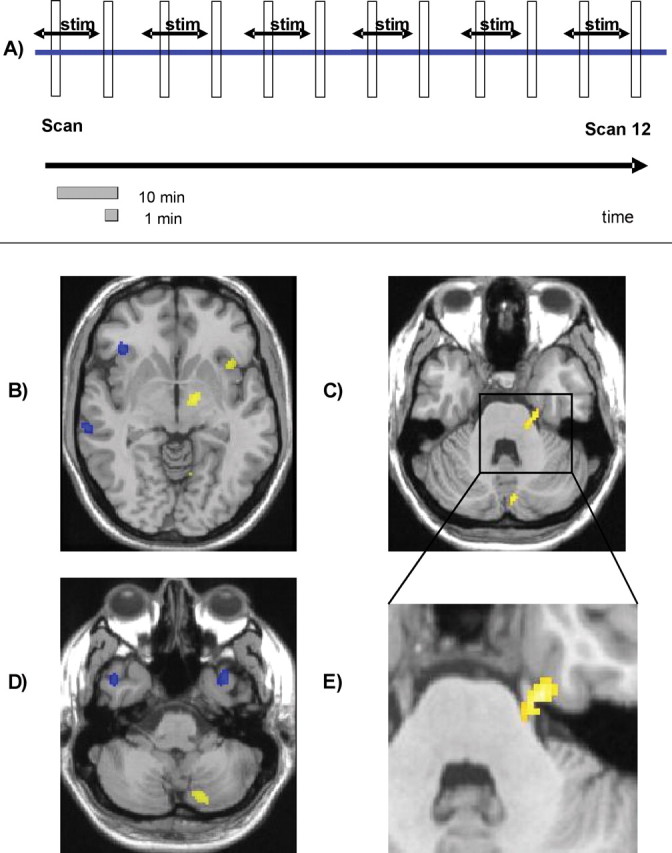
Recently, functional imaging data have underscored the crucial role the hypothalamus plays in cluster headache, one of the most severe forms of primary headache. This prompted the application of hypothalamic deep brain stimulation. Yet, it is not apparent how stimulation of an area that is thought to act as a pace-maker for acute headache attacks is able to prevent these attacks from occurring. We addressed this issue by examining 10 operated chronic cluster headache patients, using H2(15O)-positron emission tomography and alternately switching the hypothalamic stimulator on and off. The stimulation induced activation in the ipsilateral hypothalamic gray (the site of the stimulator tip), the ipsilateral thalamus, somatosensory cortex and praecuneus, the anterior cingulate cortex, and the ipsilateral trigeminal nucleus and ganglion. We additionally observed deactivation in the middle temporal gyrus, posterior cingulate cortex, and contralateral anterior insula. Both activation and deactivation are situated in cerebral structures belonging to neuronal circuits usually activated in pain transmission and notably in acute cluster headache attacks. Our data argue against an unspecific antinociceptive effect or pure inhibition of hypothalamic activity. Instead, the data suggest a hitherto unrecognized functional modulation of the pain processing network as the mode of action of hypothalamic deep brain stimulation in cluster headache.
Figures
Similar articles
-
Hypothalamic deep-brain stimulation: target and potential mechanism for the treatment of cluster headache.Cephalalgia. 2008 Jul;28(7):799-803. doi: 10.1111/j.1468-2982.2008.01629.x. Cephalalgia. 2008. PMID: 18547217
-
Deep brain stimulation in cluster headache: hypothalamus or midbrain tegmentum?Curr Pain Headache Rep. 2010 Apr;14(2):151-9. doi: 10.1007/s11916-010-0099-5. Curr Pain Headache Rep. 2010. PMID: 20425205 Review.
-
Hypothalamic activation in cluster headache attacks.Lancet. 1998 Jul 25;352(9124):275-8. doi: 10.1016/S0140-6736(98)02470-2. Lancet. 1998. PMID: 9690407
-
Specific hypothalamic activation during a spontaneous cluster headache attack.Neurology. 2004 Feb 10;62(3):516-7. doi: 10.1212/wnl.62.3.516. Neurology. 2004. PMID: 14872051 No abstract available.
-
Deep brain stimulation for medically intractable cluster headache.Neurobiol Dis. 2010 Jun;38(3):361-8. doi: 10.1016/j.nbd.2009.05.020. Epub 2009 Jun 6. Neurobiol Dis. 2010. PMID: 19501166 Review.
Cited by
-
Electrophysiology and Structural Connectivity of the Posterior Hypothalamic Region: Much to Learn From a Rare Indication of Deep Brain Stimulation.Front Hum Neurosci. 2020 May 15;14:164. doi: 10.3389/fnhum.2020.00164. eCollection 2020. Front Hum Neurosci. 2020. PMID: 32670034 Free PMC article.
-
Hypothalamic regulation of headache and migraine.Cephalalgia. 2019 Nov;39(13):1710-1719. doi: 10.1177/0333102419867280. Epub 2019 Aug 29. Cephalalgia. 2019. PMID: 31466456 Free PMC article. Review.
-
The pathophysiology of the trigeminal autonomic cephalalgias, with clinical implications.Clin Auton Res. 2018 Jun;28(3):315-324. doi: 10.1007/s10286-017-0468-9. Epub 2017 Sep 23. Clin Auton Res. 2018. PMID: 28942483 Review.
-
Central generators of migraine and autonomic cephalalgias as targets for personalized pain management: Translational links.Eur J Pain. 2023 Oct;27(9):1126-1138. doi: 10.1002/ejp.2158. Epub 2023 Jul 8. Eur J Pain. 2023. PMID: 37421221 Free PMC article. Review.
-
Network effects of deep brain stimulation.J Neurophysiol. 2015 Oct;114(4):2105-17. doi: 10.1152/jn.00275.2015. Epub 2015 Aug 12. J Neurophysiol. 2015. PMID: 26269552 Free PMC article. Review.
References
-
- Afridi SK, Matharu MS, Lee L, Kaube H, Friston KJ, Frackowiak RS, Goadsby PJ (2005). A PET study exploring the laterality of brainstem activation in migraine using glyceryl trinitrate. Brain 128:932–939. - PubMed
-
- Bartsch T, Levy MJ, Knight YE, Goadsby PJ (2004). Differential modulation of nociceptive dural input to [hypocretin] orexin A and B receptor activation in the posterior hypothalamic area. Pain 109:367–378. - PubMed
-
- Benjamin L, Levy MJ, Lasalandra MP, Knight YE, Akerman S, Classey JD, Goadsby PJ (2004). Hypothalamic activation after stimulation of the superior sagittal sinus in the cat: a Fos study. Neurobiol Dis 16:500–505. - PubMed
-
- Bittar RG, Kar-Purkayastha I, Owen SL, Bear RE, Green A, Wang S, Aziz TZ (2005). Deep brain stimulation for pain relief: a meta-analysis. J Clin Neurosci 12:515–519. - PubMed
-
- Dafny N, Dong WQ, Prieto-Gomez C, Reyes-Vazquez C, Stanford J, Qiao JT (1996). Lateral hypothalamus: site involved in pain modulation. Neuroscience 70:449–460. - PubMed
Publication types
MeSH terms
LinkOut - more resources
Full Text Sources