. 2006 Apr;80(8):3701-11.
doi: 10.1128/JVI.80.8.3701-3711.2006.

Role of residues 121 to 124 of vesicular stomatitis virus matrix protein in virus assembly and virus-host interaction

Affiliations

Role of residues 121 to 124 of vesicular stomatitis virus matrix protein in virus assembly and virus-host interaction

John H Connor et al. J Virol. 2006 Apr.

Abstract

The recent solution of the crystal structure of a fragment of the vesicular stomatitis virus matrix (M) protein suggested that amino acids 121 to 124, located on a solvent-exposed loop of the protein, are important for M protein self-association and association with membranes. These residues were mutated from the hydrophobic AVLA sequence to the polar sequence DKQQ. Expression and purification of this mutant from bacteria showed that it was structurally stable and that the mutant M protein had self-association kinetics similar to those of the wild-type M protein. Analysis of the membrane association of M protein in the context of infection with isogenic recombinant viruses showed that both wild-type and mutant M proteins associated with membranes to the same extent. Virus expressing the mutant M protein did show an approximately threefold-lower binding affinity of M protein for nucleocapsid-M complexes. In contrast to the relatively minor effects of the M protein mutation on virus assembly, the mutant virus exhibited growth restriction in MDBK but not BHK cells, a slower induction of apoptosis, and lower viral-protein synthesis. Despite translating less viral protein, the mutant virus produced more viral mRNA, showing that the mutant virus could not effectively promote viral translation. These results demonstrate that the 121-to-124 region of the VSV M protein plays a minor role in virus assembly but is involved in virus-host interactions and VSV replication by augmenting viral-mRNA translation.


Figures

FIG. 1.
FIG. 1.
Mutation of aa 121 to 124 of VSV M protein does not affect self-association. (A) Image of theoretical model (based on M crystal structure; see Materials and Methods) illustrating the location of the presumptive loop region. (B) Diagram of wt M protein (top) and a mutant used in this study illustrating placement and sequence of mutations. (C) Coomassie staining of purified recombinant M proteins. Lane 1 is purified virions as a marker for the migration of wild-type M protein. The star denotes a proteolytic fragment. mut, mutant. (D) Stopped-flow analysis of self-association of M. The graph shows changes in light-scattering intensity following a shift of the salt concentration from 250 mM to either 65 or 130 mM NaCl. The traces are representative of four experiments each from two separate protein preparations. Wild-type traces are shown in black, and hydrophobic-loop mutant traces are in gray. The inset shows the first 12 seconds of each run to illustrate the lag seen in 130 mM NaCl samples.
FIG. 2.
FIG. 2.
Nucleocapsid and membrane association of wild-type and hydrophobic-loop mutant M proteins. (A) Phosphorimages of M protein exchange assays determining the association of 35S-labeled, in vitro-translated M protein with nucleocapsid-M complexes (see Materials and Methods). Shown are supernatant (S) and pellet (P) fractions for 10, 25, and 50 μg/ml of added NCM. MN1 is a negative control M protein lacking aa 4 to 21, His6-M is an N-terminally His-tagged M, and M-His6 is a C-terminally tagged M protein. (B) M protein exchange assays for wt M (c-terminally His tagged) and a hydrophobic-loop mutant using 6, 15, and 36 μg/ml added NCM complex. (C) The membrane association of radiolabeled M protein in transfected cells was determined by fractionation of cell lysates on sucrose flotation gradients. The autoradiograph shows radiolabeled M protein levels in individual fractions (1 [top] to 9 [bottom]) of the sucrose gradient. (D) Quantitation of total M protein in each fraction. The filled bars are wt M protein, and the white bars are hydrophobic-loop mutant M. The data shown are the means ± standard deviations of three separate experiments.
FIG. 3.
FIG. 3.
Characterization of recombinant M protein mutant virus. (A) Membrane association of M protein from wt VSV (black bars) or protein from hydrophobic mutant VSV (white bars) was determined by fractionation of infected-cell lysates on sucrose flotation gradients. The error bars indicate standard deviations. (B) Association of wt and hydrophobic-mutant M proteins with NCM complexes from wild-type or mutant virions. The graph represents the quantitation of three separate experiments ± standard deviations. The solid lines represent experiments in which nucleocapsid-M complex was derived from wild-type control virus, and the dotted lines represent experiments in which NCM was derived from mutant virus. Wild-type control (▪); hydrophobic mutant (○).
FIG. 4.
FIG. 4.
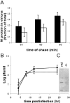
(A) Pulse-chase analysis of M protein incorporated into virions. The black bars are wild-type VSV, and the white bars are the hydrophobic-loop mutant. The bars represent the amount of radiolabeled M incorporated into virions over the amount of radiolabeled M present in virions plus cells. (B) Virus titers of recombinant wild-type VSV and recombinant VSV expressing the hydrophobic-loop mutant. Wild-type control (▪); hydrophobic mutant (○). The inset shows an SDS-PAGE gel of purified virus from media of infected cells (24 h p.i.). All experiments are the averages of three separate experiments ± standard deviations, except for B, which is five separate experiments.
FIG. 5.
FIG. 5.
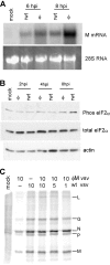
Protein synthesis changes in cells infected with recombinant wt and hydrophobic-mutant viruses. (A) Cells were mock infected or infected with recombinant wt control or hydrophobic (φM)-mutant VSV for the indicated times and labeled with 35S methionine for 10 min. The cell lysates were electrophoresed on a 10% SDS-PAGE gel. A phosphorescence image of a representative gel is shown. (B) Quantification of host protein synthesis following virus infection. The error bars indicate standard deviations. (C) Quantification of viral-protein synthesis following virus infection. (D) Cells were mock infected or infected with wt control, φM mutant, or a 2-amino-acid mutation (AVLA→AVQQ) for 8 h and then labeled and treated as described for panel A.
FIG. 6.
FIG. 6.
Analysis of RNA accumulation, eIF2α phosphorylation, and mutation dominance in hydrophobic-mutant viruses. (A) Total RNA was prepared from cells mock infected or infected with wild-type control or hydrophobic-mutant VSV. The RNA was separated on a 1% agarose gel and transferred to a nitrocellulose membrane, and mRNA was detected using a 32P-labeled probe against M protein mRNA. (B) Phosphorylation of eIF2α following virus infection. Extracts from mock- or virus-infected cells were resolved by SDS-PAGE, transferred to a nitrocellulose membrane, and probed for phospho-eIF2α (Phos eIF2α), total eIF2α, and actin. (C) Cells were infected with wild-type control, hydrophobic-mutant virus, or both at the MOIs indicated above each lane; 8 h p.i., the cells were labeled with [35S]methionine and treated as described for panel A.
FIG. 7.
FIG. 7.
Apoptosis induction in cells infected with wt and hydrophobic-mutant recombinant viruses. (A) Phase-contrast images of BHK cells infected with hydrophobic-mutant virus (left) or wt control virus (right) at an MOI of 10 for 10 h. (B) Cells were infected with wild-type control or hydrophobic-mutant virus and imaged by time-lapse microscopy (see Materials and Methods) for 48 h. Cell rounding is expressed as a fraction of the total number of cells in the field and as a function of time. (C) The cumulative fraction of cells entering apoptosis as determined by membrane blebbing and cell lysis consistent with apoptosis plotted as a function of time. The data in panels B and C are averages of three separate experiments with >150 cells per field. (D) Cells were mock infected or infected with wild-type control virus or hydrophobic-mutant virus for 8, 12, and 24 h. The cells were then lysed an analyzed for caspase 3-like activity using a fluorogenic substrate. The data are the averages of six separate experiments ± standard deviations.
FIG. 8.
FIG. 8.
Replication of wt and φ mutant virus in MDBK cells. Cells were infected with VSV or the φ mutant recombinant virus at an MOI of 10 or 0.1. At 24 h p.i., the cells were analyzed as follows. (A) Virus titers were determined from cells infected at high or low MOI. Wild-type control (▪); hydrophobic mutant (□). The titers shown are the means (plus standard deviations) of the results from three separate experiments using media from cells at 24 h p.i. (B) Phase-contrast imaging showing rwt-infected cells in the top images and cells infected by mutant virus in the bottom images.

Similar articles

Cited by

References

    1. Ahmed, M., M. O. McKenzie, S. Puckett, M. Hojnacki, L. Poliquin, and D. S. Lyles. 2003. Ability of the matrix protein of vesicular stomatitis virus to suppress beta interferon gene expression is genetically correlated with the inhibition of host RNA and protein synthesis. J. Virol. 77:4646-4657. - PMC - PubMed
    1. Black, B. L., R. B. Rhodes, M. McKenzie, and D. S. Lyles. 1993. The role of vesicular stomatitis virus matrix protein in inhibition of host-directed gene expression is genetically separable from its function in virus assembly. J. Virol. 67:4814-4821. - PMC - PubMed
    1. Connor, J. H., and D. S. Lyles. 2005. Inhibition of host and viral translation during vesicular stomatitis virus infection. eIF2 is responsible for the inhibition of viral but not host translation. J. Biol. Chem. 280:13512-13519. - PubMed
    1. Connor, J. H., and D. S. Lyles. 2002. Vesicular stomatitis virus infection alters the eIF4F translation initiation complex and causes dephosphorylation of the eIF4E binding protein 4E-BP1. J. Virol. 76:10177-10187. - PMC - PubMed
    1. Cuesta, R., Q. Xi, and R. J. Schneider. 2000. Adenovirus-specific translation by displacement of kinase Mnk1 from cap-initiation complex eIF4F. EMBO J. 19:3465-3474. - PMC - PubMed

Publication types

Substances

LinkOut - more resources