. 2006 Apr;80(8):3712-20.
doi: 10.1128/JVI.80.8.3712-3720.2006.

Evidence for a functional link between uncoating of the human immunodeficiency virus type 1 core and nuclear import of the viral preintegration complex

Affiliations

Evidence for a functional link between uncoating of the human immunodeficiency virus type 1 core and nuclear import of the viral preintegration complex

David J Dismuke et al. J Virol. 2006 Apr.

Abstract

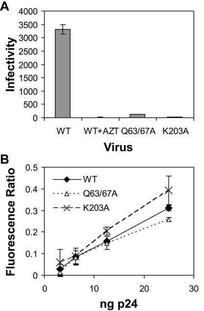
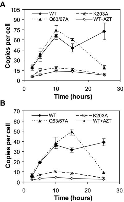
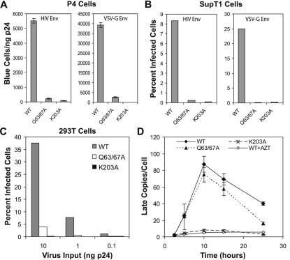
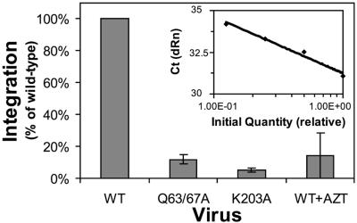
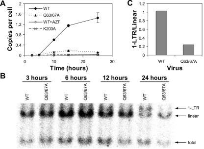
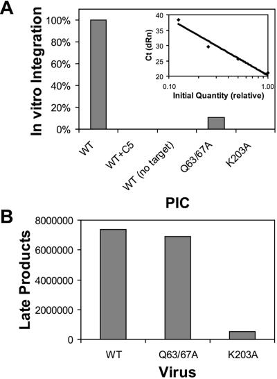
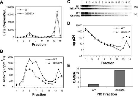
Human immunodeficiency virus type 1 (HIV-1) particles begin their replication upon fusion with the plasma membrane of target cells and release of the viral core into the host cell cytoplasm. Soon thereafter, the viral capsid, which is composed of a polymer of the CA protein, disassociates from the internal ribonucleoprotein complex. While this disassembly process remains poorly understood, the available evidence indicates that proper uncoating of the core is a key step in infection. Defects in uncoating most often lead to a failure of the virus to undergo reverse transcription, resulting in an inability to form a functional viral preintegration complex (PIC). In a previous study, we reported that an HIV-1 mutant containing two substitutions in CA (Q63A/67A) was unusual in that it was poorly infectious yet synthesized normal levels of viral DNA. Here we report that this mutant is impaired for nuclear entry. Quantitative analysis of viral DNA synthesis from infected cells by Southern blotting and real-time PCR revealed that the Q63A/Q67A mutant is impaired in the synthesis of one-long terminal repeat (1-LTR) and 2-LTR circles. Isolation of PICs from acutely infected cells revealed that the Q63A/Q67A mutant produces protein-DNA complexes similar to wild-type in yield and overall composition, but these PICs contained elevated levels of CA and were impaired for integration in vitro. These results demonstrate that mutations in CA can have deleterious effects on both nuclear targeting and integration, suggesting that these steps in the HIV-1 life cycle are dependent on proper uncoating of the viral core.


Figures

FIG. 1.
FIG. 1.
Q63/67A particles are poorly infectious but are competent for cell entry. (A) Wild type (WT) and CA mutant (Q63/67A and K203A) viruses were used to infect CD4-expressing HeLa reporter cells (P4) in a single cycle. Virus input quantities were normalized by p24 ELISA. Infected cells were identified by staining with X-Gal. Shown are the mean values for triplicate infections, with error bars representing one standard deviation. Results are representative of seven independent experiments. (B) SupT1 cells were inoculated with wild-type and CA mutant viruses containing BlaM-Vpr and were subsequently loaded with CCF2-AM substrate. Fusion was quantified by determining the extent of substrate conversion resulting from release of BlaM in the target cytoplasm and was determined by measuring the ratio of blue to green fluorescence. Each virus dilution was analyzed in triplicate. Shown are the mean values, with error bars representing one standard deviation. Results are representative of seven independent experiments.
FIG. 2.
FIG. 2.
Entry of Q63/67A mutant particles results in synthesis of normal levels of reverse transcripts. Wild-type (WT), Q63/67A, and K203A viruses were used to infect P4 cells in duplicate infections. At various times postinfection the DNA from target cells was isolated and HIV-1 early (A) and late (B) reverse transcripts were quantified by TaqMan real-time PCR for each sample in duplicate. Standards were generated by serial dilution of provirus containing plasmid DNA. For the AZT-treated control sample, cells were pretreated with drug 4 h prior to inoculation with wild-type HIV-1. The results represent the average of four determinations (duplicate measurements from two parallel samples), with error bars representing one standard deviation. Results are representative of six independent experiments.
FIG. 3.
FIG. 3.
The Q63/67A infectivity impairment is not rescued by VSV-G pseudotyping. Nonpseudotyped or VSV-G-pseudotyped viruses were used to infect P4 (A), SupT1 (B), or 293T (C) cells in single-cycle infectivity assays. Virus inocula were normalized by p24 ELISA. P4 cells were stained with X-Gal, and the infectivity was calculated as the number of blue cells per ng of p24. Shown are the mean values for triplicate infections, with error bars representing one standard deviation. Infection of 293T and SupT1 cells was quantified by flow cytometry following intracellular staining for HIV-1 Gag protein. For SupT1 cells (B), 100 ng p24 and 10 ng p24 of nonpseudotyped and VSV-G-pseudotyped viruses, respectively, were used for inoculations. WT, wild type. At various times postinfection the DNA from P4 target cells was isolated and HIV-1 late-product DNA quantified by real-time PCR (D) as previously described for Fig. 2. Results are representative of two independent experiments.
FIG. 4.
FIG. 4.
The Q63/67A HIV-1 mutant is impaired for integration in vivo. P4 cells were inoculated with VSV-G-pseudotyped viruses. Cells were cultured for 14 days to allow the degradation of nonintegrated forms of viral DNA. A nested quantitative real-time PCR assay specific for integrated HIV-1 provirus was performed on DNA isolated from approximately 5 × 10⁶ infected cells. Twofold serial dilutions of DNA from cells infected with wild-type HIV-1 were assayed to generate a standard curve (inset) and the relative integration efficiencies of the mutants determined. Shown are the mean values for triplicate determinations, with error bars representing one standard deviation. Results are representative of two independent experiments.
FIG. 5.
FIG. 5.
Q63/67A mutant particles are impaired for nuclear entry. DNA extracted from SupT1 cells infected at low multiplicity of infection (MOI) (∼1) (A) or high MOI (∼15) (B) with wild-type (WT) and mutant viruses was subjected to real-time PCR detecting 2-LTR junction sequences (A) or analyzed by Southern blotting (B). Serial dilutions of plasmid DNA containing 2-LTR junctions were used to generate a standard curve, and the levels of 2-LTR circles in experimental samples were determined by interpolation from the curve. Infections were performed in duplicate, with duplicate quantitation of each sample (A). Values shown are the means of four determinations (duplicate measurements from two parallel samples), with error bars representing one standard deviation. Southern blots were probed with a 32P-labeled probe detecting 1-LTR circles, linear proviral DNA, and total products (B) and detected on a Fujifilm FLA-2000 phosphorimager. Quantification of 1-LTR and linear product band signals was performed with ImageGauge software, and the ratios were calculated (C). Results are representative of three independent experiments.
FIG. 6.
FIG. 6.
Q63/67A mutant PICs are impaired for integration in vitro. Cytoplasmic extracts containing similar quantities of viral DNA were incubated in the presence or absence of 1 μg of φX174 DNA for 45 min at 37°C. DNA was subsequently isolated and subjected to real-time PCR to detect integrated products (A) or late products of reverse transcription (B). As a control, wild-type (WT) HIV-1 infection was also performed in cultures containing the integrase inhibitor compound 5 (C5; 1 μM). A standard curve corresponding to dilutions of an integration reaction is shown in the inset to panel A. Results are representative of three independent experiments.
FIG. 7.
FIG. 7.
Biochemical analysis of wild-type (WT) and Q63/67A mutant PICs. Cytoplasmic extracts from HIV-1-inoculated cells were fractionated by velocity sedimentation on 10 to 50% linear sucrose gradients. Fractions were collected from the top of the gradients and were analyzed for HIV-1 late-product DNA by quantitative PCR (A), reverse transcriptase activity (B), MA and IN by immunoblotting (C), and CA by ELISA (D). (E) The ratio of CA to MA was determined by dividing the CA concentration (D) in fraction 14 by the corresponding MA pixel intensity (C; quantified directly with a Li-Cor Odyssey), corresponding to the peak of viral DNA. Results are representative of two independent experiments.

Similar articles

Cited by

References

    1. Aiken, C. 1997. Pseudotyping human immunodeficiency virus type 1 (HIV-1) by the glycoprotein of vesicular stomatitis virus targets HIV-1 entry to an endocytic pathway and suppresses both the requirement for Nef and the sensitivity to cyclosporin A. J. Virol. 71:5871-5877. - PMC - PubMed
    1. Ao, Z., X. Yao, and E. A. Cohen. 2004. Assessment of the role of the central DNA flap in human immunodeficiency virus type 1 replication by using a single-cycle replication system. J. Virol. 78:3170-3177. - PMC - PubMed
    1. Bartz, S. R., M. E. Rogel, and M. Emerman. 1996. Human immunodeficiency virus type 1 cell cycle control: Vpr is cytostatic and mediates G2 accumulation by a mechanism which differs from DNA damage checkpoint control. J. Virol. 70:2324-2331. - PMC - PubMed
    1. Bouyac-Bertoia, M., J. D. Dvorin, R. A. Fouchier, Y. Jenkins, B. E. Meyer, L. I. Wu, M. Emerman, and M. H. Malim. 2001. HIV-1 infection requires a functional integrase NLS. Mol. Cell 7:1025-1035. - PubMed
    1. Briggs, J. A., T. Wilk, R. Welker, H. G. Krausslich, and S. D. Fuller. 2003. Structural organization of authentic, mature HIV-1 virions and cores. EMBO J. 22:1707-1715. - PMC - PubMed

Publication types

Substances

LinkOut - more resources