

Functional mapping of the nucleoprotein of Ebola virus
- PMID: 16571791
- PMCID: PMC1440433
- DOI: 10.1128/JVI.80.8.3743-3751.2006
Functional mapping of the nucleoprotein of Ebola virus
Abstract
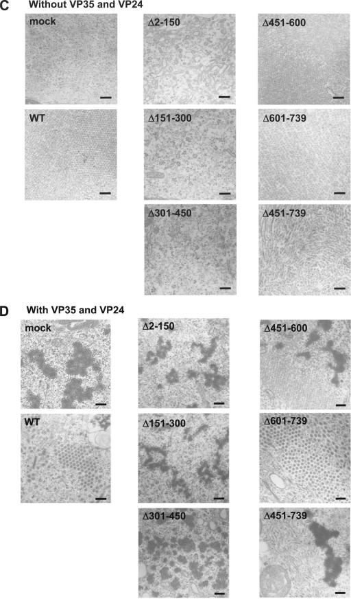
At 739 amino acids, the nucleoprotein (NP) of Ebola virus is the largest nucleoprotein of the nonsegmented negative-stranded RNA viruses, and like the NPs of other viruses, it plays a central role in virus replication. Huang et al. (Y. Huang, L. Xu, Y. Sun, and G. J. Nabel, Mol. Cell 10:307-316, 2002) previously demonstrated that NP, together with the minor matrix protein VP24 and polymerase cofactor VP35, is necessary and sufficient for the formation of nucleocapsid-like structures that are morphologically indistinguishable from those seen in Ebola virus-infected cells. They further showed that NP is O glycosylated and sialylated and that these modifications are important for interaction between NP and VP35. However, little is known about the structure-function relationship of Ebola virus NP. Here, we examined the glycosylation of Ebola virus NP and further investigated its properties by generating deletion mutants to define the region(s) involved in NP-NP interaction (self-assembly), in the formation of nucleocapsid-like structures, and in the replication of the viral genome. We were unable to identify the types of glycosylation and sialylation, although we did confirm that Ebola virus NP was glycosylated. We also determined that the region from amino acids 1 to 450 is important for NP-NP interaction (self-assembly). We further demonstrated that these amino-terminal 450 residues and the following 150 residues are required for the formation of nucleocapsid-like structures and for viral genome replication. These data advance our understanding of the functional region(s) of Ebola virus NP, which in turn should improve our knowledge of the Ebola virus life cycle and its extreme pathogenicity.
Figures

Similar articles
-
Ebola Virus Inclusion Body Formation and RNA Synthesis Are Controlled by a Novel Domain of Nucleoprotein Interacting with VP35.J Virol. 2020 Jul 30;94(16):e02100-19. doi: 10.1128/JVI.02100-19. Print 2020 Jul 30. J Virol. 2020. PMID: 32493824 Free PMC article.
-
The Integrity of the YxxL Motif of Ebola Virus VP24 Is Important for the Transport of Nucleocapsid-Like Structures and for the Regulation of Viral RNA Synthesis.J Virol. 2020 Apr 16;94(9):e02170-19. doi: 10.1128/JVI.02170-19. Print 2020 Apr 16. J Virol. 2020. PMID: 32102881 Free PMC article.
-
Reporter Assays for Ebola Virus Nucleoprotein Oligomerization, Virion-Like Particle Budding, and Minigenome Activity Reveal the Importance of Nucleoprotein Amino Acid Position 111.Viruses. 2020 Jan 15;12(1):105. doi: 10.3390/v12010105. Viruses. 2020. PMID: 31952352 Free PMC article.
-
[The properties of Ebola virus proteins].Vopr Virusol. 2006 Nov-Dec;51(6):4-10. Vopr Virusol. 2006. PMID: 17214074 Review. Russian.
-
Filovirus helical nucleocapsid structures.Microscopy (Oxf). 2023 Jun 8;72(3):178-190. doi: 10.1093/jmicro/dfac049. Microscopy (Oxf). 2023. PMID: 36242583 Review.
Cited by
-
Three minimal sequences found in Ebola virus genomes and absent from human DNA.Bioinformatics. 2015 Aug 1;31(15):2421-5. doi: 10.1093/bioinformatics/btv189. Epub 2015 Apr 2. Bioinformatics. 2015. PMID: 25840045 Free PMC article.
-
Curcumin and Natural Derivatives Inhibit Ebola Viral Proteins: An In silico Approach.Pharmacognosy Res. 2017 Dec;9(Suppl 1):S15-S22. doi: 10.4103/pr.pr_30_17. Pharmacognosy Res. 2017. PMID: 29333037 Free PMC article.
-
Impact of Měnglà Virus Proteins on Human and Bat Innate Immune Pathways.J Virol. 2020 Jun 16;94(13):e00191-20. doi: 10.1128/JVI.00191-20. Print 2020 Jun 16. J Virol. 2020. PMID: 32295912 Free PMC article.
-
A Yeast BiFC-seq Method for Genome-wide Interactome Mapping.Genomics Proteomics Bioinformatics. 2022 Aug;20(4):795-807. doi: 10.1016/j.gpb.2021.02.008. Epub 2021 Jul 24. Genomics Proteomics Bioinformatics. 2022. PMID: 34314873 Free PMC article.
-
Profiling the native specific human humoral immune response to Sudan Ebola virus strain Gulu by chemiluminescence enzyme-linked immunosorbent assay.Clin Vaccine Immunol. 2012 Nov;19(11):1844-52. doi: 10.1128/CVI.00363-12. Epub 2012 Sep 19. Clin Vaccine Immunol. 2012. PMID: 22993411 Free PMC article.
References
-
- Bankamp, B., S. M. Horikami, P. D. Thompson, M. Huber, M. Billeter, and S. A. Moyer. 1996. Domains of the measles virus N protein required for binding to P protein and self-assembly. Virology 216:272-277. - PubMed
-
- Bhella, D., A. Ralph, L. B. Murphy, and R. P. Yeo. 2002. Significant differences in nucleocapsid morphology within the Paramyxoviridae. J. Gen. Virol. 83:1831-1839. - PubMed
-
- Brownlee, M. 2001. Biochemistry and molecular cell biology of diabetic complications. Nature 414:813-820. - PubMed
Publication types
MeSH terms
Substances
Grants and funding
LinkOut - more resources
Full Text Sources
Medical
Miscellaneous
