. 2006 Apr;80(8):3823-32.
doi: 10.1128/JVI.80.8.3823-3832.2006.

Intrarectal immunization with rotavirus 2/6 virus-like particles induces an antirotavirus immune response localized in the intestinal mucosa and protects against rotavirus infection in mice

Affiliations

Intrarectal immunization with rotavirus 2/6 virus-like particles induces an antirotavirus immune response localized in the intestinal mucosa and protects against rotavirus infection in mice

Davide Agnello et al. J Virol. 2006 Apr.

Abstract

Rotavirus (RV) is the main etiological agent of severe gastroenteritis in infants, and vaccination seems the most effective way to control the disease. Recombinant rotavirus-like particles composed of the viral protein 6 (VP6) and VP2 (2/6-VLPs) have been reported to induce protective immunity in mice when administered by the intranasal (i.n.) route. In this study, we show that administration of 2/6-VLPs by the intrarectal (i.r.) route together with either cholera toxin (CT) or a CpG-containing oligodeoxynucleotide as the adjuvant protects adult mice against RV infection. Moreover, when CT is used, RV shedding in animals immunized by the i.r. route is even reduced in comparison with that in animals immunized by the i.n. route. Humoral and cellular immune responses induced by these immunization protocols were analyzed. We found that although i.r. immunization with 2/6-VLPs induces lower RV-specific immunoglobulin G (IgG) and IgA levels in serum, intestinal anti-RV IgA production is higher in mice immunized by the i.r. route. Cellular immune response has been evaluated by measuring cytokine production by spleen and Peyer's patch cells (PPs) after ex vivo restimulation with RV. Mice immunized by the i.n. and i.r. routes display higher gamma interferon production in spleen and PPs, respectively. In conclusion, we demonstrate that i.r. immunization with 2/6-VLPs protects against RV infection in mice and is more efficient than i.n. immunization in inducing an anti-RV immune response in intestinal mucosa.


Figures

FIG. 1.
FIG. 1.
Serum anti-RV antibody levels induced by i.n. and i.r. immunization with 2/6-VLPs. Mice were immunized three times at 2-week intervals with 5 μg of 2/6-VLPs in the presence of 1 μg of CT or 25 μg of CpG ODN 1826 by the i.n. route or with 5 μg of 2/6-VLPs together with 10 μg of CT or 250 μg of CpG ODN 1826 by the i.r. route. A fifth group of control mice was mock immunized with 5 μg of OVA and 10 μg of CT by the i.r. route. Four weeks after the last immunization, blood samples were taken and serum anti-RV antibodies were measured by ELISA. The levels of total anti-RV IgG (A) and IgA (B) are shown. The levels of RV-specific IgG1 and IgG2a subclasses in mice immunized with 2/6-VLPs by the i.n. (C) and i.r. (D) routes are also reported (the ratios between IgG1 and IgG2a levels are reported at the bottom of the graph). Data are displayed as mean ± standard error (SE) (six to seven mice/group). **, Significantly higher than levels of the groups of mice immunized by the i.r. route (P < 0.01 by the Kruskal-Wallis test). *, Significantly higher than levels of the VLP-CpG ODN i.r. group (P < 0.05 by the Kruskal-Wallis test).
FIG. 2.
FIG. 2.
Fecal anti-RV IgA titers induced by i.n. and i.r. immunization with 2/6-VLPs. Mice were immunized three times at 2-week intervals with 5 μg of 2/6-VLPs together with 1 μg of CT or 25 μg of CpG ODN 1826 by the i.n. route or with 5 μg of 2/6-VLPs together with 10 μg of CT or 250 μg of CpG ODN 1826 by the i.r. route. A fifth group of control mice was mock immunized with 5 μg of OVA and 10 μg of CT by the i.r. route. Four weeks after the last immunization, stool samples were collected and anti-RV IgA levels were measured by ELISA. Data are means ± SEs (six to seven mice/group). **, Significantly higher than levels of the VLP-CT i.n. group (P < 0.01 by the Kruskal-Wallis test). *, Significantly higher than levels of the VLP-CpG ODN i.n group (P < 0.05 by the Kruskal-Wallis test).
FIG. 3.
FIG. 3.
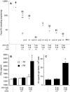
Protection from RV infection in mice immunized with 2/6-VLPs by the i.n and i.r. routes. Mice were immunized three times at 2-week intervals with 5 μg of 2/6-VLPs together with 1 μg of CT or 25 μg of CpG ODN 1826 by the i.n. route or with 5 μg of 2/6-VLPs together with 10 μg of CT or 250 μg of CpG ODN 1826 by the i.r. route. A fifth group of control mice was mock immunized with 5 μg of OVA and 10 μg of CT by the i.r. route. Six weeks after the last immunization, mice were orally challenged with murine RV (strain EDIM), and viral shedding was measured in stool samples at 3 d.p.c. (A), 5 d.p.c. (B), and 8 d.p.c. (C) by quantifying genomic RNA by real-time RT-PCR. Results are given in copies of viral genome per mg of stool. Bars indicate the median RV shedding level for each group of animals (13 to 14 animals/group in two separate experiments), and diamonds indicate the amount of RV shed by each individual animal. The numbers of mice for which RV shedding was undetectable (<2 copies/mg; indicated by arrows) are also reported at the bottom of the graph. **, Significantly lower than levels of the VLP-CT i.n. group (P < 0.01 by the Kruskal-Wallis test). *, Significantly lower than levels of the VLP-CT i.n. group (P < 0.05 by the Kruskal-Wallis test). §§, Significantly lower than levels of the OVA-CT i.r. group (P < 0.01 by the Kruskal-Wallis test).
FIG. 4.
FIG. 4.
Cytokine production by splenocytes of mice immunized with 2/6-VLPs by the i.n and i.r. routes. Mice were immunized three times at 2-week intervals with 5 μg of 2/6-VLPs together with 1 μg of CT or 25 μg of CpG ODN 1826 by the i.n. route or with 5 μg of 2/6-VLPs together with 10 μg of CT or 250 μg of CpG ODN 1826 by the i.r. route. A fifth group of control mice was mock immunized with 5 μg of OVA and 10 μg of CT by the i.r. route. Four weeks after the last immunization, three mice/group were sacrificed and their spleens were collected. Splenocytes obtained from mice of the same group were pooled and cultured in the presence of uninfected (−RV; white columns) or RV-infected (+RV; black columns) MA104 cell supernatant. One week later, IFN-γ (A) and IL-5 (B) levels were measured in culture supernatants by ELISA. Data are means ± SEs of three independent experiments. *, Significantly higher than cytokine levels produced by restimulated splenocytes from mice immunized with 2/6-VLPs and the same adjuvant by the i.r. route (P < 0.05 by the Kruskal-Wallis test).
FIG. 5.
FIG. 5.
Cytokine production by PP cells of mice immunized with 2/6-VLPs by the i.n and i.r. routes. Mice were immunized three times at 2-week intervals with 5 μg of 2/6-VLPs together with 1 μg of CT or 25 μg of CpG ODN 1826 by the i.n. route or with 5 μg of 2/6-VLPs together with 10 μg of CT or 250 μg of CpG ODN 1826 by the i.r. route. A fifth group of control mice was mock immunized with 5 μg of OVA and 10 μg of CT by the i.r. route. Four weeks after the last immunization, three mice/group were sacrificed and their PPs were collected. PP cells obtained from mice of the same group were pooled and cultured in the presence of uninfected (−RV; white columns) or RV-infected (+RV; black columns) MA104 cell supernatant. One week later, IFN-γ levels were measured in culture supernatants by ELISA (A). Six weeks after the last immunization, mice were orally challenged with murine RV (strain EDIM). At 3 d.p.c., animals were sacrificed and IFN-γ production by PP cells was measured as described above (B). Data are means ± SEs of three independent experiments. *, Significantly higher than IFN-γ levels produced by restimulated PP cells of mice immunized with 2/6-VLPs and the same adjuvant by the i.n. route (P < 0.05 by the Kruskal-Wallis test).
FIG. 6.
FIG. 6.
Anti-RV IgA production by PP cells of mice immunized with 2/6-VLPs by the i.n and i.r. routes. Mice were immunized three times at 2-week intervals with 5 μg of 2/6-VLPs together with 1 μg of CT or 25 μg of CpG ODN 1826 by the i.n. route or with 5 μg of 2/6-VLPs together with 10 μg of CT or 250 μg of CpG ODN 1826 by the i.r. route. A fifth group of control mice was mock immunized with 5 μg of OVA and 10 μg of CT by the i.r. route. Four weeks after the last immunization, two mice/group were sacrificed and their PPs were collected. PP cells obtained from mice of the same group were pooled, and the numbers of RV-specific IgA ASCs were determined by ELISPOT assay (A). Six weeks after the last immunization, mice were orally challenged with murine RV (strain EDIM). At 3 d.p.c., animals were sacrificed and the numbers of RV-specific ASCs were determined in PPs as described above (B). Data are means ± SEs of quadruplicate wells (these experiments were repeated twice with similar results). *, Significantly higher than the ASC number found in PPs of mice immunized with 2/6-VLPs and the same adjuvant by the i.n. route (P < 0.05 by the Kruskal-Wallis test).
FIG. 7.
FIG. 7.
Protection from RV infection and intestinal immune responses in mice immunized with 2/6-VLPs and different doses of CT by the i.n and i.r. routes. Mice were immunized three times at 2-week intervals with 5 μg of 2/6-VLPs together with 1, 3, or 5 μg of CT by the i.n. route or with 5 μg of 2/6-VLPs together with 1, 3, 5, or 10 μg of CT by the i.r. route. A fifth group of control mice was left untreated. Four weeks after the last immunization, mice were orally challenged with murine RV (strain EDIM), and viral shedding was determined in stool samples at 3 d.p.c. by quantifying genomic RNA by real-time RT-PCR (A). Results are given in copies of viral genome per mg of stool. Bars indicate the median RV shedding for each group of animals (four to five animals/group), and diamonds indicate the levels of RV shed by each individual animal. The numbers of mice for which RV shedding was undetectable (<2 copies/mg; indicated by arrows) are also reported at the bottom of the graph. At 3 d.p.c., control mice and mice immunized with 2/6-VLPs and 3 μg of CT by the i.r. or i.n. route were sacrificed and PPs removed. IFN-γ production was measured after 1 week of culturing in the presence of uninfected (−RV; white columns) or RV-infected (+RV; black columns) MA104 cell supernatant (B) and the numbers of RV-specific IgA ASCs were determined by ELISPOT assay (C). Data shown in panels B and C are means ± SEs of quadruplicate wells. § and §§, Significantly lower than levels of control mice (P < 0.05 and P < 0.01, respectively, by the Kruskal-Wallis test). *, Significantly higher than ASC numbers or IFN-γ levels produced by PP cells of mice immunized by the i.n. route (P < 0.05 by the Kruskal-Wallis test).

Similar articles

Cited by

References

    1. Abbas, A. K., K. M. Murphy, and A. Sher. 1996. Functional diversity of helper T lymphocytes. Nature 383:787-793. - PubMed
    1. Angel, J., M. A. Franco, H. B. Greenberg, and D. Bass. 1999. Lack of a role for type I and type II interferons in the resolution of rotavirus-induced diarrhea and infection in mice. J. Interferon Cytokine Res. 19:655-659. - PubMed
    1. Banos, D. M., S. Lopez, C. F. Arias, and F. R. Esquivel. 1997. Identification of a T-helper cell epitope on the rotavirus VP6 protein. J. Virol. 71:419-426. - PMC - PubMed
    1. Beagley, K. W., J. H. Eldridge, H. Kiyono, M. P. Everson, W. J. Koopman, T. Honjo, and J. R. McGhee. 1988. Recombinant murine IL-5 induces high rate IgA synthesis in cycling IgA-positive Peyer's patch B cells. J. Immunol. 141:2035-2042. - PubMed
    1. Belyakov, I. M., J. D. Ahlers, B. Y. Brandwein, P. Earl, B. L. Kelsall, B. Moss, W. Strober, and J. A. Berzofsky. 1998. The importance of local mucosal HIV-specific CD8(+) cytotoxic T lymphocytes for resistance to mucosal viral transmission in mice and enhancement of resistance by local administration of IL-12. J. Clin. Investig. 102:2072-2081. - PMC - PubMed

Publication types